. 2013;8(3):e55667.
doi: 10.1371/journal.pone.0055667. Epub 2013 Mar 1.

Tissue inhibitor of metalloproteinases-3 peptides inhibit angiogenesis and choroidal neovascularization in mice

Affiliations

Tissue inhibitor of metalloproteinases-3 peptides inhibit angiogenesis and choroidal neovascularization in mice

Jian Hua Qi et al. PLoS One. 2013.

Abstract

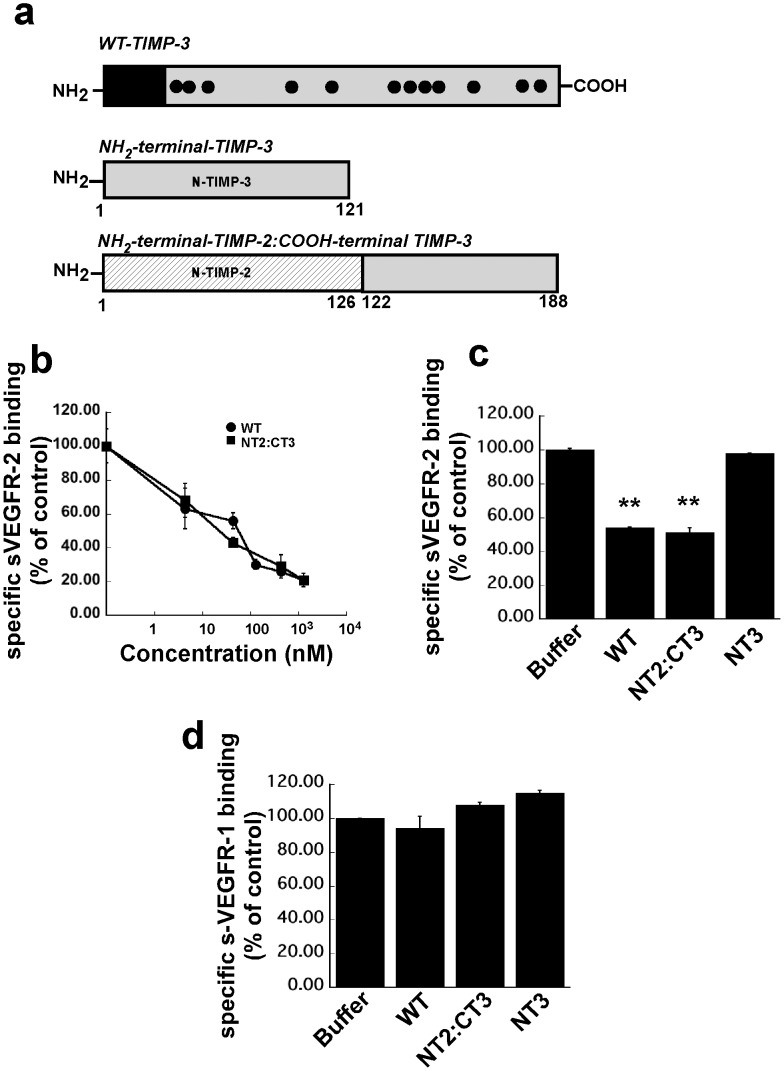
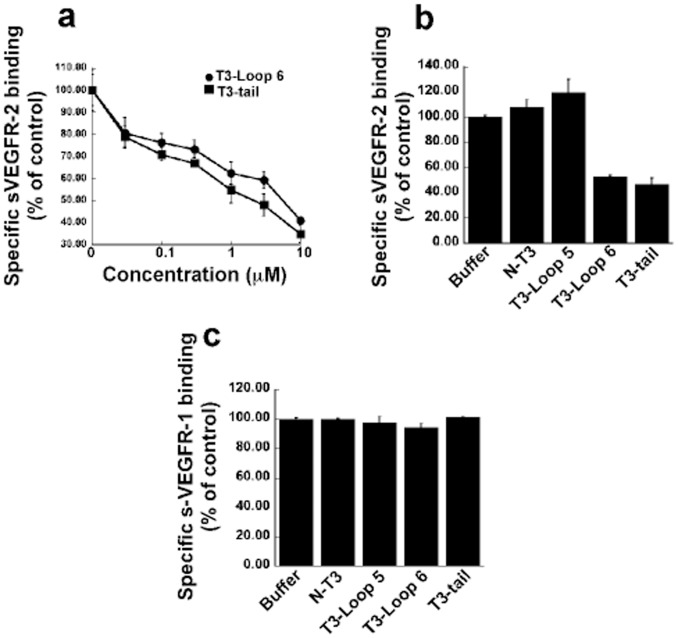
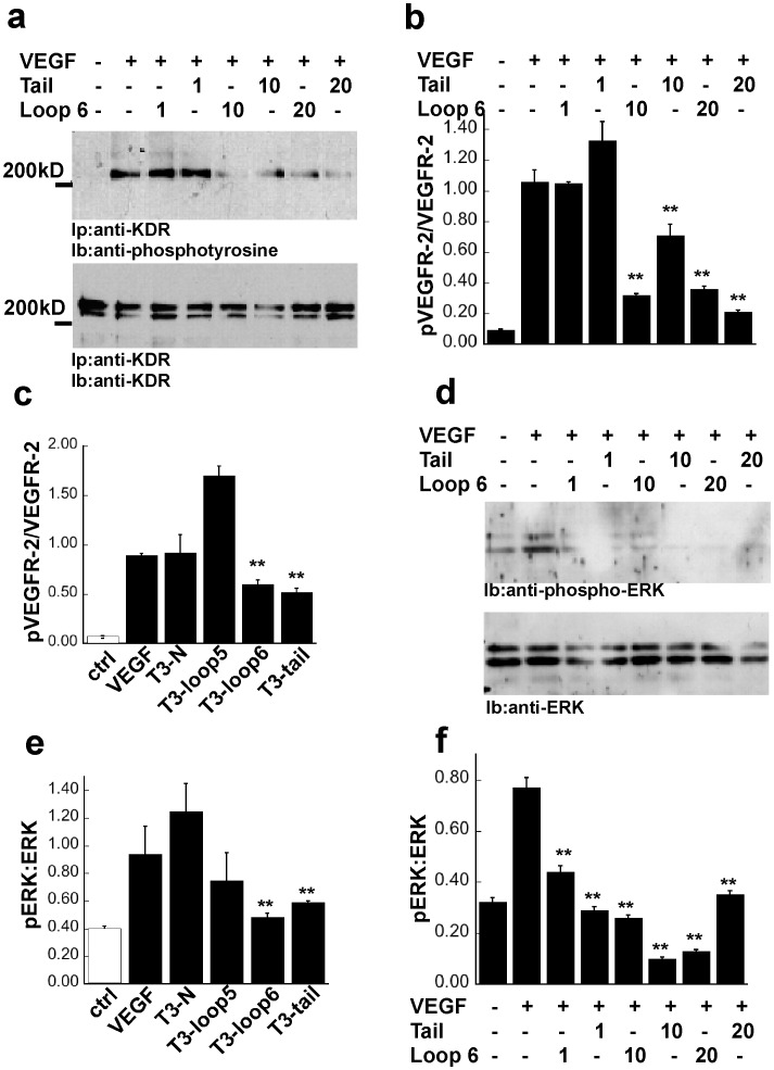
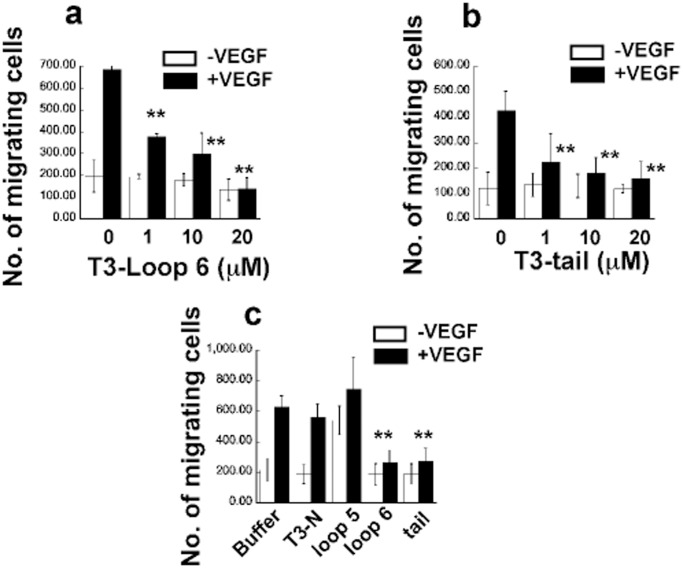
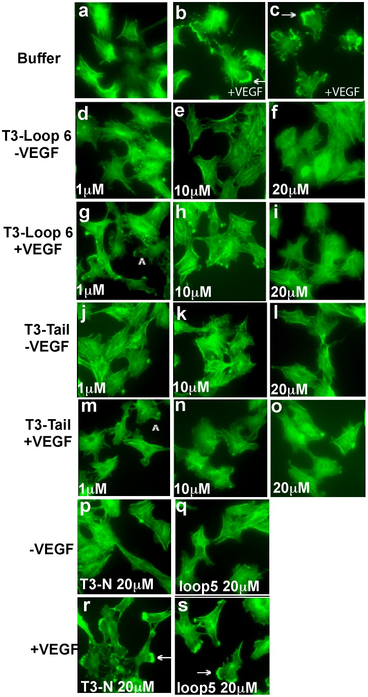
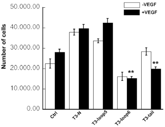
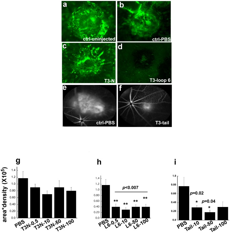
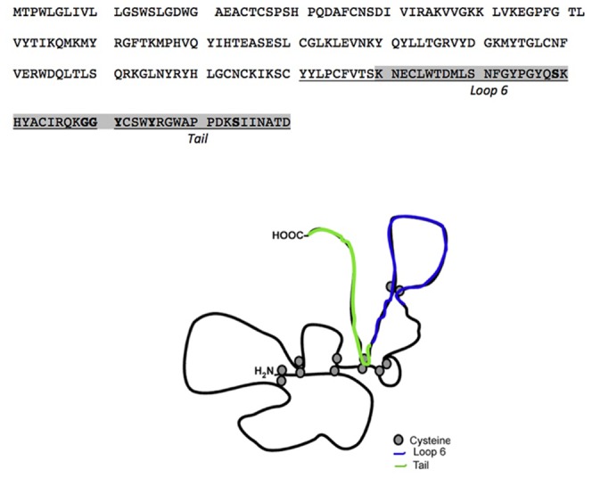
Tissue inhibitors of metalloproteinases (TIMPs) while originally characterized as inhibitors of matrix metalloproteinases (MMPs) have recently been shown to have a wide range of functions that are independent of their MMP inhibitory properties. Tissue inhibitor of metalloproteinases-3 (TIMP-3) is a potent inhibitor of VEGF-mediated angiogenesis and neovascularization through its ability to block the binding of VEGF to its receptor VEGFR-2. To identify and characterize the anti-angiogenic domain of TIMP-3, structure function analyses and synthetic peptide studies were performed using VEGF-mediated receptor binding, signaling, migration and proliferation. In addition, the ability of TIMP-3 peptides to inhibit CNV in a mouse model was evaluated. We demonstrate that the anti-angiogenic property resides in the COOH-terminal domain of TIMP-3 protein which can block the binding of VEGF specifically to its receptor VEGFR-2, but not to VEGFR-1 similar to the full-length wild-type protein. Synthetic peptides corresponding to putative loop 6 and tail region of TIMP-3 have anti-angiogenic properties as determined by inhibition of VEGF binding to VEGFR-2, VEGF-induced phosphorylation of VEGFR-2 and downstream signaling pathways as well as endothelial cell proliferation and migration in response to VEGF. In addition, we show that intravitreal administration of TIMP-3 peptide could inhibit the size of laser-induced choroidal neovascularization lesions in mice. Thus, we have identified TIMP-3 peptides to be efficient inhibitors of angiogenesis and have a potential to be used therapeutically in diseases with increased neovascularization.


Conflict of interest statement

Competing Interests: The authors have read the journal's policy and have the following conflicts. They declare that Bela Anand-Apte has an awarded patent (No US 7,183,256B2) titled TIMP-3 as VEGF inhibitor. The authors confirm that this does not alter their adherence to all the PLOS ONE policies on sharing data and materials, as detailed online in the PLOS ONE guide for authors.

Figures

Figure 1
Figure 1. The effects of recombinant wild type TIMP-3 (WT-TIMP-3), N-terminal TIMP2: C-terminal TIMP-3 chimera (NT2: CT3) and the NH3-terminal domain of TIMP-3 (NT3) on binding of 50 ng/ml soluble(s) VEGFR-1 or VEGFR-2 to immobilized VEGF.
A competitive ELISA assay was used as described under “Materials and Methods”. (a) description of WT-TIMP-3, N-T3 and NT2:CT3 (b). WT-TIMP-3 and NT2: CT3 inhibit sVEGFR-2-VEGF complexes in a concentration-dependent manner. (c). N-T3 has no effect on sVEGFR-2-VEGF complexes. (d). WT-TIMP-3, NT2 and N-T3 are unable to block sVEGFR-1-VEGF complexes.
Figure 2
Figure 2. The effect of synthetic TIMP-3 peptides on binding of 50 ng/ml soluble (s) VEGFR-1 or –2 to immobilized VEGF.
A competitive ELISA assay was used as described under “Materials and Methods” (a). Loop 6 and tail peptides inhibit sVEGFR-2-VEGF complexes in a concentration-dependent manner. (b) Loop5 and N-peptide have no effect on sVEGFR-2-VEGF complexes. (c) Loop 5, loop 6, tail and N-peptide are unable to inhibit sVEGFR-1-VEGF complexes.
Figure 3
Figure 3. Effects of TIMP-3 peptides on VEGF signaling in endothelial cells.
(a) TIMP-3 loop 6 and tail inhibit VEGF-mediated phosphorylation of VEGFR-2 in PAE/KDR cells in a concentration dependent manner. Prior to treatment with indicated concentrations of TIMP-3 peptides for 30 min, the serum-starved cells were stimulated or not with 50 ng/ml VEGF for 10 min in the presence or absence of the same concentration of peptide. Cell lysates were separated by SDS-PAGE (10%) and analyzed by immunoprecipitation with an anti-KDR anti-body followed by immunoblotting with an anti-phosphotyrosine antibody, 4G10 (top panel). KDR protein was analyzed by immunoprecipitation and immunoblotting of cell lysates using anti-KDR antibody (bottom panel). (b) Densitometric quantitation of 210-kDa bands for phospho- or total mature form of KDR. (c) TIMP-3 loop 6 and tail but not loop 5 and N-peptide inhibit VEGF-mediated phosphorylation of KDR in PAE/KDR cells. (d) TIMP-3 loop 6 and tail inhibit ERK phosphorylation. Phosphorylation of ERK1 and ERK2 in response to VEGF was detected by immunoblotting with phosphospecific MAP kinase antibodies (top panel). Total protein levels of ERK was determined by immunoblotting with anti- MAP kinase antibodies (bottom panel). (e) TIMP-3 loop 6 and tail but not loop 5 nor N-peptide inhibit VEGF-mediated phosphorylation of ERK in PAE/KDR cells.(f) Densitometric quantitation of pERK (top panel of d) and ERK bands (bottom panel of d) for phospho- or total ERK proteins. **significantly different from control (without peptides+VEGF), p<0.01 (Student’s t test).
Figure 4
Figure 4. Effects of TIMP-3 peptides on VEGF-mediated EC migration.
TIMP-3 loop 6 and tail but not loop 5 and N-peptide inhibit VEGF-induced migration in PAE/KDR cells. Cells were analyzed for migration towards 50 ng/ml VEGF in the presence or absence of increasing concentrations of loop 6 (a) and tail peptides (b) or 20 µM loop 5 or N-peptide (c), respectively, using a mini-Boyden chamber incubated at 37°C for 4 h. Migrating cell number is expressed as means± SEM of quadruplicate samples. **significantly different from control (without peptides), p<0.01 (Student’s t test).
Figure 5
Figure 5. Effects of TIMP-3 peptides on VEGF-mediated actin reorganization.
TIMP-3 loop 6 and tail but not loop 5 and N-peptide inhibit VEGF-induced actin reorganization in PAE/KDR cells. Quiescent cells were stimulated with (b,c,g,h,i,m,n,o,r,s) or without (a,d,e,f,j,k,l,p,q) 50 ng/ml VEGF in the presence or absence of increasing concentrations of loop 6 (d-i) and tail peptides (j-o) or 20 µM loop 5 (q,s) or N-peptides (p,r) for 30 min at 37°C. Cells were permeabilized and stained with fluorescein isothiocyanate-phalloidin, as described under “Materials and Methods”. The arrows indicate membrane edge ruffling in the stimulated cells. Magnification, ×1000.
Figure 6
Figure 6. Effects of TIMP-3 peptides on VEGF-mediated EC proliferation.
TIMP-3 loop 6 and tail but not loop 5 and N-peptide inhibit VEGF-induced proliferation in PAE/KDR cells. Serum-starved cells were incubated with or without VEGF (5 ng/ml) in the presence or absence of the 1 µM of TIMP-3 peptides for 5 days. Cell numbers were counted using a Coulter particle counter. **significantly different from control (with VEGF), p<0.01 (Student’s t test).
Figure 7
Figure 7. Inhibition of laser-induced CNV by TIMP-3 peptides.
TIMP-3 loop 6 and tail but not N-peptide inhibit laser-induced CNV in a mouse model. Representative images of mouse choroids at 14 days following laser photocoagulation in C57BL6 mice (a) untreated/uninjected (b) injected with PBS control (c) T3N (d) T3-Loop6 (e) injected with PBS contro (f) T3-tail. Effect of Tail peptides on CNV was evaluated by Fluorescein angiogram and Scanning Laser Ophthalmoscopy (SLO). Peptides were injected intravitreally immediately following laser burn. CNV lesion size (area X density) was calculated for each dose of T3N-peptide (Fig. 7g), Loop6-peptide (Fig. 7h) and Tail-peptide (fig. 7i).
Figure 8
Figure 8. Location of Loop 6 and Tail TIMP-3 peptides on TIMP-3 protein.

References

    1. Amour A, Knight CG, Webster A, Slocombe PM, Stephens PE, et al. (2000) The in vitro activity of ADAM-10 is inhibited by TIMP-1 and TIMP-3. FEBS Lett 473: 275–279. - PubMed
    1. Amour A, Slocombe PM, Webster A, Butler M, Knight CG, et al. (1998) TNF-alpha converting enzyme (TACE) is inhibited by TIMP-3. FEBS Lett 435: 39–44. - PubMed
    1. Hashimoto G, Aoki T, Nakamura H, Tanzawa K, Okada Y (2001) Inhibition of ADAMTS4 (aggrecanase-1) by tissue inhibitors of metalloproteinases (TIMP-1, 2, 3 and 4). FEBS Lett 494: 192–195. - PubMed
    1. Jacobsen J, Visse R, Sorensen HP, Enghild JJ, Brew K, et al. (2008) Catalytic properties of ADAM12 and its domain deletion mutants. Biochemistry 47: 537–547. - PubMed
    1. Kashiwagi M, Tortorella M, Nagase H, Brew K (2001) TIMP-3 is a potent inhibitor of aggrecanase 1 (ADAM-TS4) and aggrecanase 2 (ADAM-TS5). J Biol Chem 276: 12501–12504. - PubMed

Publication types

MeSH terms

Substances