. 2013;8(3):e57732.
doi: 10.1371/journal.pone.0057732. Epub 2013 Mar 1.

A new oxidative stress model, 2,2-azobis(2-amidinopropane) dihydrochloride induces cardiovascular damages in chicken embryo

Affiliations

A new oxidative stress model, 2,2-azobis(2-amidinopropane) dihydrochloride induces cardiovascular damages in chicken embryo

Rong-Rong He et al. PLoS One. 2013.

Abstract

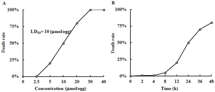
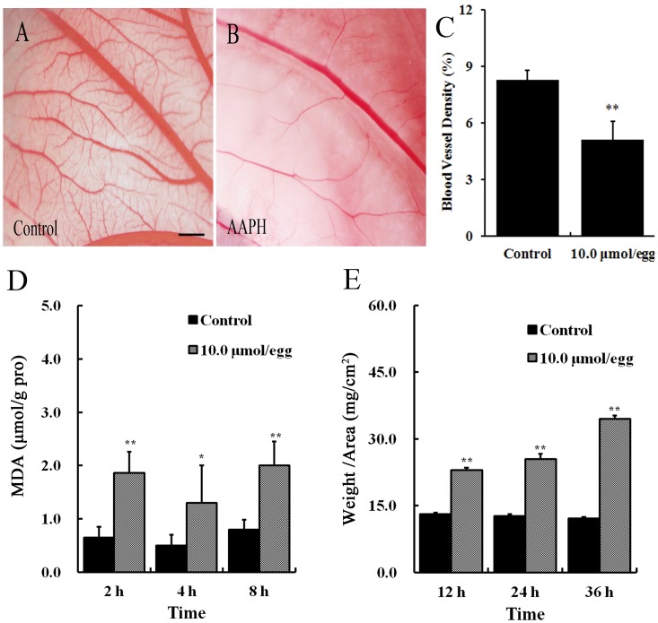
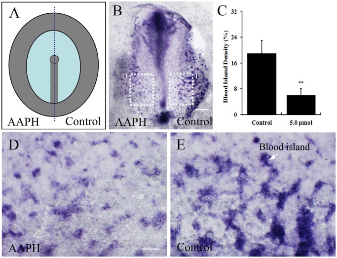
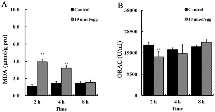
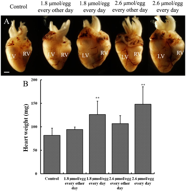
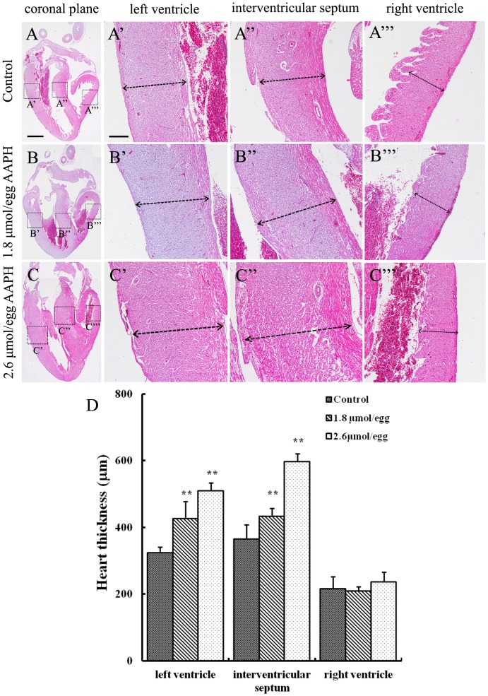
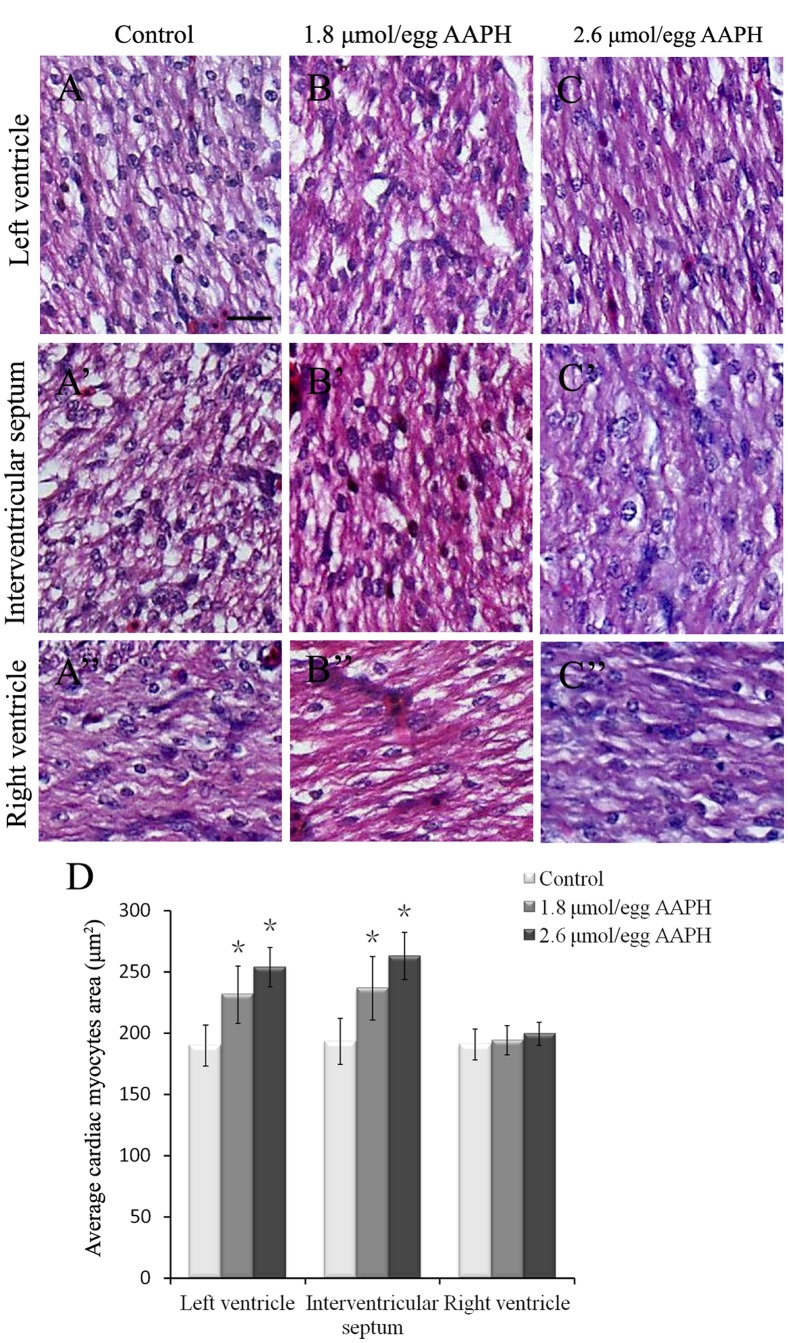
It is now well established that the developing embryo is very sensitive to oxidative stress, which is a contributing factor to pregnancy-related disorders. However, little is known about the effects of reactive oxygen species (ROS) on the embryonic cardiovascular system due to a lack of appropriate ROS control method in the placenta. In this study, a small molecule called 2,2-azobis(2-amidinopropane) dihydrochloride (AAPH), a free radicals generator, was used to study the effects of oxidative stress on the cardiovascular system during chick embryo development. When nine-day-old (stage HH 35) chick embryos were treated with different concentrations of AAPH inside the air chamber, it was established that the LD50 value for AAPH was 10 µmol/egg. At this concentration, AAPH was found to significantly reduce the density of blood vessel plexus that was developed in the chorioallantoic membrane (CAM) of HH 35 chick embryos. Impacts of AAPH on younger embryos were also examined and discovered that it inhibited the development of vascular plexus on yolk sac in HH 18 embryos. AAPH also dramatically repressed the development of blood islands in HH 3+ embryos. These results implied that AAPH-induced oxidative stress could impair the whole developmental processes associated with vasculogenesis and angiogenesis. Furthermore, we observed heart enlargement in the HH 40 embryo following AAPH treatment, where the left ventricle and interventricular septum were found to be thickened in a dose-dependent manner due to myocardiac cell hypertrophy. In conclusion, oxidative stress, induced by AAPH, could lead to damage of the cardiovascular system in the developing chick embryo. The current study also provided a new developmental model, as an alternative for animal and cell models, for testing small molecules and drugs that have anti-oxidative activities.


Conflict of interest statement

Competing Interests: The authors have declared that no competing interests exist.

Figures

Figure 1
Figure 1. Embryo mortality rate in presence of different AAPH concentration.
(A) AAPH (0–40 µmol/egg) were injected into the air chamber of eggs containing 9-day-old (stage HH 35) chick embryos and incubated for 24 hours. (B) Stage HH 35 chick embryos were treated with AAPH (10 µmol/egg) and cultured for 2–48 hours. The experiments were performed in triplicates with 20 eggs assigned for each group. Embryos without heart beat were deemed dead.
Figure 2
Figure 2. Effect of AAPH on angiogenesis in the CAM of chick embryos.
(A) Representative appearance of CAM treated with saline for 24 h. (B) Representative appearance of CAM treated with 10 µmol/egg of AAPH for 24 h. (C) Statistical chart showing the blood vessel density of CAM from AAPH treated and untreated embryos. (D) The CAMs of embryos were treated with 10 µmol/egg of AAPH for 2–8 hours and then the MDA content of embryos were measured. (E) The thickness of CAM after 12–36 h exposure to AAPH. CAM thickness was calculated as the ratio of weight to area of CAM (mg/cm2). The results are presented as mean ± S.D (n = 10). Statistical significances were evaluated using SPSS13.5 software, presented as *p<0.05, **p<0.01 in comparison with control group. Scale bar = 1 mm.
Figure 3
Figure 3. Effects of AAPH on yolk-sac blood vessels of chick embryo.
(A) Representative appearance of yolk-sac blood vessels of HH 18 embryo treated with saline, (B) 4 µmol AAPH and (C) 5 µmol AAPH for 12 hours. The silastic rings in A–C had inside diameter at 9 mm and outside diameter at 11 mm. The yolk-sac blood vessel images were taken by a stereomicroscope (Olympus MVX10 with OPTPRO 2007 image acquisition system) with resolution ratio at 1024×768 (Scale bar = 1 mm). (D) Statistical chart showing the yolk-sac blood vessel density from AAPH treated and untreated embryos. The results represent the mean ± S.D (n = 10). Statistical significances were determined using SPSS13.5 software, **p<0.01 compared with control group.
Figure 4
Figure 4. Effect of AAPH on blood islands formation in early chick embryos.
(A) Schematic illustration of primitive streak stage chick embryos treated with AAPH (left side of embryo) and saline (right side of embryo). (B) In situ hybridization of whole-mount chick embryo revealing VE-Cadherin expression and extent of blood island formation. (C) Statistical chart showing the blood islands density of AAPH (5.0 µmol) treated and untreated sides of embryos. Results presented as mean ± S.D (n = 10). Data analyzed using SPSS13.5 software, **p<0.01 compared with control. (D) Magnified appearance of blood islands (white arrow) of left side of embryos treated with AAPH and (E) right side with saline. Scale bars = 1 mm in B and 100 µm in D–E.
Figure 5
Figure 5. AAPH induces oxidative stress in the heart of chick embryo.
(A) Statistical charts of MDA and (B) ORAC level in the heart of embryos treated with AAPH (10 µmol/egg) and saline. Results represent mean ± S.D (n = 10). Analyzed by SPSS13.5 software, **p<0.01 compared with control.
Figure 6
Figure 6. Effect of AAPH exposure on heart size and weight of chick embryos.
(A) Representative appearance of hearts harvested from 14-day-old (HH 40) embryos treated with saline, 1.8 and 2.6 µmol of AAPH. (B) Statistical chart showing the embryonic heart weight after AAPH exposure. Results presented as mean ± S.D. (n = 10). Analyzed by SPSS13.5 software, **p<0.01 compared with control. Abbreviation: LV, left ventricle; RV, right ventricle. Scale bar = 1 mm.
Figure 7
Figure 7. Effect of AAPH exposure on wall thickness of chick embryonic heart.
(A–C) Haematoxylin- and eosin-stained histological vertical sections of embryonic hearts treated with (A) saline, (B) 1.8 µmol AAPH and (C) 2.6 µmol AAPH over 5 days. All histologic sections are presented with the atria on top and the left ventricle on the left. High magnification of (A’, B’ and C’) left ventricle, (A’’, B’’ and C’’) interventricular septum, and (A’’’, B’’’ and C’’’) right ventricle. (D) Statistical chart showing the thickness of the left and right ventricular walls and thickness of the interventricular septum. Results presented as mean ± S.D (n = 10). Data analyzed using SPSS13.5 software, **p<0.01 compared with control. Scale bars = 1mm in A–C and 500 µm in A’–A’’’, B’-B’’’ and C’–C’’’.
Figure 8
Figure 8. Effect of AAPH on cardiac myocytes size of chick embryo.
(A–C) H&E-staining of heart sections obtained from left ventricle of HH 40 stage chick embryo of (A) control, (B) 1.8 µmol/egg AAPH and (C) 2.6 µmol/egg AAPH group. (A’–C’) H&E-staining of interventricular septum from HH 40 stage chick embryo of (A’) control, (B’) 1.8 µmol/egg AAPH and (C’) 2.6 µmol/egg AAPH group. (A’’–C’’) H&E-staining of right ventricle from HH 40 stage chick embryo of (A’’) control, (B’’) 1.8 µmol/egg AAPH and (C’’) 2.6 µmol/egg AAPH group. (D) Statistical data of average cardiac myocytes surface area. Results presented as mean ± S.D (n = 10) calculated by SPSS13.5 software, *p<0.05. Scale bar = 20 µm in A–C, A’–C’, A’’–C’’.

References

    1. Valko M, Leibfritz D, Moncol J, Cronin MTD, Mazur M, et al. (2007) Free radicals and antioxidants in normal physiological functions and human disease. Int J Biochem Cell Biol 39: 44–84. - PubMed
    1. Abramov JP, Wells PG (2012) Embryoprotective role of endogenous catalase in acatalasemic and human catalase-expressing mouse embryos exposed in culture to developmental and phenytoin-enhanced oxidative stress. Toxicol Sci 120: 428–438. - PubMed
    1. Wan J, Winn LM (2006) In utero–initiated cancer: The role of reactive oxygen species. Birth Defects Res C Embryo Today 78: 326–332. - PubMed
    1. Dennery PA (2007) Effects of oxidative stress on embryonic development. Birth Defects Res C Embryo Today 81: 155–162. - PubMed
    1. Covarrubias L, Hernández-García D, Schnabel D, Salas-Vidal E, Castro-Obregón S (2008) Function of reactive oxygen species during animal development: Passive or active? Dev Biol 320: 1–11. - PubMed

Publication types

MeSH terms

LinkOut - more resources