. 2013;8(3):e57838.
doi: 10.1371/journal.pone.0057838. Epub 2013 Mar 1.

Chimeric antigen receptor (CAR)-specific monoclonal antibody to detect CD19-specific T cells in clinical trials

Affiliations

Chimeric antigen receptor (CAR)-specific monoclonal antibody to detect CD19-specific T cells in clinical trials

Bipulendu Jena et al. PLoS One. 2013.

Abstract

Clinical trials targeting CD19 on B-cell malignancies are underway with encouraging anti-tumor responses. Most infuse T cells genetically modified to express a chimeric antigen receptor (CAR) with specificity derived from the scFv region of a CD19-specific mouse monoclonal antibody (mAb, clone FMC63). We describe a novel anti-idiotype monoclonal antibody (mAb) to detect CD19-specific CAR(+) T cells before and after their adoptive transfer. This mouse mAb was generated by immunizing with a cellular vaccine expressing the antigen-recognition domain of FMC63. The specificity of the mAb (clone no. 136.20.1) was confined to the scFv region of the CAR as validated by inhibiting CAR-dependent lysis of CD19(+) tumor targets. This clone can be used to detect CD19-specific CAR(+) T cells in peripheral blood mononuclear cells at a sensitivity of 1∶1,000. In clinical settings the mAb is used to inform on the immunophenotype and persistence of administered CD19-specific T cells. Thus, our CD19-specific CAR mAb (clone no. 136.20.1) will be useful to investigators implementing CD19-specific CAR(+) T cells to treat B-lineage malignancies. The methodology described to develop a CAR-specific anti-idiotypic mAb could be extended to other gene therapy trials targeting different tumor associated antigens in the context of CAR-based adoptive T-cell therapy.


Conflict of interest statement

Competing Interests: Dr. Laurence J.N. Cooper is the founder of InCellerate, Inc. However, this does not alter the authors’ adherence to all the PLOS ONE policies on sharing data and materials.

Figures

Figure 1
Figure 1. Schematic of DNA plasmids used for electroporation and expression of transgenes.
(A) Constructs showing components of CD19-specific CAR (CD19RCD28). The scFv is derived from mAb clone FMC63 that binds human CD19 and was generated by fusing the VL and VH regions via a “Whitlow” linker peptide. The scFv was attached to modified human IgG4 hinge and CH2-CH3 regions that was fused to the CD28 (transmembrane and cytoplasmic) and CD3ζ (cytoplasmic) domains (B) DNA plasmid design CD19scFv-mCD8α used to express the transgene on L cells for immunization and screening. The CD19-specific scFv was generated as described above, mCD8α EC and TM represents mouse CD8α extracellular and transmembrane domains respectively. (C) Map of destination vector pIRESneo2 (Clontech) where the cDNA for the fusion protein CD19scFv-mCD8α is cloned into NheI and NotI restriction enzyme sites. Expression cassette shows ColE1, colicin E1 (origin of replication); pCMV IE, human cytomegalovirus promoter/enhancer; ECMV IRES, encephalomyocarditis virus internal ribosome entry site which permits translation of two open reading frames from one messenger RNA, and poly A (polyadenylation) signal of the bovine growth hormone. Plasmid confers resistance through ampicillin and after electroporation the cells expressing the transgene of interest are selected on cytocidal concentration of drug neomycin sulfate (G418).
Figure 2
Figure 2. Specificity of anti-CD19scFv mAb.
(A) Solid phase ELISA shows specificity of mAb (clone no. 136.20.1) as it binds to parental monoclonal antibody (FMC63) with background binding to other antibodies (e.g., CD20-specific mAb or a different CD19-specific mAb). Purified human IgG served as a negative control. (B) Western blot shows clone 136.20.1 detects of CAR protein in T cells genetically modified to express CD19RCD28. Lane A: Unmodified control T cells show endogenous CD3ζ (14 kDa) as detected by commercial CD3ζ-specific antibody and absence of CAR. Lane B: CAR+ T cells show CAR-specific band at 75 kDa as detected by mAb (clone 136.20.1). Lane C: CAR+ T cells show absence of CAR-specific band when the blot is treated with primary antibody after blocking (clone 136.20.1 was blocked with molar excess (1∶5) of parental antibody FMC63). Lane D: CAR+ T cells show the presence of CAR (∼75 kDa) and endogenous CD3ζ (14 KDa) as detected by commercial CD3ζ-specific antibody.
Figure 3
Figure 3. Detection of CD19-specific CAR on the surface of genetically modified cells.
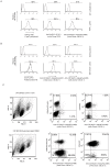
(A) CAR+ T cells were electroporated with Sleeping Beauty system and propagated on aAPC. Upper row: Unmodified T cells as back ground control; Bottom row: CD19RCD28+ T cells (labeled as CAR+ T cells) detected by flow cytometry using clone no. 136.20.1, a commercially-available antibody (clone H10104, Invitrogen) that binds to the CAR hinge/Fc scaffold, and our Fc scaffold-specific mAb (clone 2D3). (B) Jurkat cells were genetically modified to express CD19RCD28 was detected by clone no. 136.20.1 conjugated to Alexa-Fluor 488 and Alexa-Fluor 647 similar to commercial antibody (clone H10104). (C) Multi-parameter flow cytometry analysis of T cells genetically modified to express CD19RCD28. Sequential staining was performed using anti-CD19scFv mAb and/or anti-CD4, anti-CD8 mAb in combination.
Figure 4
Figure 4. Sensitivity of clone no. 136.20.1 to detect CD19-specific CAR+ T cells in PBMC.
CD19-specific T cells expressing CD19RCD28 were serially diluted in PBMC (1∶10 to 1∶10,000). (A) The upper panel shows background level signal in PBMC control. (B) Middle panel shows staining of CAR+ T cells in undiluted sample. (C) Bottom panel shows lowest detection level (1∶1,000) of CAR+ T cells in PBMC. Data acquisition by FACSCaliber and plots were analyzed using FlowJo software. Shown in picture are representative flow cytometry data obtained from two independent experiments.
Figure 5
Figure 5. Localization of CD19RCD28 CAR on the surface of genetically modified T cells.
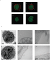
(A) Genetically modified CAR+ T cells (expressing CD19RCD28) and unmodified control T cells were fixed using paraformaldehyde, stained with AlexaFluor 647-conjugated mAb clone no. 136.20.1 and then spread on glass slides using cytospin. Confocal images were acquisitioned by Leica microscope (60X magnification). Upper panels (i and ii) showed surface distribution of CAR molecules and bottom panels (iii and iv) showed no staining in unmodified control T cells. Nuclear staining was by DAPI (pseudo-color green). The bar (1 µm) on images indicates scale. (B) TEM images showing staining of gold-labeled nanoparticles conjugated to mAb clone no. 136.20.1. Upper row: Unmodified control T cells. (i) 120 nm thin section; 15 k magnification stained with uranyl acetate. (ii) 120 nm thin section; 75 k magnification, and (iii) 120 nm thin section; 200 k magnification. No gold particles were appreciated in these CARneg T cells. Bottom row: CAR+ T cells with enforced expression of CD19RCD28. (iv) 120 nm thin section stained with uranyl acetate; 20 k magnification. (v) 120 nm thin section; 200 k magnification, and (vi) 80 nm thin section 200K magnification. Arrow heads on plates v and vi indicate surface distribution of gold-labeled nanoparticles attached to CAR molecules. The bar on images indicates scale.
Figure 6
Figure 6. Clone no. 136.20.1 inhibits CAR+ T-cell effector function.
(A) Data from CRA shows inhibition of tumor cell lysis in CD19+ EL4 cells when CD19-specific CAR+T cells were blocked with clone no. 136.20.1 (B) Dose response curve showing clone no. 136.20.1-mediated inhibition of lysis of CD19+ EL4 target cells. (C) Images obtained from VTLM showing CD19-specific CAR+ T cells co-cultured with CD19+ Daudiβ2m expressing EGFP and stained with either clone no. 136.20.1 conjugated to AlexaFluor-647 or PE-conjugated anti-Fc mAb that recognizes CAR scaffold. Images show two separate focal planes (at 2 and 90 minutes respectively). Upper panels (i-iii) reveal coculture of Daudiβ2m target cells with CAR+ T cells in the presence of Fc-specific antibody; Lower panels (iv-vi) reveal coculture of Daudiβ2m target cells with CAR+ T cells in the presence of clone no. 136.20.1. Panels (i-iv) show phase contrast images of CAR+ T cells along with Daudiβ2m. Panels (ii-v) show formation of immunological synapse (yellow arrow heads) when CD19-specfiic CAR+ T cells attack tumor cells. Panel (iii) shows killing of CD19+ tumor cell by CD19-specific CAR+ T cell as observed by formation of green fluorescence blub. Panel (vi) shows intact Daudiβ2m cell when the CAR+ T cells were blocked by clone no. 136.20.1 and no killing was observed even when effector T cells engaged the tumor cells for more than 6 hours. Shown in figure are data from 3 independent experiments. Movies are available online as supplementary files (Movie S1 and Movie S2).

References

    1. Till BG, Jensen MC, Wang J, Chen EY, Wood BL, et al. (2008) Adoptive immunotherapy for indolent non-Hodgkin lymphoma and mantle cell lymphoma using genetically modified autologous CD20-specific T cells. Blood 112: 2261–2271. - PMC - PubMed
    1. Pule MA, Savoldo B, Myers GD, Rossig C, Russell HV, et al. (2008) Virus-specific T cells engineered to coexpress tumor-specific receptors: persistence and antitumor activity in individuals with neuroblastoma. Nat Med 14: 1264–1270. - PMC - PubMed
    1. Morgan RA, Dudley ME, Rosenberg SA (2010) Adoptive cell therapy: genetic modification to redirect effector cell specificity. Cancer J 16: 336–341. - PMC - PubMed
    1. Kochenderfer JN, Wilson WH, Janik JE, Dudley ME, Stetler-Stevenson M, et al. (2010) Eradication of B-lineage cells and regression of lymphoma in a patient treated with autologous T cells genetically engineered to recognize CD19. Blood 116: 4099–4102. - PMC - PubMed
    1. Kalos M, Levine BL, Porter DL, Katz S, Grupp SA, et al. (2011) T cells with chimeric antigen receptors have potent antitumor effects and can establish memory in patients with advanced leukemia. Sci Transl Med 3: 95ra73. - PMC - PubMed

Publication types

MeSH terms