. 2013;7(2):e2087.
doi: 10.1371/journal.pntd.0002087. Epub 2013 Feb 28.

Proteomic selection of immunodiagnostic antigens for human African trypanosomiasis and generation of a prototype lateral flow immunodiagnostic device

Affiliations

Proteomic selection of immunodiagnostic antigens for human African trypanosomiasis and generation of a prototype lateral flow immunodiagnostic device

Lauren Sullivan et al. PLoS Negl Trop Dis. 2013.

Abstract

Background: The diagnosis of Human African Trypanosomiasis relies mainly on the Card Agglutination Test for Trypanosomiasis (CATT). While this test is successful, it is acknowledged that there may be room for improvement. Our aim was to develop a prototype lateral flow test based on the detection of antibodies to trypanosome antigens.

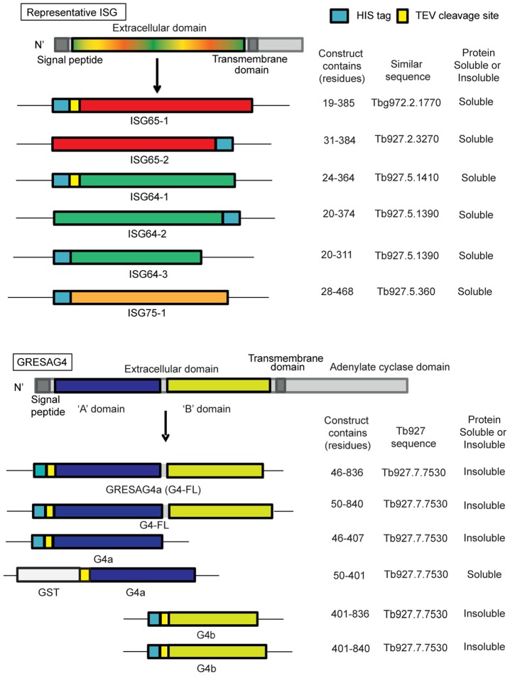
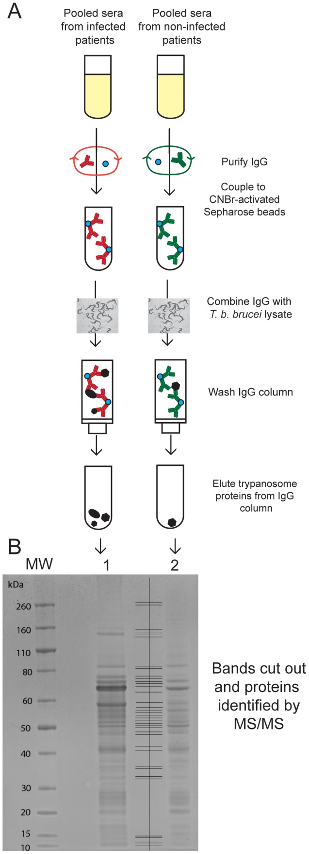
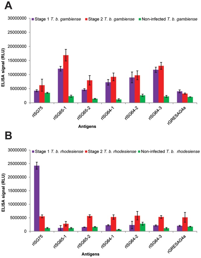
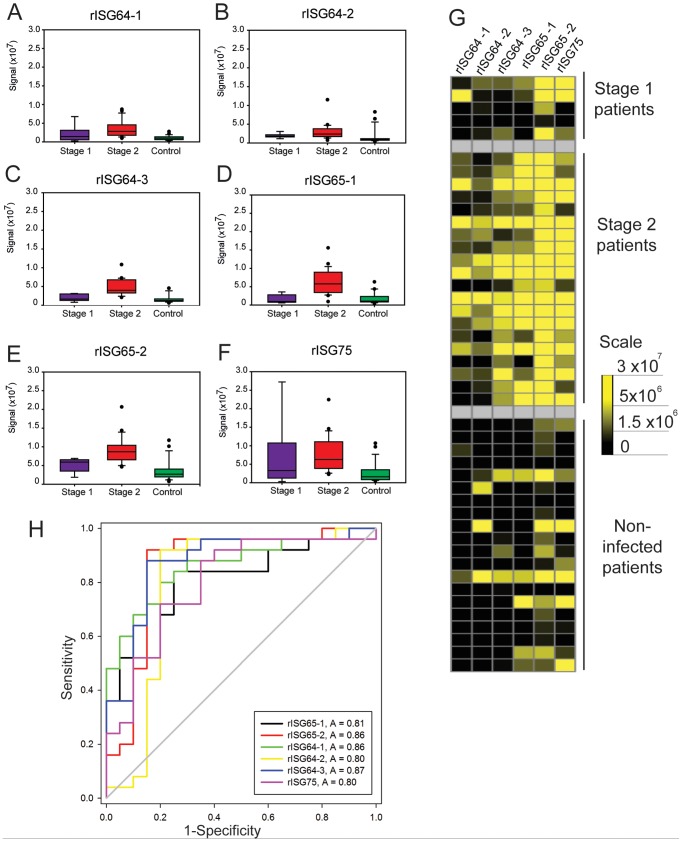
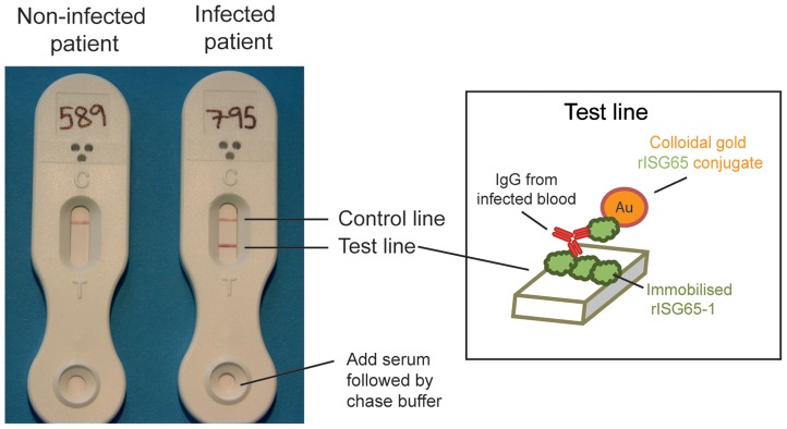
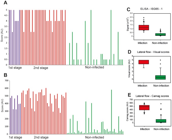
Methodology/principal findings: We took a non-biased approach to identify potential immunodiagnostic parasite protein antigens. The IgG fractions from the sera from Trypanosoma brucei gambiense infected and control patients were isolated using protein-G affinity chromatography and then immobilized on Sepharose beads. The IgG-beads were incubated with detergent lysates of trypanosomes and those proteins that bound were identified by mass spectrometry-based proteomic methods. This approach provided a list of twenty-four trypanosome proteins that selectively bound to the infection IgG fraction and that might, therefore, be considered as immunodiagnostic antigens. We selected four antigens from this list (ISG64, ISG65, ISG75 and GRESAG4) and performed protein expression trials in E. coli with twelve constructs. Seven soluble recombinant protein products (three for ISG64, two for ISG65 and one each for ISG75 and GRESAG4) were obtained and assessed for their immunodiagnostic potential by ELISA using individual and/or pooled patient sera. The ISG65 and ISG64 construct ELISAs performed well with respect to detecting T. b. gambiense infections, though less well for detecting T. b. rhodesiense infections, and the best performing ISG65 construct was used to develop a prototype lateral flow diagnostic device.

Conclusions/significance: Using a panel of eighty randomized T. b. gambiense infection and control sera, the prototype showed reasonable sensitivity (88%) and specificity (93%) using visual readout in detecting T. b. gambiense infections. These results provide encouragement to further develop and optimize the lateral flow device for clinical use.


Conflict of interest statement

The authors have declared that no competing interests exist.

Figures

Figure 1
Figure 1. Recombinant protein antigens used in this study.
A generic representation of the ISGs is shown at the top and a representation of GRESAG4 is shown at the bottom. All have cleavable N-terminal signal peptides and internal transmembrane domains, typical of type-1 membrane proteins. The constructs prepared and expressed and the soluble proteins successfully purified, are indicated.
Figure 2
Figure 2. Immuno-affinity chromatography and identification of potential diagnostic antigens.
(A) Schematic representation of the preparation of IgG-Sepharose from T. b. gambiense infection and non-infection (control) sera, the immune-affinity capture of trypanosome antigens from a whole detergent lysate and their subsequent elution and concentration by ethanol precipitation. (B) Colloidal Comassie blue stained SDS-PAGE gel of the proteins eluted from infection IgG-Sepharose (lane 1) and non-infection (control) IgG-Sepharose (lane 2). The gel lanes were excised in 18 slices per lane, as indicated between lanes 1 and 2, and analysed by LC-MS/MS after reduction, S-alkylation and tryptic digestion. The positions of molecular weight markers are indicated on the left.
Figure 3
Figure 3. ELISA results with pooled human sera.
(A) Pooled human sera representing stage 1 T. b. gambiense infections (pool of 10 sera), stage 2 T. b. gambiense infections (pool of 40 sera) and matched uninfected controls (pool of 50 sera) were diluted 1∶1000 and used in triplicate on ELISA plates coated with the rISG75, rISG65-1, rISG65-2, rISG64-1, rISG64-2, rISG64-3 and rGRESAG4a recombinant proteins described in (Figure 2). The mean ELISA signals ± SEM are plotted against the recombinant protein used in the ELISA. (B) As panel A but using pooled human sera representing stage 1 T. b. rhodesiense infections (pool of 5 sera), stage 2 T. b. rhodesiense infections (pool of 20 sera) and matched uninfected controls (pool of 25 sera).
Figure 4
Figure 4. ELISA results using individual T. b. gambiense infection and matched control sera.
(A–F) Box plots (generated by Cleveland method) represent the 25th percentile to the 75th percentile boundaries in the box with the median line within the box, the whiskers indicate the 10th and 90th percentiles. The box plots show ELISA signals for each recombinant protein ELISA plate: (A) rISG64-1, (B) rISG64-2, (C) rISG64-3, (D) rISG65-1, (E) rISG65-2 and (F) rISG75) tested against individual sera diluted 1∶1000 from stage 1 T. b. gambiense infections (n = 10), stage 2 T. b. gambiense infections (n = 40) and matched uninfected controls (n = 50). (G) Heat maps of the same data for the individual sera versus the recombinant protein ELISA plates. (H) Receiver operating characteristics (ROC) plots of the same data. The output statistics for sensitivity and specificity are shown in (Table 2).
Figure 5
Figure 5. ELISA results using individual T. b. rhodesiense infection and matched control sera.
(A–F) Box plots (generated by Cleveland method) represent the 25th percentile to the 75th percentile boundaries in the box with the median line within the box, the whiskers indicate the 10th and 90th percentiles. The box plots represent the ELISA signals for each recombinant protein ELISA plate: (A) rISG64-1, (B) rISG64-2, (C) rISG64-3, (D) rISG65-1, (E) rISG65-2 and (F) rISG75) tested against individual sera diluted 1∶1000 from stage 1 T. b. rhodesiense infections (n = 5), stage 2 T. b. rhodesiense infections (n = 20) and matched uninfected controls (n = 20). (G) Heat maps of the same data for the individual sera versus the recombinant protein ELISA plates. (H) Receiver operating characteristics (ROC) plots of the same data. The output statistics for sensitivity and specificity are shown in (Table 2).
Figure 6
Figure 6. Prototype lateral flow device for detecting antibodies to rISG65-1 protein.
Representative results using serum samples from a matched uninfected patient (left) and a stage 2 T. b. gambiense infected patient (right). The visual scores for these test lines were 0 and 5, respectively, and the CAMAG densitometry measurements were 24.2 and 597.4, respectively. The inset shows the principle of detection, with patient antibody to ISG65 forming a bridge between rISG56-1 immobilised on the nitrocellulose strip and the colloidal gold-coupled rISG65-1 picked up from the sample pad.
Figure 7
Figure 7. Performance of the prototype lateral flow device in a blinded study with eighty randomised serum samples.
(A) Visual scores of test line density from rISG65-1 prototype lateral flow devices (scored in increments of 1 from 0 to 5, with very faint test line shadows represented as 0.5) are plotted against the subsequently decoded patient status (stage 1 T. b. gambiense infections (n = 8), stage 2 T. b. gambiense infections (n = 32) and matched uninfected controls (n = 40). (B) The same test strips were removed from the devices and scanned by CAMAG densitometer. The data are plotted directly below the results for the visual scores for the same samples. The R2 of a scatter plot was 0.96, showing very good correlation between visual score and CAMAG reading. (C–E) Box plots of the results for the same serum samples analysed by (C) rISG65-1 ELISA, (D) rISG65-1 lateral flow prototype with visual scoring and (E) rISG65-1 lateral flow prototype with CAMAG scanner scoring.

References

    1. Balmer O, Beadell JS, Gibson W, Caccone A (2011) Phylogeography and taxonomy of Trypanosoma brucei . PLoS Neglected Tropical Diseases 5 (2) e961. - PMC - PubMed
    1. Malvy D, Chappuis F (2011) Sleeping sickness. Clinical Microbiology and Infection 17: 986–995. - PubMed
    1. Jackson AP, Sanders M, Berry A, McQuillan J, Aslett MA, et al. (2010) The genome sequence of Trypanosoma brucei gambiense, causative agent of chronic human African Trypanosomiasis. PLoS Neglected Tropical Diseases 4 (4) e658. - PMC - PubMed
    1. Simarro PP, Diarra A, Ruiz Postigo JA, Franco JF, Jannin JG (2011) The Human African Trypnaosomiasis Control and Surveillance Program of the World Health Organization 2000–2009: The Way Forward. PLoS Neglected Tropical Diseases 5 (2) e1007. - PMC - PubMed
    1. Simarro PP, Cecchi G, Paone M, Franco JR, Diarra A, et al. (2010) The Atlas of human African trypanosomiasis: A contribution to global mapping of neglected tropical diseases. International Journal of Health Geographics 9: e57. - PMC - PubMed

Publication types

LinkOut - more resources