. 2013 Jun;125(5):756-65.
doi: 10.1111/jnc.12231. Epub 2013 Apr 2.

The role of MAC1 in diesel exhaust particle-induced microglial activation and loss of dopaminergic neuron function

Affiliations

The role of MAC1 in diesel exhaust particle-induced microglial activation and loss of dopaminergic neuron function

Shannon Levesque et al. J Neurochem. 2013 Jun.

Abstract

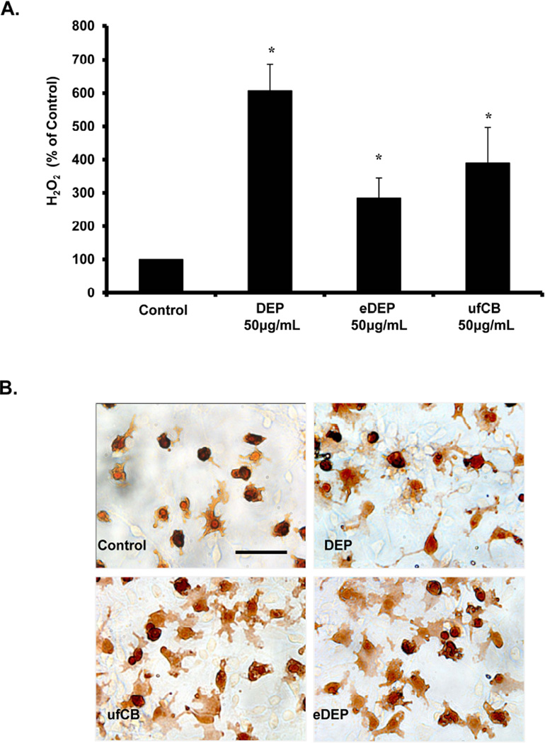
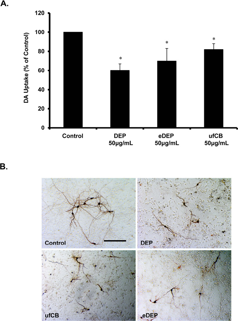
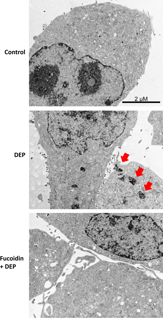
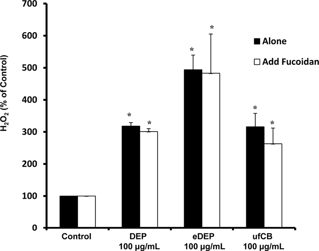
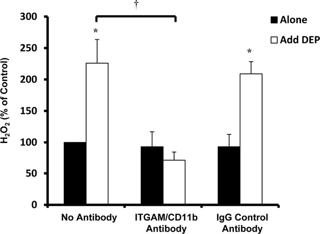
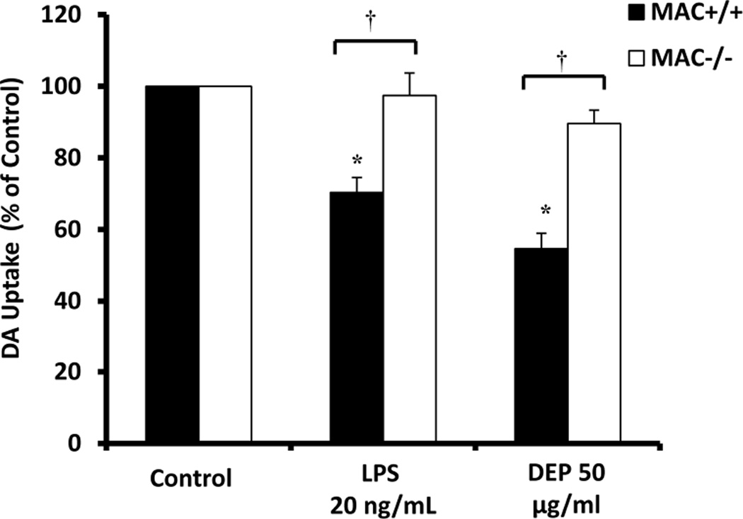
Increasing reports support that air pollution causes neuroinflammation and is linked to central nervous system (CNS) disease/damage. Diesel exhaust particles (DEP) are a major component of urban air pollution, which has been linked to microglial activation and Parkinson's disease-like pathology. To begin to address how DEP may exert CNS effects, microglia and neuron-glia cultures were treated with either nanometer-sized DEP (< 0.22 μM; 50 μg/mL), ultrafine carbon black (ufCB, 50 μg/mL), or DEP extracts (eDEP; from 50 μg/mL DEP), and the effect of microglial activation and dopaminergic (DA) neuron function was assessed. All three treatments showed enhanced ameboid microglia morphology, increased H2 O2 production, and decreased DA uptake. Mechanistic inquiry revealed that the scavenger receptor inhibitor fucoidan blocked DEP internalization in microglia, but failed to alter DEP-induced H2 O2 production in microglia. However, pre-treatment with the MAC1/CD11b inhibitor antibody blocked microglial H2 O2 production in response to DEP. MAC1(-/-) mesencephalic neuron-glia cultures were protected from DEP-induced loss of DA neuron function, as measured by DA uptake. These findings support that DEP may activate microglia through multiple mechanisms, where scavenger receptors regulate internalization of DEP and the MAC1 receptor is mandatory for both DEP-induced microglial H2 O2 production and loss of DA neuron function.


Conflict of interest statement

The authors report no conflicts of interest.

Figures

Figure 1
Figure 1. Components of diesel exhaust particles (DEP) activate microglia
(A) HAPI microglia cells were treated with DEP (50µg/ml), DEP extract (eDEP, from 50µg/ml DEP), and carbon black (ufCB, 50 µg/ml). The production of hydrogen peroxide (H2O2) was measured by the catalase-inhibitable fluorescence. Samples were run in triplicates and the data are the result of 4 independent experiments (n=4). Results are expressed as percent of control and represent the mean ± SEM. The raw data (fluorescence) for the control treatment range from 580 – 906 across experimental replicates. An asterisks indicates a significant difference from control (1 Way ANOVA, p<0.05). (B) Primary neuron-glia cultures were treated with DEP (50µg/ml), eDEP (from 50µg/ml DEP), and ufCB (50 µg/ml) for 9 hr and stained with the IBA-1 antibody. Microglial activation in response to the DEP components is depicted by an increase in number of stained cells, enlarged size of stained cells, and irregular amoeboid morphology. Representative images from the culture are shown from three independent experiments (n=3). Images were taken at 400× and the scale bar depicts 20µM.
Figure 2
Figure 2. Components of diesel exhaust particles (DEP) cause a loss of dopaminergic neuron function
Primary mesencephalic neuron-glia cultures were treated with DEP (50µg/ml), DEP extract (eDEP, from 50µg/ml DEP), and carbon black (ufCB, 50 µg/ml). (A) Dopaminergic (DA) neuron function was measured in neuron-glia cultures 7–9 d after treatment with the [3H] DA uptake assay. Samples were run in triplicates and the data are the result of 5 independent experiments (n=5). Results are expressed as percent of control and are the mean ± SEM. The raw data (counts per second) for control values range from 8686– 5129 across experimental replicates. An asterisks indicates a significant difference from control (1 Way ANOVA, p<0.05). (B) Cultures were treated for 7–9 days and stained with the TH antibody. Dopaminergic neuron damage in response to the DEP components is depicted by shorter processes and fewer processes connecting the neurons. Representative images from the culture are shown from three independent experiments (n=3). Images were taken at 100× and the scale bar depicts 100µM.
Figure 3
Figure 3. Scavenger receptors mediate diesel exhaust particle (DEP) internalization
Microglial cells were pretreated with the scavenger receptor inhibitor fucoidan (100µg/ml) for 30 min followed by DEP (100µg/ml) treatment. Electron micrographs representative of the HAPI microglia culture at 3 hr post-DEP treatment from 3 independent experiments are shown (n=3). Images were taken at 6000× to visualize internalization and the scale bar depicts 2µM. Red arrows indicate punctuate compartments in microglia containing particulate matter following DEP treatment. Fucoidan blocked DEP internalization, indicating a role for scavenger receptors in this process.
Figure 4
Figure 4. Scavenger receptors do not mediate diesel exhaust particle (DEP)-induced H2O2 production in microglia
HAPI microglia cells were pretreated with the scavenger receptor inhibitor fucoidan (100µg/ml) for 30 min followed by DEP (100 µg/ml) DEP extract (eDEP, from 100 µg/ml DEP), and carbon black (ufCB, 100 µg/ml) treatment. The production of hydrogen peroxide (H2O2) was measured by the catalase-inhibitable fluorescence at 3 hr post-treatment. Fucoidan failed to affect DEP, eDEP, or ufCB -induced H2O2 production. Samples were run in triplicates and the data are the result of 3 independent experiments (n=3). Results are expressed as percent of control and are the mean ± SEM. The raw data (fluorescence) for the control treatment range from 601 –986 across experimental replicates. An asterisks indicates a significant difference from control (2 Way ANOVA, p<0.05; fucoidin treatment main effect, p<0.05; DEP treatment main effect, p<0.05).
Figure 5
Figure 5. MAC1 mediates diesel exhaust particle (DEP)-induced H2O2 production in microglia
HAPI microglial cells were pretreated with the MAC1/CD11b inhibitor antibody (20 µg/ml) or mouse IgG control antibody (20 µg/ml) for 30 min followed by DEP (50 µg/ml) or LPS (200 ng/ml) treatment. The production of hydrogen peroxide (H2O2) was measured by the catalase-inhibitable fluorescence at 3 hr post-treatment. The MAC1/CD11b antibody inhibited DEP-induced H2O2 production. Samples were run in triplicates and the data are the result of three independent experiments (n=3). Results are expressed as percent of control and are the mean ± SEM. The raw data (fluorescence) for the control treatment range from 580 – 906 across experimental replicates. An asterisks indicates a significant difference from control (p<0.05) and a “†” indicates a significant difference between the mouse strains (2 Way ANOVA, p<0.05; strain by treatment interaction, p<0.05).
Figure 6
Figure 6. MAC1 mediates diesel exhaust particle (DEP)-induced loss of dopaminergic neuron function
The effect of diesel exhaust particles on loss of DA neuron function was compared in mesencephalic neuron-glia cultures from MAC1−/− and MAC1+/+ mice. Lipopolysaccharide (LPS) was used as a positive control for microglia-induced loss of neuronal function and MAC1-specific DA neuron damage. Loss of DA neuron function was measured at 7 days post treatment using the [3H] DA uptake assay. Results are expressed as percent of control and are the mean ± SEM. Samples were run in triplicates and the data are the result of 3 independent experiments (n=3). The raw data (counts per second) for control values range from 1429– 4811 across experimental replicates. An asterisks indicates a significant difference from control (p<0.05) and a “†” indicates a significant difference between the mouse strains (2 Way ANOVA, p<0.05; strain × treatment interaction, p<0.05).

References

    1. Akiyama H, McGeer PL. Brain microglia constitutively express beta-2 integrins. J Neuroimmunol. 1990;30:81–93. - PubMed
    1. Block ML, Calderon-Garciduenas L. Air pollution: mechanisms of neuroinflammation and CNS disease. Trends Neurosci. 2009;32:506–516. - PMC - PubMed
    1. Block ML, Wu X, Pei Z, et al. Nanometer size diesel exhaust particles are selectively toxic to dopaminergic neurons: the role of microglia, phagocytosis, and NADPH oxidase. FASEB J. 2004;18:1618–1620. - PubMed
    1. Block ML, Zecca L, Hong JS. Microglia-mediated neurotoxicity: uncovering the molecular mechanisms. Nat Rev Neurosci. 2007;8:57–69. - PubMed
    1. Bolton JL, Smith SH, Huff NC, Gilmour MI, Foster WM, Auten RL, Bilbo SD. Prenatal air pollution exposure induces neuroinflammation and predisposes offspring to weight gain in adulthood in a sex-specific manner. FASEB J. 2012;26:4743–4754. - PubMed

Publication types