. 2013 May;182(5):1595-606.
doi: 10.1016/j.ajpath.2013.01.013. Epub 2013 Mar 5.

Bifidobacteria stabilize claudins at tight junctions and prevent intestinal barrier dysfunction in mouse necrotizing enterocolitis

Affiliations

Bifidobacteria stabilize claudins at tight junctions and prevent intestinal barrier dysfunction in mouse necrotizing enterocolitis

Kelly R Bergmann et al. Am J Pathol. 2013 May.

Abstract

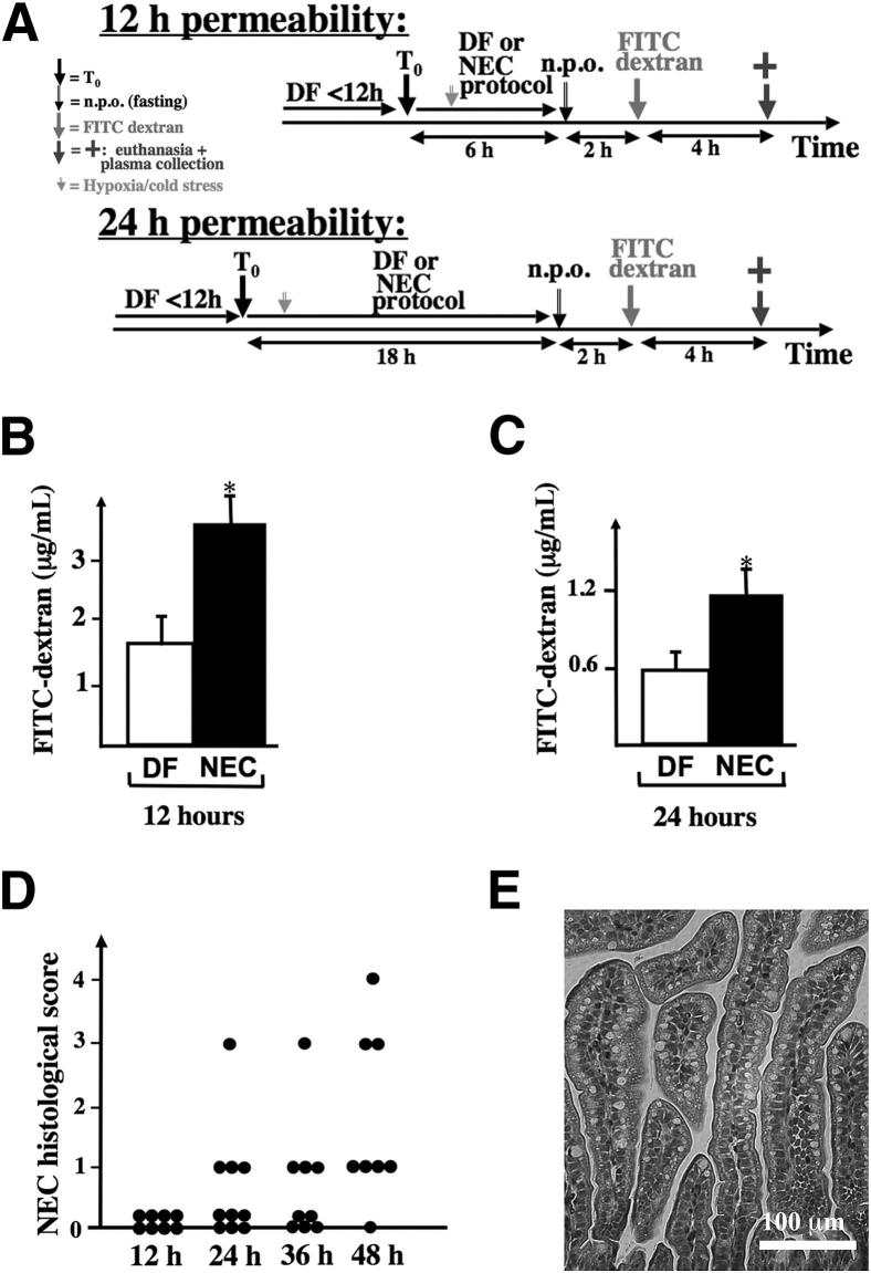
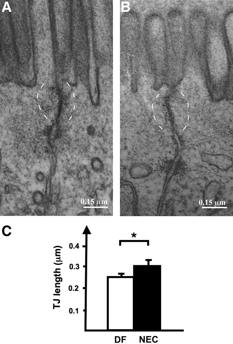
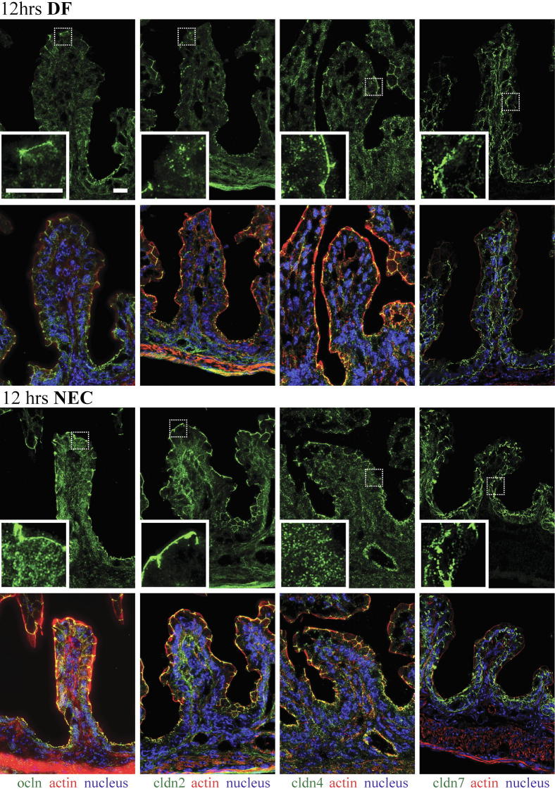
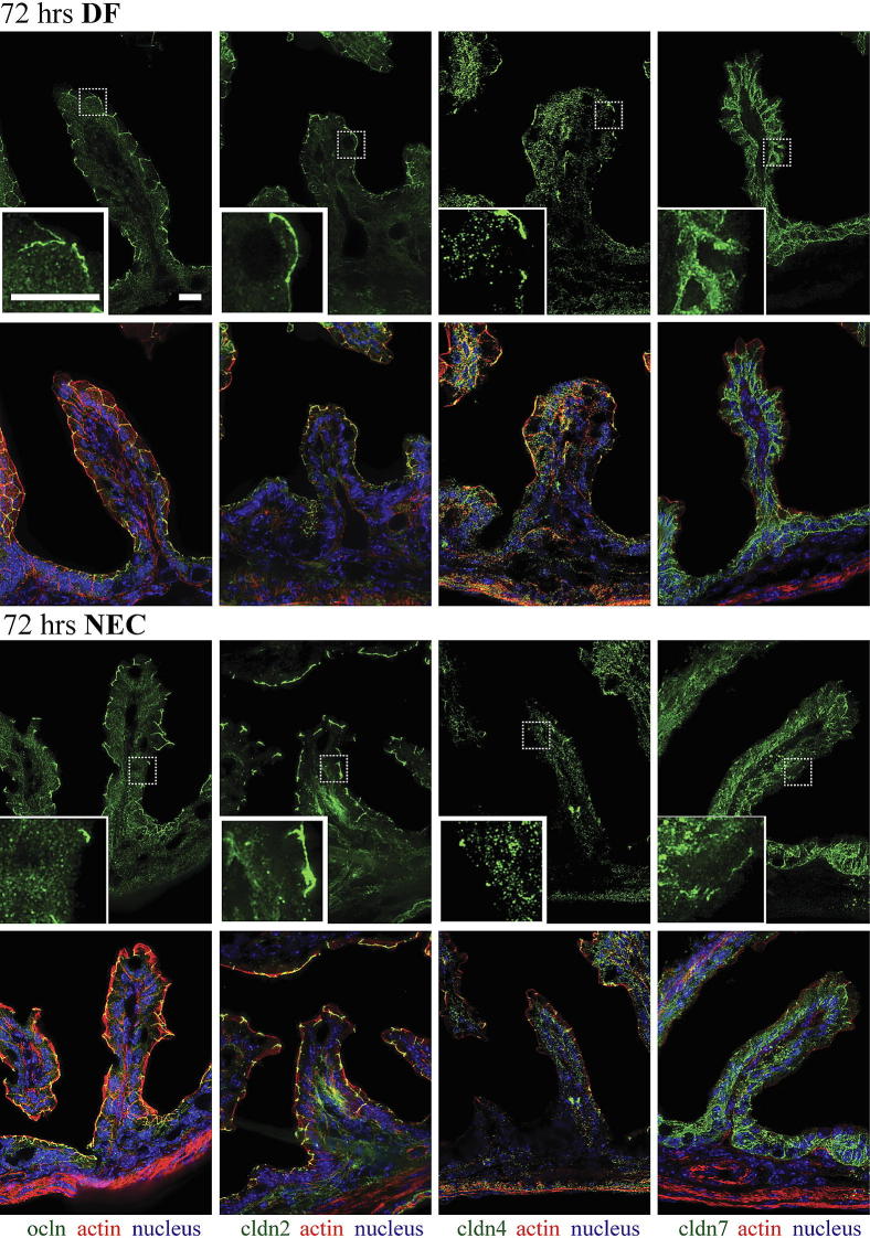
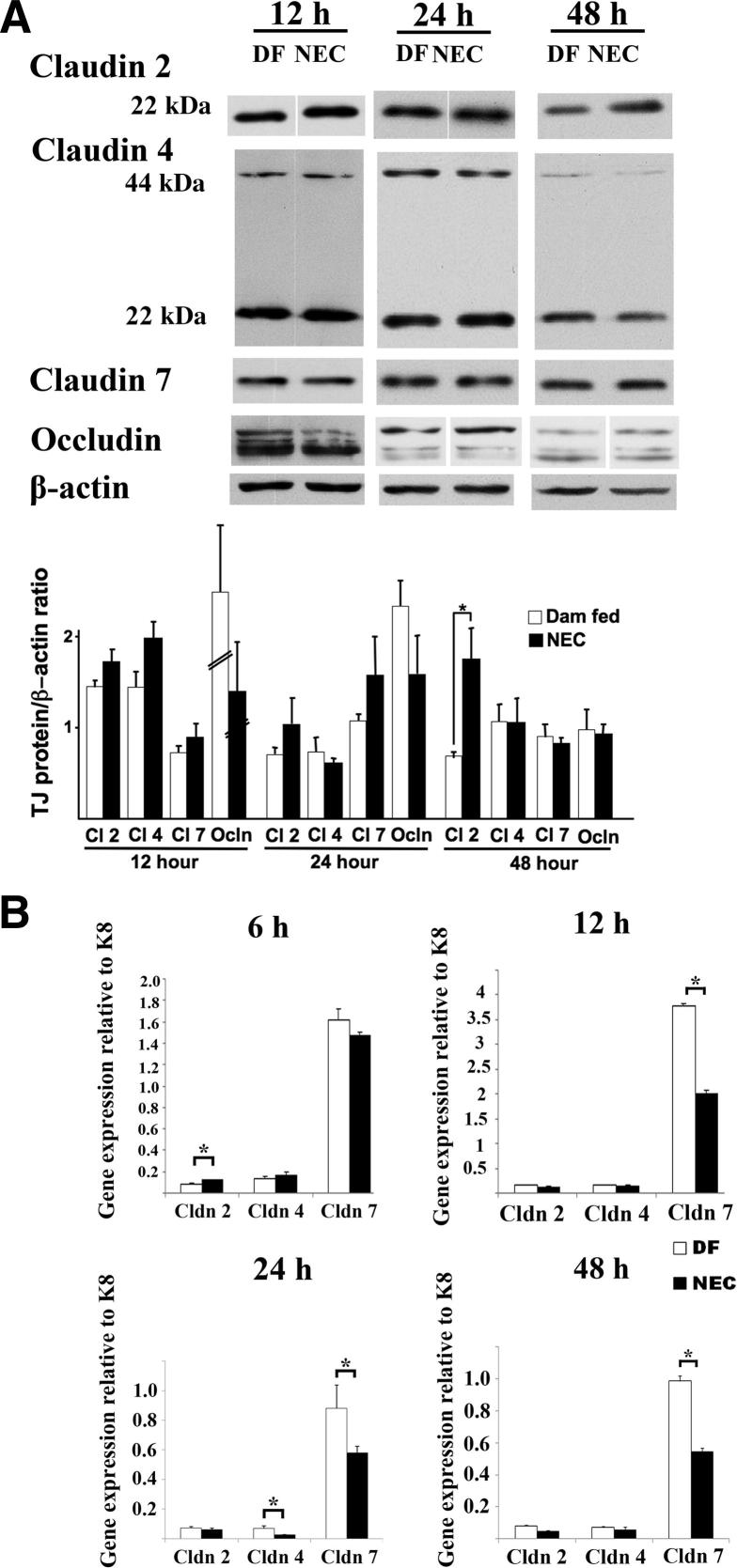
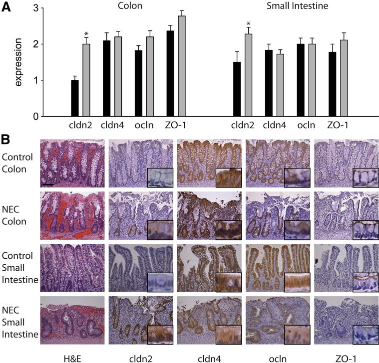
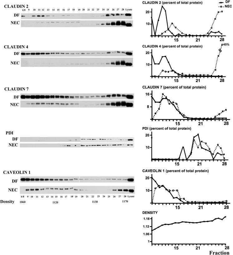
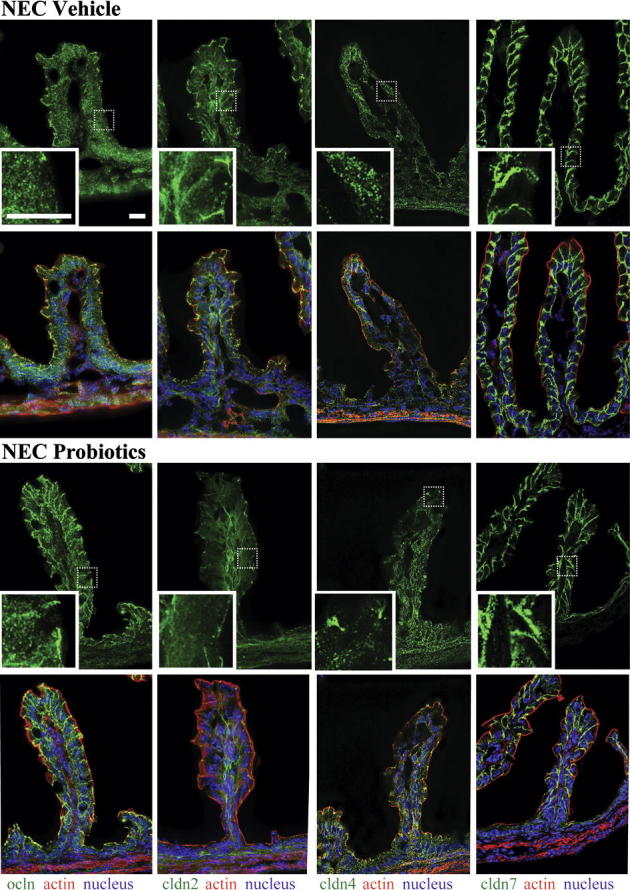
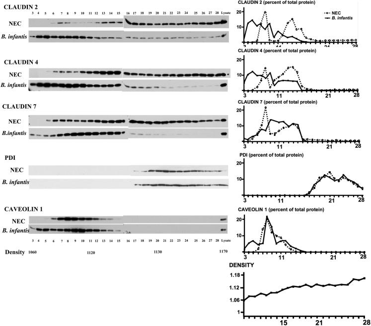
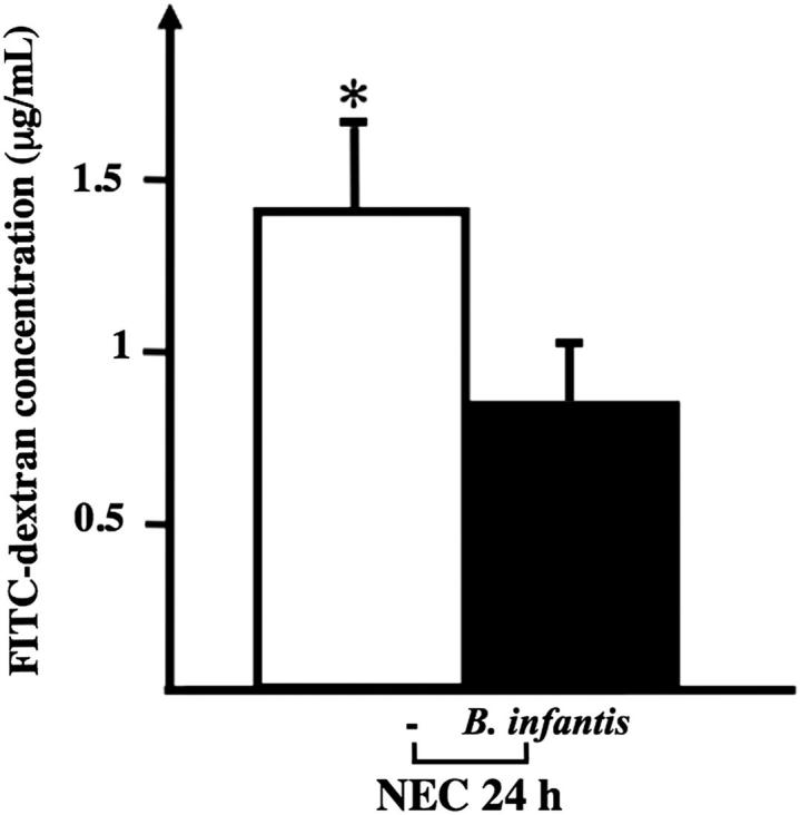
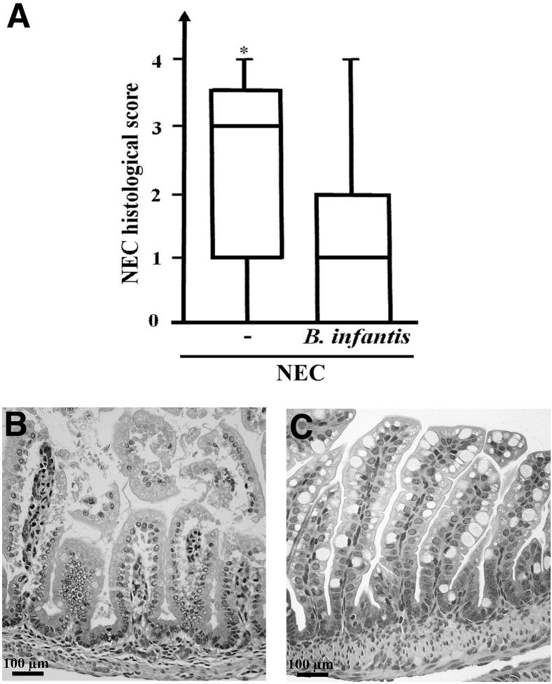
Whether intestinal barrier disruption precedes or is the consequence of intestinal injury in necrotizing enterocolitis (NEC) remains unknown. Using a neonatal mouse NEC model, we examined the changes in intestinal permeability and specific tight-junction (TJ) proteins preceding NEC and asked whether these changes are prevented by administration of Bifidobacterium infantis, a probiotic known to decrease NEC incidence in humans. Compared with dam-fed controls, pups submitted to the NEC protocol developed i) significantly increased intestinal permeability at 12 and 24 hours (as assessed by 70-kDa fluorescein isothiocyanate-dextran transmucosal flux); ii) occludin and claudin 4 internalization at 12 hours (as assessed by immunofluorescence and low-density membrane fraction immunoblotting); iii) increased claudin 2 expression at 6 hours and decreased claudin 4 and 7 expression at 24 hours; and iv) increased claudin 2 protein at 48 hours. Similar results were seen in human NEC, with claudin 2 protein increased. In mice, administration of B. infantis micro-organisms attenuated increases in intestinal permeability, preserved claudin 4 and occludin localization at TJs, and decreased NEC incidence. Thus, an increase in intestinal permeability precedes NEC and is associated with internalization of claudin 4 and occludin. Administration of B. infantis prevents these changes and reduces NEC incidence. The beneficial effect of B. infantis is, at least in part, due to its TJ and barrier-preserving properties.


Figures

Figure 1
Figure 1
Intestinal permeability is increased in NEC at 12 and 24 hours, before histological changes of NEC are detected. A: Pups were randomized into two groups at time T0: group 1 (DF) was left with the dams as controls, and group 2 (NEC) was stressed according to the NEC protocol. Six hours before euthanasia, pups were fasted for 2 hours, and then 750 mg/kg (diluted in 25 μL of 0.9% NaCl) FITC–dextran (70 kDa) was administered through an orogastric tube. B and C: At the time of euthanasia, 50 μL of whole blood was collected, and serum FITC–dextran concentration was determined at 12 hours (B) and 24 hours (C). D: Histological grade of the small intestine of mice submitted to the NEC protocol and euthanized at different time points (every 12 hours). E: Typical appearance of the intestine after 24 hours on the NEC protocol, showing absence of villous injury. Data in B and C are expressed as means ± SEM. n = 7 to 11 per group. ∗P < 0.05. Scale bar = 100 μm.
Figure 2
Figure 2
Small intestine epithelial cell TJ regions in controls and stressed pups. A and B: At 24 hours, electron micrographs reveal intact TJs (delineated by dashed brackets) in both DF controls (A) and pups stressed on the NEC protocol (B). The apical-to-basolateral TJ length was longer in pups stressed for 24 hours (B) than in DF controls (A). C: Mean TJ length was measured by a pathologist (C.R.W.) masked to group assignment. Data are expressed as means ± SEM. n = 16 (DF control) or 13 (stressed pups). ∗P < 0.05. Scale bar = 0.15 μm.
Figure 3
Figure 3
Internalization of claudin 4 and occludin in enterocytes of stressed pups at 12 hours. Frozen sections of small intestines of pups submitted to the NEC protocol or DF controls were examined for occludin (ocln, green) and claudins (cldn, green) 2, 4, and 7 by immunofluorescence (top row in each group). Boxed regions correspond to higher-magnification images in the insets. Merged images (bottom row in each group) also show β-actin (red) and DAPI nuclear staining (blue). In DF mice, claudin 2, claudin 4, and occludin were present mainly at TJs, and claudin 7 was detected along cellular membranes. In pups stressed for 12 hours, occludin and claudin 4 were found throughout the cytoplasm. Claudin 2 was largely associated with TJs while claudin 7 was found along the cellular membranes as in dam fed controls. n = 3 to 6 per group. Scale bar = 40 μm.
Figure 4
Figure 4
Internalization of claudins 2, 4, and 7 and occludin in enterocytes of stressed pups at 72 hours. Frozen sections of small intestines of pups submitted to the NEC protocol or DF controls were examined for occludin (green) and claudins (green) 2, 4, and 7 by immunofluorescence (top row in each group). Merged images (bottom row in each group) also show β-actin (red) and DAPI nuclear staining (blue). In DF mice, claudin 2, 4, and occludin were present mainly at TJs, and claudin 7 was detected along cellular membranes. In pups stressed for 72 hours, occludin and claudins 2, 4, and 7 were found throughout the cytoplasm; the association of claudins 2 and 4 with TJs was decreased, whereas the association of occludin with TJs was only focally decreased in villi and a small amount was found throughout the cytoplasm. n = 3 to 6 per group. Scale bar = 40 μm.
Figure 5
Figure 5
Claudin 2 protein levels are increased in the small intestine of stressed pups at 48 hours, claudin 2 gene expression is up-regulated at 6 hours, and claudin 4 and 7 gene expression is down-regulated at 24 hours, compared with DF controls. A: Presence of occludin and claudins 2, 4, and 7 in the small intestine of NEC stressed mouse pups versus DF controls at 12, 24, and 48 hours was analyzed by Western blotting. Membranes were then stripped and immunoblotted for β-actin, and the results were quantified. Similar results were obtained in two independent experiments. Nonadjacent lanes from the same blot are separated by a fine space. B: The gene expression profile of claudins 2, 4, and 7 relative to K8 was obtained by qPCR in DF versus stressed pups at 6, 12, 24, and 48 hours. Data are expressed as means ± SEM. n = 4 samples per group (A) or 3 to 8 samples per group, run in duplicate (B). ∗P < 0.05. Cl, claudin.
Figure 6
Figure 6
Claudin 2 expression is elevated in human NEC. A: Immunohistochemical staining in human control and NEC tissues was scored semiquantitatively from 0 to 3. B: In the crypts, the expression of claudin 2 was low in control colon and small intestine, but was increased in NEC tissues. In contrast, the expression of claudin 4, occludin, and ZO-1 was similar between control and NEC tissues. ∗P ≤ 0.05; n = 9 to 11 per group. Scale bars: 100 μm (main images); 20 μm (insets).
Figure 7
Figure 7
Redistribution of TJ proteins from membrane to high-density fractions in the small intestine of pups submitted to the NEC protocol for 24 hours. Subcellular fractions from small intestines of DF controls and stressed pups were separated by centrifugation on a discontinuous sugar density gradient and submitted to Western blotting and quantification of claudin proteins and specific subcellular markers. Protein disulfide isomerase (PDI) was used as an endoplasmic reticulum marker and caveolin 1 as a lipid raft marker. Results are representative of two independent experiments, run in triplicate.
Figure 8
Figure 8
Administration of B. infantis preserved claudin 4 and occludin localization at TJs. Frozen sections of small intestines of pups treated with B. infantis (Probiotics group) or vehicle control (NEC group) and submitted to the NEC protocol for 12 hours were examined for claudins 2, 4, and 7 (green) and for occludin (green) by immunofluorescence (top row in each group). Boxed regions correspond to higher-magnification images in the insets. Merged images (bottom row in each group) also show β-actin (red) and DAPI nuclear staining (blue). In vehicle-treated pups, occludin and claudin 4 were found throughout the cytoplasm, whereas in B. infantis–treated mice, claudin 4 remained localized at TJs and occludin was found both in the cytoplasm and at the TJ. n = 4 per group. Scale bar = 40 μm.
Figure 9
Figure 9
The localization of claudins 2, 4, and 7 to low-density fractions is preserved in B. infantis–treated pups, as indicated by Western blotting and quantification of claudin proteins and specific subcellular markers in small intestinal tissue fractions of stressed pups (24 hours) treated with B. infantis or vehicle control (NEC). Results are representative of two independent experiments, run in triplicate.
Figure 10
Figure 10
Administration of B. infantis attenuated the increase in intestinal permeability in pups stressed for 24 hours. Serum FITC–dextran concentration 4 hours after orogastric administration was lower in mice submitted to the NEC protocol for 24 hours and treated with B. infantis, compared with vehicle control (−). Data are expressed as means ± SEM. n = 11 to 13 per group. ∗P < 0.05.
Figure 11
Figure 11
Administration of B. infantis significantly decreased the incidence of NEC in a mouse model. A: NEC histological scores of mice submitted to the NEC protocol for 72 hours and treated with B. infantis or vehicle alone (−). B and C: Typical appearance of the small intestine of an untreated pup (B) showing evidence of grade 2 NEC, compared with the small intestine of a pup treated with B. infantis (C) and showing intact villi. Data are expressed as box-and-whisker plots, indicating median, interquartile range, and minimum and maximum values. n = 26 or 27 per group. ∗P < 0.05.

References

    1. Lin P.W., Stoll B.J. Necrotising enterocolitis. Lancet. 2006;368:1271–1283. - PubMed
    1. Piena-Spoel M., Albers M.J., ten Kate J., Tibboel D. Intestinal permeability in newborns with necrotizing enterocolitis and controls: does the sugar absorption test provide guidelines for the time to (re-)introduce enteral nutrition? J Pediatr Surg. 2001;36:587–592. - PubMed
    1. Clark J.A., Doelle S.M., Halpern M.D., Saunders T.A., Holubec H., Dvorak K., Boitano S.A., Dvorak B. Intestinal barrier failure during experimental necrotizing enterocolitis: protective effect of EGF treatment. Am J Physiol Gastrointest Liver Physiol. 2006;291:G938–G949. - PubMed
    1. Feng J., El Assal O.N., Besner G.E. Heparin-binding epidermal growth factor-like growth factor decreases the incidence of necrotizing enterocolitis in neonatal rats. J Pediatr Surg. 2006;41:144–149. - PubMed
    1. Arrieta M.C., Bistritz L., Meddings J.B. Alterations in intestinal permeability. Gut. 2006;55:1512–1520. - PMC - PubMed

Publication types

MeSH terms