. 2013 Aug;19(15-16):1733-53.
doi: 10.1089/ten.TEA.2012.0385. Epub 2013 Apr 6.

Matrix-embedded cytokines to simulate osteoarthritis-like cartilage microenvironments

Affiliations

Matrix-embedded cytokines to simulate osteoarthritis-like cartilage microenvironments

Sumit Murab et al. Tissue Eng Part A. 2013 Aug.

Abstract

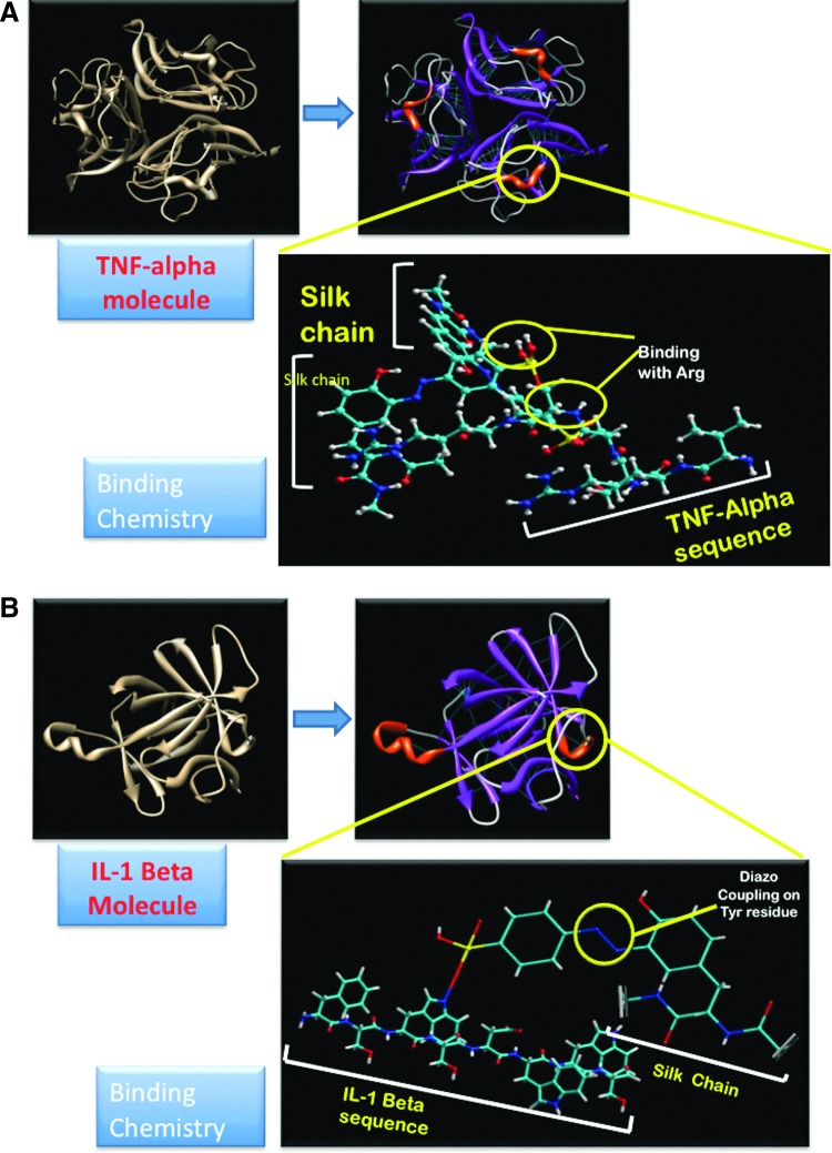
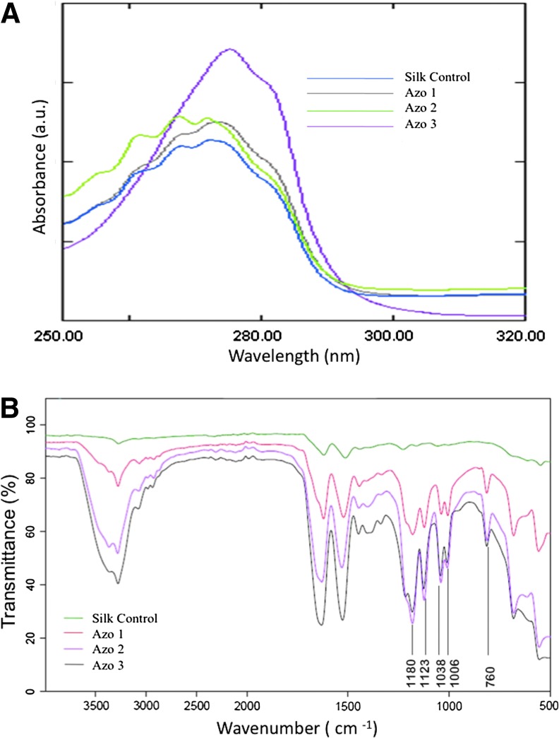
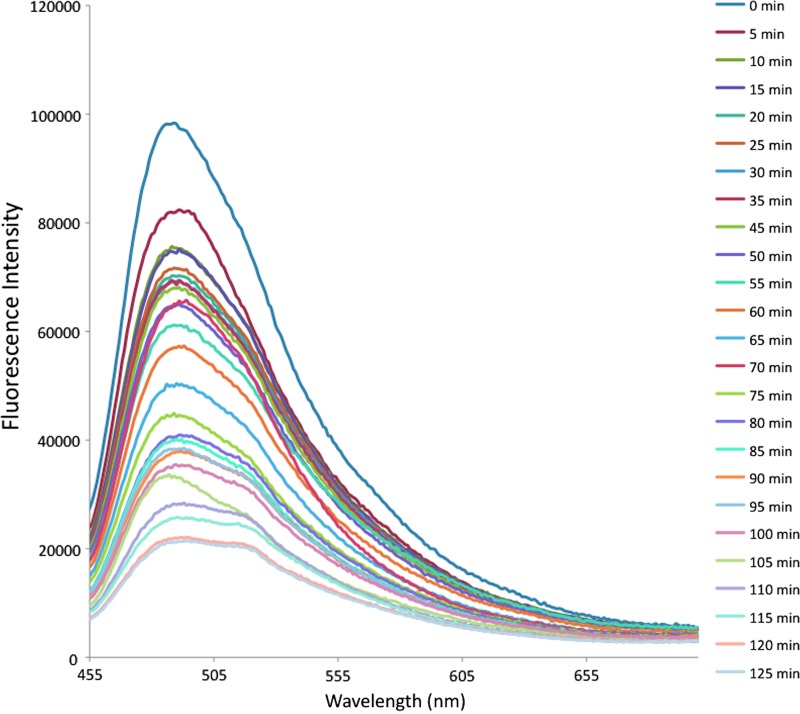
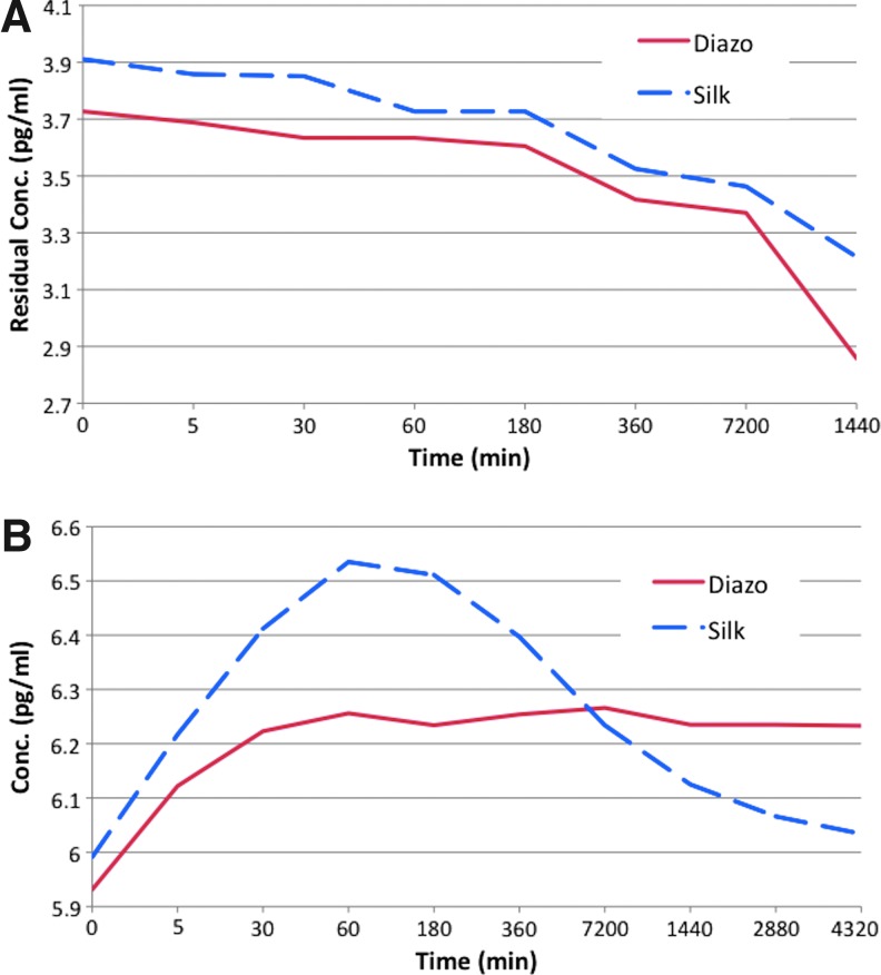
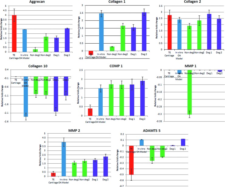
In vivo, cytokines noncovalently bind to the extracellular matrix (ECM), to facilitate intimate interactions with cellular receptors and potentiate biological activity. Development of a biomaterial that simulates this type of physiological binding and function is an exciting proposition for designing controlled advanced delivery systems for simulating in vivo conditions in vitro. We have decorated silk protein with sulfonated moieties through diazonium coupling reactions to noncovalently immobilize pro-inflammatory cytokines interleukin-1 beta (IL-1β) and tumor necrosis factor alpha (TNF-α) in such a biomimetic manner. After adsorption of the cytokines to the diazonium-modified silk matrix, constant release of cytokines up to at least 3 days was demonstrated, as an initial step to simulate an osteoarthritic (OA) microenvironment in vitro. Matrix-embedded cytokines induced the formation of multiple elongated processes in chondrocytes in vitro, akin to what is seen in OA cartilage in vivo. Gene expression profiles with this in vitro tissue model of OA showed significant similarities to profiles from explanted OA cartilage tissues collected from patients who underwent total knee replacement surgery. The common markers of OA, including COL, MMP, TIMP, ADAMTS, and metallothioneins, were upregulated at least 35-fold in the in vitro model when compared to the control-non-OA in vitro generated tissue-engineered cartilage. The microarray data were validated by reverse transcriptase-polymerase chain reaction. Mechanistically, protein interaction studies indicated that TNF-α and IL-1β synergistically controlled the equilibrium between MMPs and their inhibitors, TIMPs, resulting in ECM degradation through the MAPK pathway. This study offers a promising initial step toward establishing a relevant in vitro OA disease model, which can be further modified to assess signaling mechanisms, responses to cell or drug treatments and patient-specific features.


Figures

FIG. 1.
FIG. 1.
(A) Binding of tumor necrosis factor alpha (TNF-α) to silk polymer chain: the VRSSSR sequence is highlighted in red color. (B) Binding of interleukin-1 beta (IL-1β) to silk polymer chain: the Ser-Asp-Trp-Trp-Ser sequence is highlighted in red color. Color images available online at www.liebertpub.com/tea
FIG. 2.
FIG. 2.
(A) Comparison of the UV-Vis Spectra of unmodified silk and sulfonic acid azosilks modified with increasing equivalents of diazonium salt. (B) ATR-FTIR Spectra showing increase in modification of tyrosine residues with increase in azo concentration. Color images available online at www.liebertpub.com/tea
FIG. 3.
FIG. 3.
Binding kinetics of IL-1β on diazotized silk film. Color images available online at www.liebertpub.com/tea
FIG. 4.
FIG. 4.
(A) Adsorption and (B) release kinetics of cytokines from the silk film as determined by ELISA. Color images available online at www.liebertpub.com/tea
FIG. 5.
FIG. 5.
Morphology of chondrocytes on (A) silk film and (B) cytokine immobilized diazotized silk film within 8 h, (C) extensive expression of cellular extensions on cytokine immobilized silk film after 1 day, (D) similar extensions noticed on cytokine-sequestered Matrigel layer, (E) chondrocytes on Matrigel-coated coverslip without cytokines after 1 day (arrows indicate smaller cytoplasmic projections from the periphery of the cells on silk films, compared to cytokine immobilized silk films and Matrigel).
FIG. 6.
FIG. 6.
(A) Hematoxylin and eosin (H&E)–stained section of tissue-engineered cartilage showing rounded cell morphology. (B) H&E-stained section of in vitro osteoarthritic (OA) model showing mixed population of rounded and elongated cells (marked by arrows). (C) Safranin-O staining of tissue-engineered cartilage showing the presence of glycosaminoglycans (GAG). (D) Safranin-O staining showing the absence of GAG. (E) H&E-stained cartilage biopsy tissue showing histological features of osteoarthritis such as fibrillation and erosion of GAG deposition, (F) clustered chondrocytes, (G) tide mark and subchondral bone cyst (marked by arrow), and (H) signs of angiogenic invasion in cartilage (marked by arrow). Color images available online at www.liebertpub.com/tea
FIG. 7.
FIG. 7.
(A) Modulation of gene expression in biopsies from degraded lesion, in vitro OA model, cartilage from intact region, and tissue-engineered cartilage. (B) Extent of the up- or downregulation of each gene in tissue-engineered cartilage, degraded lesion of OA biopsies, intact portion of OA biopsies, and in vitro OA model. (C) Variation among biopsies. (D) Relative expression of vimentin. Color images available online at www.liebertpub.com/tea
FIG. 8.
FIG. 8.
Protein interaction network analysis (confidence view) for (A) upregulated genes in diseased condition, (B) matrix-related proteins, (C) receptors binding, and (D) apoptotic process. Color images available online at www.liebertpub.com/tea
FIG. 9.
FIG. 9.
Comparative gene ontology of OA biopsies versus tissue-engineered in vitro disease model system, compared to tissue-engineered cartilage. Color images available online at www.liebertpub.com/tea
FIG. 10.
FIG. 10.
Relative expression of ADAMTS5, aggrecan, COMP1, collagen 1, collagen 2, collagen 10, MMP1, and MMP2 using tissue-engineered cartilage, engineered in vitro OA-like model, biopsies from nondegraded region (intact cartilage biopsy), and degraded regions from two patients. Color images available online at www.liebertpub.com/tea

References

    1. Anderson D.D. Chubinskaya S. Guilak F. Martin J.A. Oegema T.R. Olson S.A., et al. Post-traumatic osteoarthritis: Improved understanding and opportunities for early intervention. J Orthop Res. 2011;29:802. - PMC - PubMed
    1. Li Y. Xu L. Olsen B.R. Lessons from genetic forms of osteoarthritis for the pathogenesis of the disease. Osteoarthritis Cartilage. 2007;15:1101. - PMC - PubMed
    1. Martin J.A. Buckwalter J.A. Telomere erosion and senescence in human articular cartilage chondrocytes. J Gerontol A Biol Sci Med Sci. 2001;56:B172. - PubMed
    1. Aigner T. Rose J. Martin J. Buckwalter J. Aging theories of primary osteoarthritis: from epidemiology to molecular biology. Rejuvenation Res. 2004;7:134. - PubMed
    1. Valdes A.M. Loughlin J. Oene M.V. Chapman K. Surdulescu G.L. Doherty M., et al. Sex and ethnic differences in the association of ASPN, CALM1, COL2A1, COMP, and FRZB with genetic susceptibility to osteoarthritis of the knee. Arthritis Rheum. 2007;56:137. - PubMed

Publication types

MeSH terms