. 2013 Jul;20(7):869-77.
doi: 10.1038/cdd.2013.16. Epub 2013 Mar 8.

The loss of the BH3-only Bcl-2 family member Bid delays T-cell leukemogenesis in Atm-/- mice

Affiliations

The loss of the BH3-only Bcl-2 family member Bid delays T-cell leukemogenesis in Atm-/- mice

S Biswas et al. Cell Death Differ. 2013 Jul.

Abstract

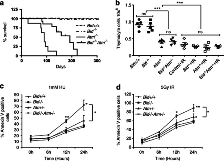
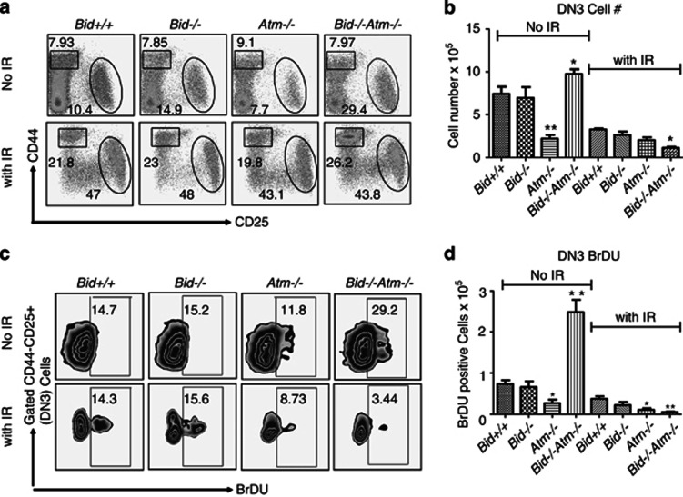
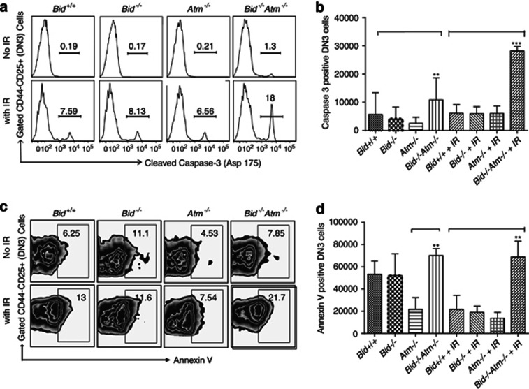
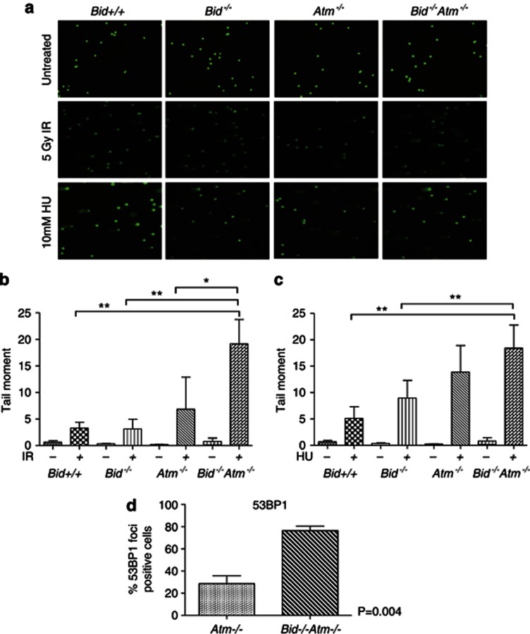
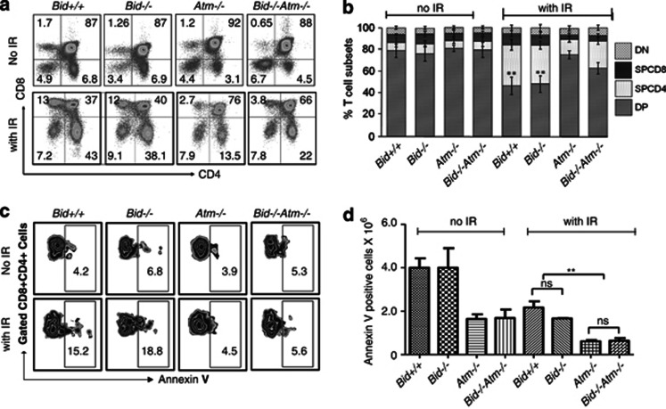
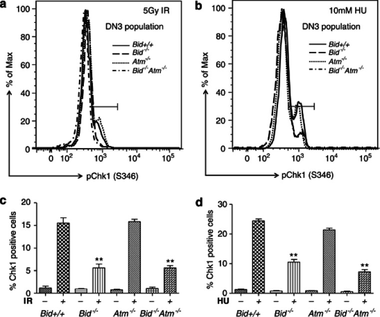
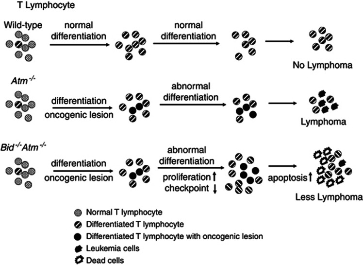
Multicellular organisms maintain genomic integrity and resist tumorigenesis through a tightly regulated DNA damage response (DDR) that prevents propagation of deleterious mutations either through DNA repair or programmed cell death. An impaired DDR leads to tumorigenesis that is accelerated when programmed cell death is prevented. Loss of the ATM (ataxia telangiectasia mutated)-mediated DDR in mice results in T-cell leukemia driven by accumulation of DNA damage accrued during normal T-cell development. Pro-apoptotic BH3-only Bid is a substrate of Atm, and Bid phosphorylation is required for proper cell cycle checkpoint control and regulation of hematopoietic function. In this report, we demonstrate that, surprisingly, loss of Bid increases the latency of leukemogenesis in Atm-/- mice. Bid-/-Atm-/- mice display impaired checkpoint control and increased cell death of DN3 thymocytes. Loss of Bid thus inhibits T-cell tumorigenesis by increasing clearance of damaged cells, and preventing propagation of deleterious mutations.


Figures

Figure 1
Figure 1
Loss of Bid attenuates T-cell lymphoblastic leukemia/lymphoma development in Atm−/− mice. (a) Kaplan–Meier survival curves for cohorts of Bid+/+, Bid−/−, Atm−/−, and Bid−/−Atm−/− mice. (b) The total cellularity of control, Bid+/+, Bid−/−, Atm−/−, and Bid−/−Atm−/− thymocytes 48 h after 5 Gy IR treatment. (c, d). Thymocytes from Bid+/+, Bid−/−, Atm−/−, and Bid−/−Atm−/− mice were treated with (c) 1 mM hydroxyurea, or (d) 5 Gy ionizing radiation. Cell death was measured by annexin V staining with propidium iodine exclusion followed by flow cytometry. Data are representative of three independent experiments. *P<0.05, **P<0.01 and ***P<0.001.
Figure 2
Figure 2
Bid−/−Atm−/− mice accumulate DN3 thymocytes. (a) Immunophenotype of double negative populations, DN1 (CD4CD8CD25CD44) and DN3 (CD4CD8CD25+CD44). Bid −/−Atm−/− mice accumulate DN3 cells. (b) DN3 cell number of untreated and IR-treated mice of the indicated genotypes. (c) BrdU incorporation of DN3 populations of untreated and IR-treated mice of the indicated genotypes. The BrdU-positive DN3 subsets cells were detected by intracellular staining with FITC-conjugated anti-BrdU antibody (BD Pharmingen) (d), quantification of data in (c). *P<0.05 and **P<0.01
Figure 3
Figure 3
Bid−/−Atm−/− DN3 cells are more sensitive to IR. (a) Relative expression of intracellular activated caspase-3 in DN3 cells from Bid+/+, Bid−/−, Atm−/−, and Bid−/−Atm−/− mice by flow cytometric analysis. Mice of the indicated genotypes were treated with 5 Gy IR and thymocytes were harvested after 12 h. After surface staining with anti-CD4, anti-CD8α, anti-CD44, and anti-Cd25, thymocytes were fixed, permeabilized, and stained with anti-cleaved Caspase-3 (Cell Signaling) followed by staining with Alexa Fluor 488-conjugated anti-rabbit secondary antibody. (b) Caspase-3-positive subsets of DN3 cells were measured by flow cytometry and quantitative analysis was performed. Data are from three sets of mice. (c) Flow cytometry analysis of apoptosis of the DN3 population by annexin V staining. (d) Quantification of apoptosis by annexin V staining of DN3 subsets from the indicated genotypes of mice. *P<0.05, **P<0.01 and ***P<0.001
Figure 4
Figure 4
Bid−/−Atm−/− thymocytes display significantly increased DNA damage after IR and HU and Bid−/−Atm−/− DN thymocytes display increased DNA damage at baseline. Micrographs showing comet assays of thymocytes from the indicated genotypes treated with 5 Gy IR or 10 mM HU, and harvested after 24 h. The untreated and treated cells were collected in ice-cold PBS and an alkaline comet assay (Trevigen) was performed. The samples were run in alkaline electrophoresis solution at 21 V for 30 min. After staining with SYBR Green I, samples were examined using a Leica DM IRBE inverted wild-field microscope. (b, c) At least 75 randomly chosen cells per sample without or with (b) IR and, (c) HU treatment were analyzed using CometScore Program version 1.5. Atm−/− and Bid−/−Atm−/− DN thymocytes were isolated by magnetic bead sorting and stained with anti-53BP1. The percentage of cells with at least five 53BP1 positive nuclear foci was determined. At least 75 randomly chosen cells were evaluated per sample. *P<0.05 and **P<0.01.
Figure 5
Figure 5
Both Atm−/− and Bid−/−Atm−/− DP populations are more resistant to IR. (a) Flow cytometry analysis of T-cell subsets from thymocytes of mice untreated and irradiated with 5 Gy IR harvested after 48 h. After IR treatment, the Atm−/− and Bid−/−Atm−/− DP populations are protected from cell death. (b) Three sets of irradiated and untreated mice of each genotype were analyzed and the percentage of the indicated thymocyte subsets are presented. Error bars denote S.D. (c) Flow cytometry analysis of apoptosis of the DP population by annexin V staining. (d) Quantification of the data in (c). **P<0.01
Figure 6
Figure 6
Bid−/− and Bid−/−Atm−/− DN3 thymocytes display decreased Chk1 phosphorylation following IR and HU. (a, b) Total thymocytes were treated with (a) 5 Gy IR and harvested after 4 h or, (b) 10 mM hydroxyurea and harvested after 2 h. Cells were stained with anti-CD4, anti-CD8, anti-CD25, and anti-CD44 for 30 min. Chk1 phosphorylation was detected by intracellular staining with anti-pChk1 (S345) antibody (Cell signaling, no. 2348) and Alexa Fluor 488-conjugated Goat anti-Rabbit IgG antibody (Invitrogen). (c) Quantitative analysis of data in (a), showing the percentage of pChk1-positive DN3 subsets from three independent experiments. (d) Quantitative analysis of data in (b), showing the percentage of pChk1-positive DN3 subsets from three independent experiments. **P<0.01
Figure 7
Figure 7
Model for the role of Bid in T-cell lymphoblastic leukemia/lymphoma. Atm−/− mice develop T-cell lymphoblastic leukemia/lymphoma within 3 months, due to an aberrant DNA damage response to DNA strand breaks accrued during T-cell development. Bid−/−Atm−/− mice display increased proliferation and cell death in the DN3 thymocyte progenitor population, and increased latency of tumorigenesis. We propose a model in which loss of Bid further impairs the DNA damage response in Atm−/− thymocytes, and increases the DNA damage and cell death of a vulnerable population of cells, increasing tumor latency

Comment in

References

    1. Soria G, Polo SE, Almouzni G. Prime, repair, restore: the active role of chromatin in the DNA damage response. Mol Cell. 2012;46:722–734. - PubMed
    1. Roos WP, Kaina B.DNA damage-induced apoptosis: From specific DNA lesions to the DNA damage response and apoptosis Cancer Lett 2012. doi:10.1016/j.canlet.2012.01.007 - DOI - PubMed
    1. Shiloh Y. ATM and related protein kinases: safeguarding genome integrity. Nat Rev Cancer. 2003;3:155–168. - PubMed
    1. Bartek J, Lukas C, Lukas J. Checking on DNA damage in S phase. Nat Rev Mol Cell Biol. 2004;5:792–804. - PubMed
    1. Ciccia A, Elledge SJ. The DNA damage response: making it safe to play with knives. Mol Cell. 2010;40:179–204. - PMC - PubMed

Publication types

MeSH terms