

Commissioning and implementation of an implantable dosimeter for radiation therapy
- PMID: 23470929
- PMCID: PMC5714364
- DOI: 10.1120/jacmp.v14i2.3989
Commissioning and implementation of an implantable dosimeter for radiation therapy
Abstract
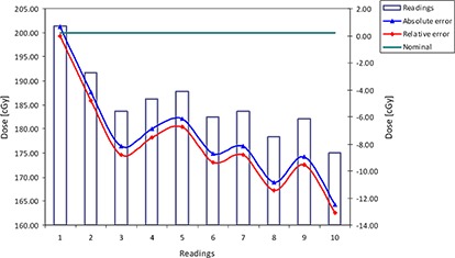
In this article we describe commissioning and implementation procedures for the Dose Verification System (DVS) with permanently implanted in vivo wireless, telemetric radiation dosimeters for absolute dose measurements. The dosimeter uses a semiconductor device called a metal-oxide semiconductor field-effect transistor (MOSFET) to measure radiation dose. A MOSFET is a transistor that is generally used for amplifying or switching electronic signals. The implantable dosimeter was implemented with the goal of verifying the dose delivered to radiation therapy patients. For the purpose of acceptance testing, commissioning, and clinical implementation and to evaluate characteristics of the dosimeter, the following tests were performed: 1) temperature dependence, 2) reproducibility,3) field size dependence, 4) postirradiation signal drift, 5) dependence on average dose rate, 6) linearity test, 7) angular dependence (different gantry angle position), 8) angular dependence (different DVS angle position), 9) dose rate dependence,10) irradiation depth dependence, 11) effect of cone-beam exposure to the dosimeter, and 12) multiple reading effect. The dosimeter is not currently calibrated for use in the kV range; nonetheless, the effect of the cone-beam procedure on the MOSFET dosimeter was investigated. Phantom studies were performed in both air and water using an Elekta Synergy S Beam-Modulator linear accelerator. Commissioning and clinical implementation for prostate cancer patients receiving external-beam radiation therapy were performed in compliance with the general recommendations given for in vivo dosimetry devices. The reproducibility test in water at human body temperature (37°C) showed a 1.4% absolute difference, with a standard deviation of 5.72 cGy (i.e., SD = 2.9%). The constancy test shows that the average readings at room temperature were 3% lower compared to the readings at human body temperature, with a SD = 2%. Measurements were not dependent upon field size. Due to postirradiation signal drift, the following corrections are suggested: -2.8%, -2%, 0.5%, and 2.5% for the readings taken after 0.5, 1, 5, or 10 min, respectively. Different gantry angles did not influence the readings. The maximum error was less than 1% with a maximum SD = 3.61 cGy (1.8%) for the gantry angle of 45°. However, readings are dependent on the dosimeter orientation. The average dose reading was 7.89 cGy (SD = 1.46 cGy) when CBCT imaging was used for the pelvis protocol, and when postirradiation measurement was taken at 2.5 min (expected 2-3 cGy). The clinical implementation of the implantable MOSFET dosimeters for prostate cancer radiation therapy is described. Measurements performed for commissioning show that the dosimeter, if used within specifications, provides sufficient accuracy for its intended use in clinical procedures. The postradiation signal drift, temperature dependence, variation of reproducibility, and rotational isotropy could be encountered if the dosimeter is used outside the manufacturer's specifications. The dosimeter can be used as a tool for quantifying dose at depth, as well as to evaluate adherence between planned doses and the delivered doses. Currently, the system is clinically implemented with ± 7% tolerance.
Figures


References
-
- Cilla S, Macchia G, Digesù C, et al. Endocavitary in vivo dosimetry for IMRT treatments of gynecologic tumors. Med Dosimet. 2011;36(4):455–62. - PubMed
-
- Francois P, Boissard P, Berger L, Mazal A. In vivo dose verification from back projection of a transit dose measurement on the central axis of photon beams. Phys Med. 2011;27(1):1–10. - PubMed
-
- Haughey A, Coalter G, Mugabe K. Evaluation of linear array MOSFET detectors for in vivo dosimetry to measure rectal dose in HDR brachytherapy. Austral Phys Eng Sci Med. 2011;34(3):361–66. - PubMed
-
- Kadesjö N, Nyholm T, Olofsson J. A practical approach to diode based in vivo dosimetry for intensity modulated radiotherapy. Radiother Oncol. 2011;98(3):378–81. - PubMed
MeSH terms
LinkOut - more resources
Full Text Sources
Other Literature Sources
