. 2013 Mar 8;339(6124):1216-9.
doi: 10.1126/science.1231097.

Evidence for a common mechanism of SIRT1 regulation by allosteric activators

Affiliations

Evidence for a common mechanism of SIRT1 regulation by allosteric activators

Basil P Hubbard et al. Science. .

Abstract

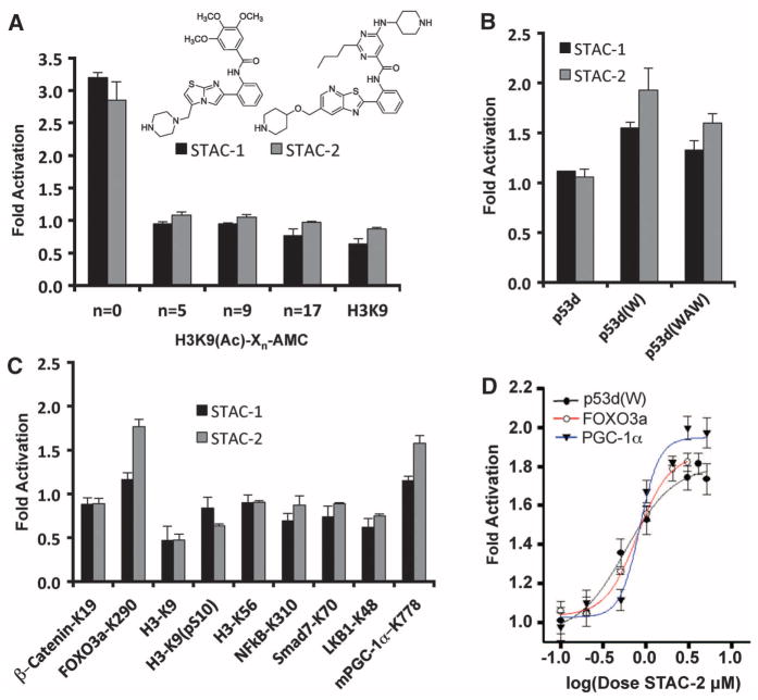
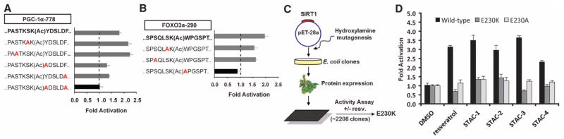
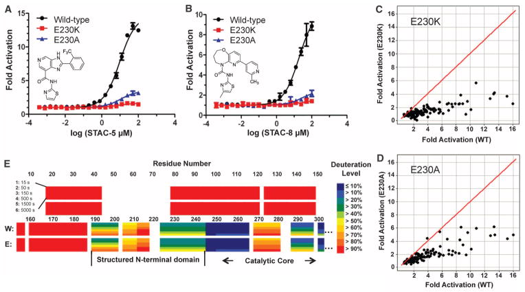
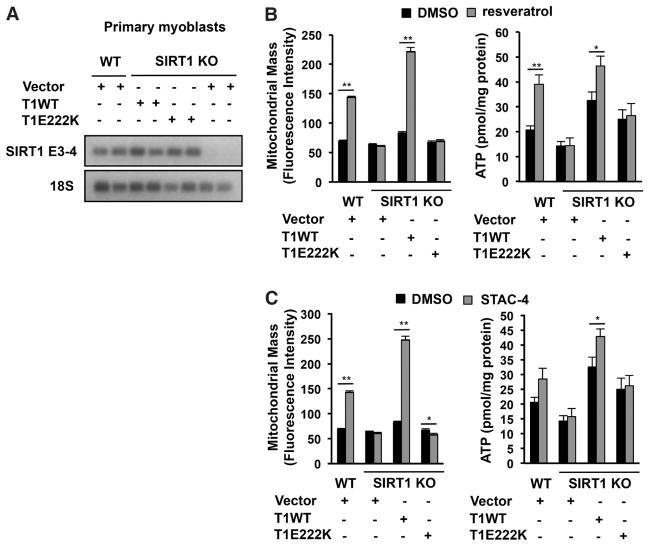
A molecule that treats multiple age-related diseases would have a major impact on global health and economics. The SIRT1 deacetylase has drawn attention in this regard as a target for drug design. Yet controversy exists around the mechanism of sirtuin-activating compounds (STACs). We found that specific hydrophobic motifs found in SIRT1 substrates such as PGC-1α and FOXO3a facilitate SIRT1 activation by STACs. A single amino acid in SIRT1, Glu(230), located in a structured N-terminal domain, was critical for activation by all previously reported STAC scaffolds and a new class of chemically distinct activators. In primary cells reconstituted with activation-defective SIRT1, the metabolic effects of STACs were blocked. Thus, SIRT1 can be directly activated through an allosteric mechanism common to chemically diverse STACs.


Figures

Fig. 1
Fig. 1
SIRT1 activation by STACs on native peptide sequences. (A) SIRT1 activation by 50 μM STAC-1 or 5 μM STAC-2 with peptides bearing an AMC moiety at the indicated positions, where Xn represents the number of amino acids between the acetylated lysine and the AMC. (B) SIRT1 activation by STACs on hydrophobic patch peptides. Complete amino acid sequences of peptides bearing tryptophan (W) or tryptophan and alanine substitutions (WAW) are provided in the supplementary materials. (C) SIRT1 activation on native peptide sequences of known targets (detailed in the supplementary materials). (D) Dose-response curves for STAC-2 as measured by PNC1-OPT assay; data are means ± SEM (n = 3).
Fig. 2
Fig. 2
Substrate sequence requirements and regions on SIRT1 necessary for activation. (A and B) SIRT1 activation by STAC-2 on peptides derived from PGC-1α–K778 (A) and FOXO3a-K290 (B) as measured by PNC1-OPT assay; data are means ± SEM (n = 3). (C) Biochemical screen for activation-compromised mutants. A bacterial expression plasmid (pET28a) carrying the SIRT1 gene was mutagenized and used to generate recombinant SIRT1 proteins that were screened for activity in the presence or absence of resveratrol using an AMC-based assay. (D) Activation of wild-type SIRT1, E230K, and E230A mutants by 40 μM resveratrol, 50 μM STAC-1, 5 μM STAC-2, 5 μM STAC-3, and 10 μM STAC-4 as measured by an AMC assay; data are means ± SD (n = 3). Dimethyl sulfoxide (DMSO) was used as a control.
Fig. 3
Fig. 3
Effects of SIRT1-E230 substitutions on activation and identification of an ordered activation domain. (A and B) Dose-response titrations of STAC-5 (A) and STAC-8 (B) on the activity of wild-type SIRT1 and E230 mutants with the Trp 5-mer peptide serving as the substrate, as measured by mass spectrometry–based OAcADPR assay. The sequence of the Trp 5-mer peptide is included in the supplementary materials. data are means ± SD (n = 3). (C and D) Relative activation by a chemically diverse, 117-compound set (25 μM) using the Trp 5-mer substrate for wild-type versus E230K (C) or wild-type versus E230A (D), as measured by OAcADPR assay (n = 2). The red line represents y = x correlation. (E) HDXMS heat map of deuteration levels of wild-type (W) and SIRT1-E230K (E) N termini at six different time points (15 to 5000 s).
Fig. 4
Fig. 4
mSIRT1-E222K–dependent effects of STACs on mitochondrial-related parameters in cells. (A) Full-length murine SIRT1 (mSIRT1) transcripts in wild-type and primary myoblasts reconstituted with wild-type mSIRT1 or mSIRT1-E222K. The SIRT1 exon 3-4 junction (SIRT1 E3-4) and 18S ribsosomal RNA, as an internal control for loading, were detected by reverse transcription polymerase chain reaction. (B and C) Effect of 25 μM resveratrol (B) or 1 μM STAC-4 (C) on mitochondrial mass and ATP in primary myoblasts; data are means ± SEM (n = 6). *P < 0.05, ** P < 0.01 (t test versus DMSO control).

Comment in

References

    1. Sebastian C, Satterstrom KF, Haigis MC, Mostoslavsky R. J Biol Chem. 2012;287:42444. - PMC - PubMed
    1. Haigis MC, Sinclair DA. Annu Rev Pathol. 2010;5:253. - PMC - PubMed
    1. Chalkiadaki A, Guarente L. Nat Rev Endocrinol. 2012;8:287. - PubMed
    1. Howitz KT, et al. Nature. 2003;425:191. - PubMed
    1. Milne JC, et al. Nature. 2007;450:712. - PMC - PubMed

Publication types

MeSH terms