Review
. 2013;3(3):223-38.
doi: 10.7150/thno.5409. Epub 2013 Mar 1.

Gold nanorods based platforms for light-mediated theranostics

Affiliations
Review

Gold nanorods based platforms for light-mediated theranostics

Zhenjiang Zhang et al. Theranostics. 2013.

Abstract

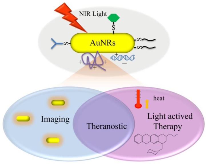
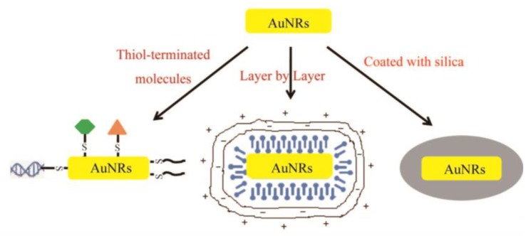
Due to their tunable surface plasmon and photothermal effects, gold nanorods (AuNRs) have proved to be promising in a wide range of biomedical applications such as imaging, hyperthermia therapy and drug delivery. All these applications can be remotely controlled by near infrared (NIR) light which can penetrate deep into human tissues with minimal lateral invasion. AuNRs thus hold the potential to combine both imaging diagnosis and therapeutic treatment into one single system and function as a NIR light-mediated theranostic platform. Herein we review recent progress in diagnostic and therapeutic applications of AuNRs with a highlight on combined applications for theranostic purposes.

Keywords: Gold nanorods; cancer therapy; drug delivery.; hyperthermia; imaging; theranostics.


Conflict of interest statement

Conflict of Interest: The authors have declared that no conflict of interest exists.

Figures

Fig 1
Fig 1
Illustration of various light-mediated biomedical applications of AuNRs.
Fig 2
Fig 2
Three most widely used methods for surface modification and functionalization of AuNRs.
Fig 3
Fig 3
(A) Two-photon luminescence and merged image with differential interference contrast (DIC) of chitosan oligosaccharide-modified AuNRs with and without anti-EGFR conjugation in Cal 27cells. Cells were incubated with 0.1 μg gold nanorods for 4 hours. Scale bar, 10 μm. Adapted with permission from (B) In vivo backscatter mode ultrasound (gray scale) and PAT (red) images of the intramuscular injection of a negative control (0 nM Au@SiO2, no cells), and 800 000 Au@SiO2-labeled MSCs all in 50% matrigel/PBS into hind limb muscle of an athymic mouse. The “b” in panels indicates bone and the red dashed circle highlights the injection. Adapted with permission from . (C) Cross-sectional photothermal OCT images obtained at different depths below the surface by slicing the 3D data cube (shown in (a)) acquired from the SLN at the time point of (b) 12 and (c) 96 h after AuNRs injection. Adapted with permission from . (D) In vivo X-ray imaging of mice after subcutaneous injection without and with folic acids-conjugated Au@SiO2 at different time points. (a) The photograph of mice; the X-ray images at (b) 0 h, (c) 2 h, (d) 6 h, (e) at 8 h, and (f) 24 h. Adapted with permission from .
Fig 4
Fig 4
Membrane blebbing is induced by Ca2+ influx during AuNRs-mediated photothermolysis. (A,B) Cells with membrane-bound AuNRs (red) in PBS containing 0.9 mM Ca2+ exhibited blebbing after exposure to fs-pulsed laser irradiation at 3 mW for 61.5 s. (C) Incubation with 2.5 μM EB (red) and 2 μM Oregon Green 488 for 20 min indicated a compromise in membrane integrity and an elevation in intracellular Ca2+ , respectively. Scale bar = 10 μm. Adapted with permission from .
Fig 5
Fig 5
Overview of selective release. Laser irradiation of DNA-conjugated nanocapsules (blue ovals) and nanobones (red bones) are exposed to 800 irradiation (left), which melts the nanocapsules and selectively releases the conjugated DNA (labeled by FAM (green triangles)). Exposure to 1100 irradiation (right) melts the nanobones, selectively releasing the conjugated DNA (labeled by TMR (orange stars)) .
Fig 6
Fig 6
GNR-5′PPP-ssRNA enhances IFN-β and RIG-I expression and inhibits replication of 2009 pandemic H1N1 influenza viruses and Solomon Islands seasonal flu strain. A459 cells (3.5×10⁵cells/well) in a six-well tissue culture plate were mock transfected or transfected with 3 μg of RNAs complexed with 2.5 μg of GNRs per well for 48 h and then infected with A/California/08/09 or A/Solomon Islands/03/06 at an MOI of 1. Lysates to determine mRNA levels by qRT-PCR (A and B). The viral titers were determined from the supernatants collected 24 h later (C and D) . (GNRs: gold nanorods; CIAP: calf intestinal alkaline phosphatase)
Fig 7
Fig 7
The AuNRs-HIV Env plasmid complex formed and the effects of AuNRs on the immune response and dendritic cell maturation. (A) AuNRs with different surface coatings mixed with Env plasmid. (B) IFN-γ analyzed by ELISPOT. (C) CD3+CD8+ T cells proliferation. (D) The Env 69 specific antibody titer measurement. Adapted with permission from .
Fig 8
Fig 8
(A) Schematic Representation of Synthesis of MMP-AuNRs for Simultaneous Imaging and Photothermal Therapy. (B) Microscopic images of HeLa cell after staining with trypan blue at different treatment conditions. (C) Optical and NIR fluorescent images of excised tumor after injection of MMP-AuNRs without and with MMP-2 inhibitor. (D) NIR fluorescent tomographic images of SCC-7 tumor-bearing mice after intratumoral injection of the MMP-AuNRs probe without (1) and with (2) inhibitor. Adapted with permission from .
Fig 9
Fig 9
Photodestruction of A549 malignant cells shown by fluorescence. ICG, Au-PSMA nanorods, and Au-PSMA-ICG nanorods a) without and b) with conjugated AbEGFR-treated A549 cells, and irradiated by an NIR laser (808 nm) at 22.5 W cm-2 power density. The dotted circles indicate the laser beam area. The cells were stained with calcein AM and incubated for 2 h in the dark after irradiation; live cells exhibited green fluorescence. Scale bar: 1 mm. Adapted with permission from .
Fig 10
Fig 10
(A) Schematic illustration of the synthetic procedure for RITC-labeled hollow/rattle-type Au@SiO2 nanocapsules and (B) application in fluorescent drug delivery for HepG-2 cancer cells. (RITC, rhodamine B isothiocyanate). Adapted with permission from .
Fig 11
Fig 11
Multifunctional platform of imaging, hyperthermia and drug delivery. (A) TOC image of the report; TEM image of (B) AuNRs and (C) Au@SiO2; (D) DOX release profiles from Au@SiO2-DOX with and without NIR laser irradiation at different pHs; (E, F) Intracellular localization of DOX (red) and Au@SiO2 (blue) with organelle-specific probes including Lyso-Tracker (green) and Mito-Tracker (green) using two-photon confocal microscope. Adapted with permission from .

References

    1. Tong L, Wei Q, Wei A. et al. Gold nanorods as contrast agents for biological imaging: optical properties, surface conjugation and photothermal effects. Photochem Photobiol. 2009;85:21–32. - PMC - PubMed
    1. Alkilany AM, Thompson LB, Boulos SP. et al. Gold nanorods: Their potential for photothermal therapeutics and drug delivery, tempered by the complexity of their biological interactions. Adv Drug Deliv Rev. 2011;64:190–9. - PubMed
    1. Ungureanu C, Kroes R, Petersen W. et al. Light interactions with gold nanorods and cells: implications for photothermal nanotherapeutics. Nano Lett. 2011;11:1887–94. - PubMed
    1. Sau TK, Murphy CJ. Seeded high yield synthesis of short Au nanorods in aqueous solution. Langmuir. 2004;20:6414–20. - PubMed
    1. Oldenburg AL, Hansen MN, Ralston TS. et al. Imaging gold nanorods in excised human breast carcinoma by spectroscopic optical coherence tomography. J Mater Chem. 2009;19:6407–11. - PMC - PubMed

Publication types

MeSH terms