Review
. 2013 Mar;77(1):1-52.
doi: 10.1128/MMBR.00043-12.

Cyclic di-GMP: the first 25 years of a universal bacterial second messenger

Affiliations
Review

Cyclic di-GMP: the first 25 years of a universal bacterial second messenger

Ute Römling et al. Microbiol Mol Biol Rev. 2013 Mar.

Abstract

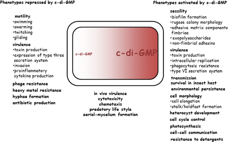
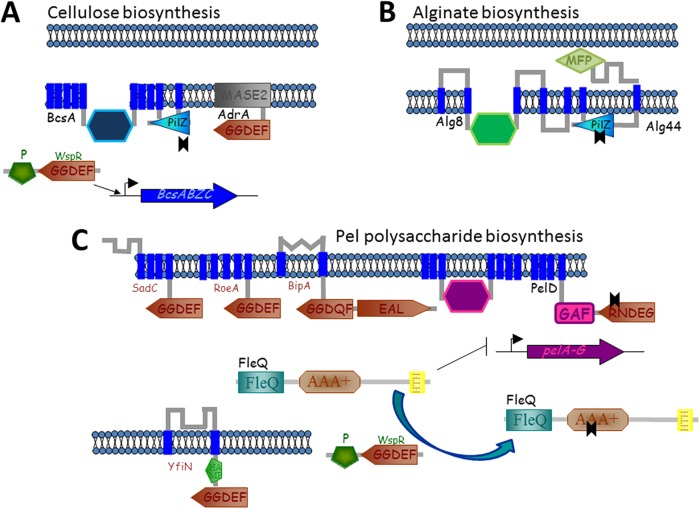
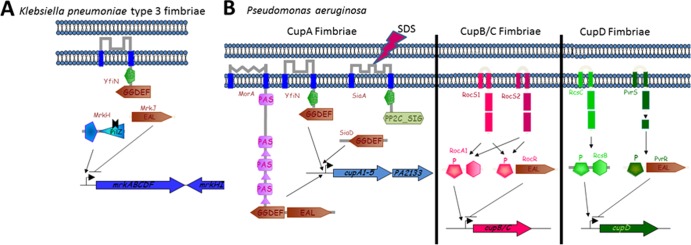
Twenty-five years have passed since the discovery of cyclic dimeric (3'→5') GMP (cyclic di-GMP or c-di-GMP). From the relative obscurity of an allosteric activator of a bacterial cellulose synthase, c-di-GMP has emerged as one of the most common and important bacterial second messengers. Cyclic di-GMP has been shown to regulate biofilm formation, motility, virulence, the cell cycle, differentiation, and other processes. Most c-di-GMP-dependent signaling pathways control the ability of bacteria to interact with abiotic surfaces or with other bacterial and eukaryotic cells. Cyclic di-GMP plays key roles in lifestyle changes of many bacteria, including transition from the motile to the sessile state, which aids in the establishment of multicellular biofilm communities, and from the virulent state in acute infections to the less virulent but more resilient state characteristic of chronic infectious diseases. From a practical standpoint, modulating c-di-GMP signaling pathways in bacteria could represent a new way of controlling formation and dispersal of biofilms in medical and industrial settings. Cyclic di-GMP participates in interkingdom signaling. It is recognized by mammalian immune systems as a uniquely bacterial molecule and therefore is considered a promising vaccine adjuvant. The purpose of this review is not to overview the whole body of data in the burgeoning field of c-di-GMP-dependent signaling. Instead, we provide a historic perspective on the development of the field, emphasize common trends, and illustrate them with the best available examples. We also identify unresolved questions and highlight new directions in c-di-GMP research that will give us a deeper understanding of this truly universal bacterial second messenger.


Figures

Fig 1
Fig 1
Three-dimensional structures of cyclic di-GMP. Carbon atoms are shown in green, nitrogen in blue, oxygen in red, and phosphorus in orange. (A and B) Cyclic di-GMP monomer (from Protein Data Bank [PDB] entry 3N3T). This form is usually seen bound to the EAL domain, e.g., in PDB entries 3GG1, 3N3T, 2W27, and 3HV8 (–65, 85). Note the characteristic 12-member ribose-phosphate ring in the center of the molecule. (C and D) Cyclic di-GMP dimer (from PDB entry 2L74). This form has been seen bound to the allosteric site of PleD (PDB entry 1W25), PilZ domains (PDB entries 2L74 and 3KYF), the transcriptional regulator VpsT (PDB entry 3KLO), and a riboswitch (PDB entry 3MUT) (, , –84).
Fig 2
Fig 2
Basic biochemistry of c-di-GMP synthesis, degradation, and c-di-GMP receptors. The diagrams show the protein domains involved in c-di-GMP metabolism and signaling. Enzymatically active GGDEF, EAL, and HD-GYP domains are shown on a white background. Enzymatically inactive domains involved in substrate binding are shown in light gray, and domains that are no longer associated with c-di-GMP are shown in dark gray. (Adapted from reference .)
Fig 3
Fig 3
Sequence conservation in cyclic di-GMP-related domains. Sequence logos of the GGDEF (A), EAL (B), HD-GYP (C), and PilZ (D) domains were generated with the WebLogo tool (457) from sequence alignments of Pfam (116) entries PF00990, PF00563, PF01966, and PF07238, respectively. Residue numbering is from Conserved Domain Database (140) entries cd01949, cd01948, cd00077, and cl01260, respectively. The height of each letter reflects the relative frequency of the corresponding amino acid at that position; the overall height of the column reflects the degree of sequence conservation at that position (measured in bits). The eponymous sequence motifs correspond to residues 79 to 83 in panel A, residues 31 to 33 in panel B, and residues 38, 39, and 101 to 103 in panel C.
Fig 4
Fig 4
Conservation of active site residues in various GGDEF and EAL domains. The residues that form the enzyme active sites and are required for the diguanylate cyclase activity of the GGDEF domain (A) or the c-di-GMP phosphodiesterase activity of the EAL domain (B) are shown in white on a red or blue background; other conserved residues in the vicinity of the active sites are shown in bold. Yellow shading in panel A indicates the residues forming the allosteric I site. The residue numbering shows positions of the respective amino acids in Conserved Domain Database (140) entries cd01949 (GGDEF) and cd01948 (EAL) and in Caulobacter crescentus PleD (UniProt entry Q9HX69), Pseudomonas aeruginosa WspR (UniProt entry Q3SJE6) and RocR (UniProt entry Q9HX69), and Thiobacillus denitrificans TBD1265 (UniProt entry Q3SJE6) (36, 65, 94, 118). (Modified from references and and based on previous data [38, 65, 94, 118, 125, 267].)
Fig 5
Fig 5
Structural organization of the active sites of cyclic di-GMP-related molecules. The upper row shows enzymes of c-di-GMP metabolism, and the lower row shows c-di-GMP-binding proteins and riboswitches. The residues highlighted in Fig. 3 and 4 are shown with the same numbers. Residue coloring is as in Fig. 1, except that carbon atoms of GTPαS and c-di-GMP are in silver, and Mg and Fe atoms are shown as pink spheres. (A) Active site of the GGDEF domain of PleD with the bound substrate analog GTPαS (PDB entry 2V0N) (86). The catalytic Asp/Glu81 residue is shown in gold, Gly79 and Gly80 of the GGDEF motif are in silver, and Arg70 and Asp73 of the RxxD motif in the allosteric inhibitory I site (36) are shown in red. (B) Active site of the EAL domain of Tbd1265 with bound c-di-GMP (PDB entry 3N3T) (65). Glu31 and Leu33 residues of the EAL motif are shown in gold. (C) Active site of the HD-GYP domain of Bd1817 with bound c-di-GMP (PDB entry 3TM8) (129). His38, Asp39, Gly101, and Pro103 of the HD and GYP motifs are shown in gold (Tyr102 is missing in Bd1817). (D) c-di-GMP binding site of the PilZ domain of PA4608 (PDB entry 2L74) (82). For simplicity, one of the c-di-GMP molecules is shown only as lines. (E) c-di-GMP bound to riboswitch I (PDB entry 3IRW) (75). (F) c-di-GMP bound to riboswitch II (PDB entry 3Q3Z) (76). (G) c-di-GMP bound to the stimulator of interferon genes STING (PDB entry 4EMT) (; see references to for further details). The figure was generated with PyMOL (Schrödinger, LLC).
Fig 6
Fig 6
Phenotypes that are regulated by cyclic di-GMP signaling. On the left are target outputs activated by low c-di-GMP or repressed by high c-di-GMP (require the absence of c-di-GMP binding to the cognate receptor for expression), and on the right are target outputs activated by high c-di-GMP (require c-di-GMP binding to the cognate receptor for expression). Some processes can be repressed and activated by c-di-GMP, depending on the bacterial species and conditions. (Adapted from reference 12 with permission of the publisher. Copyright © 2009 Karger Publishers, Basel, Switzerland.)
Fig 7
Fig 7
Regulation of exopolysaccharides by c-di-GMP signaling. (A) Bacterial cellulose biosynthesis is positively regulated by c-di-GMP signaling on the posttranslational level, as the cellulose synthase BcsA contains a PilZ domain at its C-terminal end which binds c-di-GMP (1, 52). For example, in S. enterica, the DGC AdrA provides the c-di-GMP to activate cellulose biosynthesis (30). The DGC WspR regulates transcription of cellulose biosynthesis operons upon constitutive activation in P. fluorescens (261). (B) Alginate polymerization by the alginate synthase Alg8 requires activation by the c-di-GMP receptor protein Alg44 (149). (C) The PelD protein is the c-di-GMP receptor that activates biosynthesis of the exopolysaccharide PelD on the posttranslational level, with regulation of the c-di-GMP pool through the DGCs RoeA (PA1107) and SadC (PA4332) and the PDE BifA (PA4367) (229, 230). Repression of pel transcription by the transcriptional regulator FleQ is relieved upon c-di-GMP binding with the DGCs WspR (PA3702) and YfiN (PA1120), providing the c-di-GMP (93, 168). See the text for further details.
Fig 8
Fig 8
Regulation of type 3 fimbriae and Cup fimbriae by c-di-GMP signaling. (A) Type 3 fimbriae of K. pneumoniae and Cup fimbriae of P. aeruginosa are positively regulated on the transcriptional level by c-di-GMP signaling. An entire regulatory circuit of c-di-GMP signaling has been identified for transcriptional regulation of type 3 fimbriae. The transcriptional regulator MrkH binds c-di-GMP produced by the DGC YfiN and subsequently activates transcription of the type 3 fimbria mrkABCDF operon (120, 158), while the PDE MrkJ represses mrkABCDF transcription by degrading c-di-GMP (158, 286). (B) Transcription of CupA fimbriae is activated in response to detergent (sodium dodecyl sulfate [SDS]) exposure by the DGC SiaA (PA0172) (290) or in small-colony variants of P. aeruginosa by the DGCs MorA (PA2474) and YfiN (PA1120) (97). Transcription of CupB/C fimbriae is repressed by the response regulator PDE RocR (PA3947) (282), while repression of CupD fimbriae requires the response regulator PDE PvrR (283). See the text for further explanation.
Fig 9
Fig 9
Regulation of virulence, type IV pilus motility, and biofilm formation in X. campestris. The transcriptional regulator Clp controls a virulence regulon of more than 300 genes, among them engXCA, encoding endoglucanase. Cyclic di-GMP binding to Clp prevents transcription and abolishes virulence. The c-di-GMP PDEs RavR and RpfG additively create an environment low in c-di-GMP, which is required for the activity of Clp. Hypoxia and the diffusible signaling factor DSF activate the respective sensor kinases RavS and RpfC, which phosphorylate RavR and RpfG. RpfF is required for the synthesis of DSF. Besides activation of virulence, RpfG but not RavR is required for type IV pilus-mediated motility and repression of biofilm formation. PilZ domain-containing proteins XC_2249 and XC_3221 appear to function as an adaptor in the interaction of two different protein complexes with two different ATPases. RpfG in a complex with the diguanylate cyclases XC_0249 and XC_0420 recruits the XC_2249 adaptor and interacts with the PilT/PilU ATPases required for pilus retraction (295a). This interaction stimulates motility, as does XC_3221-mediated interaction of the c-di-GMP binding protein FimX with the pilus polymerization ATPase PilB. Mechanisms of RpfG suppression of biofilm formation involve inhibition of transcription of the putative exopolysaccharide operon xagABC and activation of dispersion (222).
Fig 10
Fig 10
Cyclic di-GMP signaling circuit controlling surface location of the P. fluorescens adhesin LapA. LapA can exist in a cell surface-associated or proteolytically processed supernatant released form. Cyclic di-GMP binding to the c-di-GMP receptor LapD (Pfl01_0131) sequesters the periplasmic protease LapG and prevents LapA cleavage (67, 68). Cyclic di-GMP dedicated to binding to LapD is produced by the DGCs GcbB (Pfl01_1789) and GcbC (Pfl01_4666), while RapA (Pfl01_1678) degrades the respective c-di-GMP (167, 302). LapEBC proteins constitute components of the type I secretion system. The PhoR-PhoB two-component system responds to alterations in environmental phosphate levels to regulate LapA's surface location (302). Low phosphate activates the response regulator PhoB, which represses the expression of the LapA type I secretion system and activates expression of the c-di-GMP PDE RapA. RapA activity favors the supernatant released form of LapA and subsequently leads to biofilm dispersal.
Fig 11
Fig 11
Specificity and redundancy of c-di-GMP-mediated regulation of target output. The apparent redundancy of c-di-GMP signaling proteins in biofilm formation and other phenotypes might be caused by distinct molecular mechanisms. (A) More than one target is required for phenotype output, and individual targets are affected by distinct c-di-GMP signaling pathways consisting of a pair of c-di-GMP-metabolizing proteins and an effector protein. (B) One target causes the phenotype output. Cyclic di-GMP from several cyclic di-GMP-metabolizing proteins simultaneously contributes to target output through one effector. (C) Alternatively, target output is affected by c-di-GMP signaling on several levels through different effector proteins. Specific c-di-GMP-metabolizing proteins provide the c-di-GMP for each effector.
Fig 12
Fig 12
Regulation of biofilm formation in V. cholerae El Tor. The transcriptional regulators VpsR and VpsT activate expression of the Vps exopolysaccharide required for rugose colony morphology and biofilm formation upon c-di-GMP binding (242). The c-di-GMP signaling network that regulates biofilm formation consists of various DGCs (CdgA, CdgG, CdgH, and VpvC) and PDEs (RocS, MbaA, and CdgC). V. cholerae quorum sensing represses biofilm formation through the quorum sensing regulator HapR, which regulates c-di-GMP-metabolizing proteins, among them CdgA and CdgG, and the transcription of vpsT (62). On the other hand, HapR is repressed by c-di-GMP through VpsT and VpsR via the virulence regulator AphA (459).
Fig 13
Fig 13
Components of the c-di-GMP signaling network regulate cell cycle progression and cell differentiation. Only components proficient in metabolizing c-di-GMP and c-di-GMP-dependent processes are shown. Elevated c-di-GMP levels in cells upon G1- to S-phase transition are indicated by a gray background. PleD, which is dispersed in the cytoplasm of swarmer cells due to dephosphorylation by PleC, becomes DGC proficient upon phosphorylation and localizes to the differentiating pole (37). DgcB displays DGC activity, presumably throughout the cell cycle, whereby the PDE PdeA antagonizes DgcB activity in swarmer cells (318). Recruited to the cell pole by a c-di-GMP-independent mechanism, PopA remains localized at the differentiating pole upon swarmer- to stalked-cell transition, where it mediates S-phase entry and CtrA degradation upon c-di-GMP binding to its I site (162). DgrA and DgrB are PilZ domain c-di-GMP receptor proteins (51) which stall motility upon elevated c-di-GMP levels. DgrA and DgrB potentially act in the predivisional cell, stalling motility before the completion of cytokinesis and/or upon surface sensing, when the flagellar rotation slows down (460).
Fig 14
Fig 14
Cyclic di-GMP signaling models. (A) Spatial proximity of c-di-GMP-metabolizing proteins, the effector, and/or target output is required for effective signaling. In this case, c-di-GMP synthesis and degradation are probably low. (B) The DGC and/or PDE is distant from the effector/target. Cyclic di-GMP synthesis of the DGC is probably high. (C) Processes inhibited by c-di-GMP signaling are more likely to be affected by signaling from a distance.

References

    1. Ross P, Weinhouse H, Aloni Y, Michaeli D, Weinberger-Ohana P, Mayer R, Braun S, de Vroom E, van der Marel GA, van Boom JH, Benziman M. 1987. Regulation of cellulose synthesis in Acetobacter xylinum by cyclic diguanylic acid. Nature 325:279–281 - PubMed
    1. D'Argenio DA, Miller SI. 2004. Cyclic di-GMP as a bacterial second messenger. Microbiology 150:2497–2502 - PubMed
    1. Jenal U. 2004. Cyclic di-guanosine-monophosphate comes of age: a novel secondary messenger involved in modulating cell surface structures in bacteria? Curr. Opin. Microbiol. 7:185–191 - PubMed
    1. Jenal U, Malone J. 2006. Mechanisms of cyclic-di-GMP signaling in bacteria. Annu. Rev. Genet. 40:385–407 - PubMed
    1. Römling U, Gomelsky M, Galperin MY. 2005. c-di-GMP: the dawning of a novel bacterial signalling system. Mol. Microbiol. 57:629–639 - PubMed

Publication types

MeSH terms

Substances

LinkOut - more resources