Review
. 2013 Mar;77(1):112-43.
doi: 10.1128/MMBR.00054-12.

Molecular regulation of antibiotic biosynthesis in streptomyces

Affiliations
Review

Molecular regulation of antibiotic biosynthesis in streptomyces

Gang Liu et al. Microbiol Mol Biol Rev. 2013 Mar.

Abstract

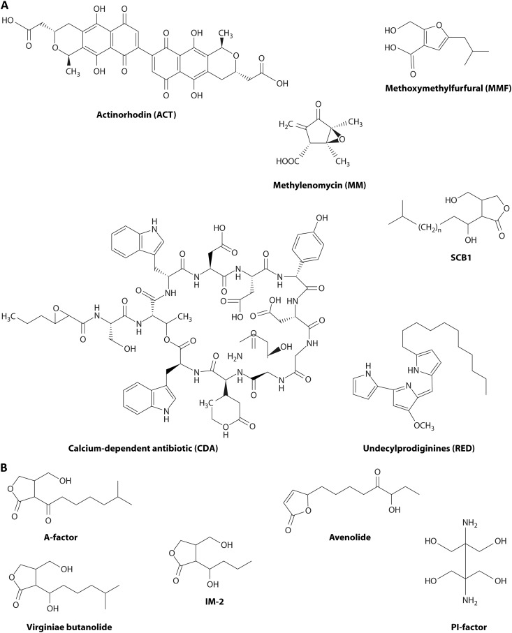
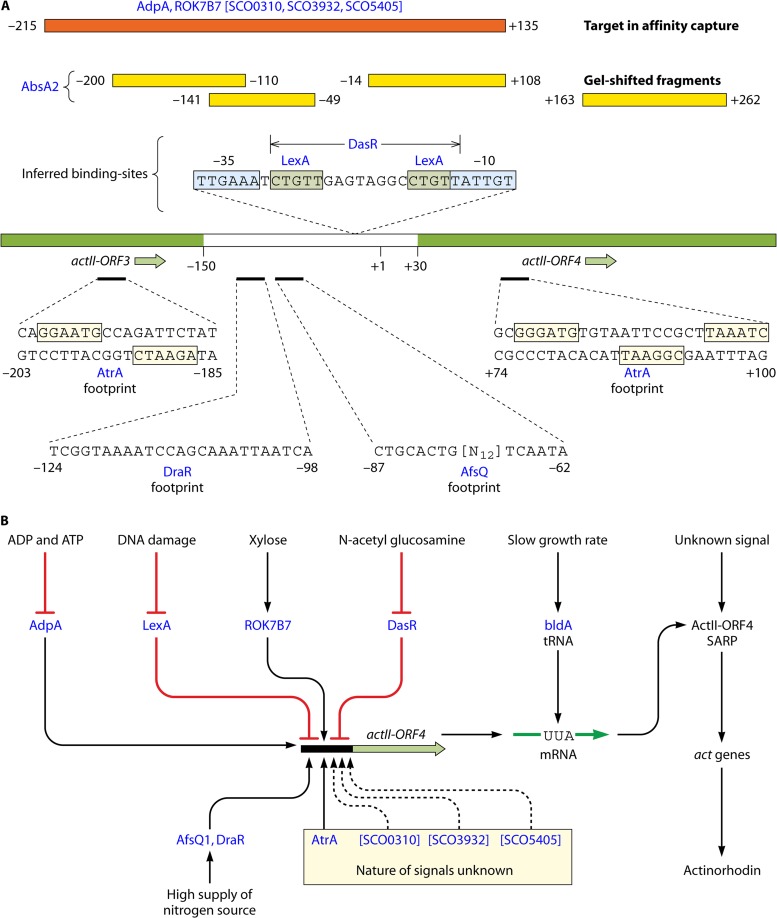
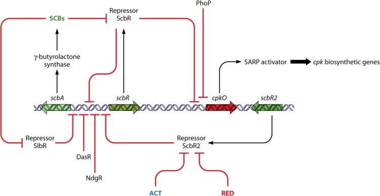
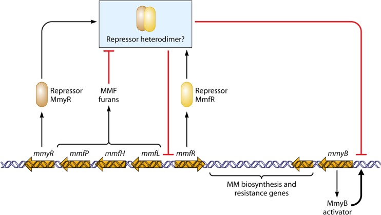
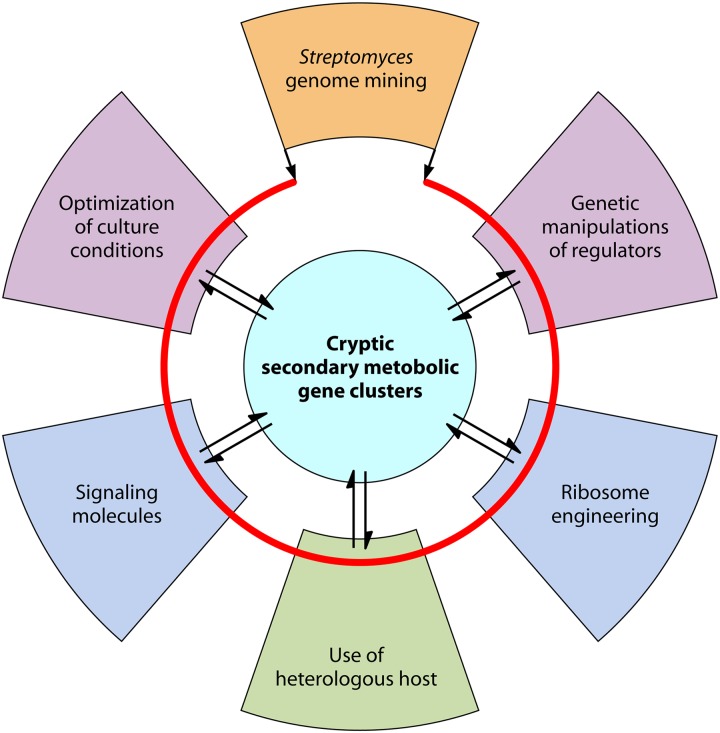
Streptomycetes are the most abundant source of antibiotics. Typically, each species produces several antibiotics, with the profile being species specific. Streptomyces coelicolor, the model species, produces at least five different antibiotics. We review the regulation of antibiotic biosynthesis in S. coelicolor and other, nonmodel streptomycetes in the light of recent studies. The biosynthesis of each antibiotic is specified by a large gene cluster, usually including regulatory genes (cluster-situated regulators [CSRs]). These are the main point of connection with a plethora of generally conserved regulatory systems that monitor the organism's physiology, developmental state, population density, and environment to determine the onset and level of production of each antibiotic. Some CSRs may also be sensitive to the levels of different kinds of ligands, including products of the pathway itself, products of other antibiotic pathways in the same organism, and specialized regulatory small molecules such as gamma-butyrolactones. These interactions can result in self-reinforcing feed-forward circuitry and complex cross talk between pathways. The physiological signals and regulatory mechanisms may be of practical importance for the activation of the many cryptic secondary metabolic gene cluster pathways revealed by recent sequencing of numerous Streptomyces genomes.


Figures

Fig 1
Fig 1
Diverse antibiotics and autoregulator molecules produced by Streptomyces coelicolor A3(2) and some other streptomycetes. (A) The compounds from S. coelicolor. Actinorhodin (ACT) is a red/blue pH-indicating benzoisochromanequinone made by a type II polyketide synthase-based pathway involving a 22-gene cluster (291). Undecylprodiginines (REDs) are red hydrophobic tripyrroles made by a fatty acid synthase-like pathway involving a 22-gene cluster (292). Methylenomycin A (MM) is an epoxycyclopentenone made by an unusual pathway encoded by 11 genes located on the linear plasmid SCP1 (57). The calcium-dependent antibiotic (CDA) is a lipopeptide made by a route involving nonribosomal peptide synthases and specified by a 48-gene cluster (293). The autoregulator SCB1 (240) is one of several gamma-butyrolactone congeners whose synthesis involves three genes linked to the 19-gene cluster for biosynthesis of a structurally incompletely characterized yellow antibiotic (S. coelicolor polyketide [CPK]) (41, 42, 294). Methylenomycin furans (MMFs) are autoregulators that control MM biosynthesis, and they are made by a pathway resembling that of gamma-butyrolactones that involves three biosynthetic and two regulatory genes next to the MM biosynthetic cluster (58). (B) Autoregulatory molecules from other streptomycetes. A-factor regulates streptomycin biosynthesis in S. griseus (295). Virginiae butanolides (VBs) induce the coordinated production of virginiamycin M and virginiamycin S, synergistically acting but biosynthetically distinct antibiotics in S. virginiae (235). IM-2 controls the production of showdomycin and minimycin in S. lavendulae (237). PI factor enhances pimaricin production in S. natalensis (239). Avenolide is required at nanomolar concentrations for the onset of avermectin biosynthesis in S. avermitilis (238) (avenolide homologs are also found in S. fradiae, Streptomyces ghanaensis, and S. griseoauranticus).
Fig 2
Fig 2
Complexity of the promoter region of actII-ORF4. (A) Binding of regulatory proteins. The central line represents the actII-ORF3 to actII-ORF4 region, with the noncoding intergenic region given in white and coding sequences in green. Numbers refer to the transcription start site (+1) defined by Gramajo et al. (296). Binding to the diverse regulators (red lettering) was defined by different routes: footprinting results are given below the promoter, while gel-shifted fragments are indicated above as yellow boxes. Brackets indicate proteins defined only by affinity capture to the orange fragment shown. Also shown are binding sites inferred by coupling of experimental evidence of interaction with the promoter region and the presence of matches to known consensus binding sites. References are given in the text. (B) General nature of signal inputs influencing the expression of actII-ORF4. See the text for further details and references.
Fig 3
Fig 3
Regulation of CPK biosynthesis involves a gamma-butyrolactone and interplay with other biosynthetic pathways. Genes associated with the cpk cluster are indicated by large open arrows. Regulatory interactions are indicated by bold arrows (activation steps) or bold lines ending with a bar (repressing or inhibitory steps). Small-molecule ligands are indicated in colored letters, with ACT and RED being the products of other pathways encoded by distant gene clusters. For further explanation and references, see the text.
Fig 4
Fig 4
Regulation of methylenomycin biosynthesis, a cascade involving furan autoregulators. Large open arrows indicate genes associated with the gene cluster for MM biosynthesis. Regulatory interactions are indicated by bold arrows (activation steps) or bold lines ending with a bar (repressing or inhibitory steps). See the text for further details and references.
Fig 5
Fig 5
RedZ and other variations on the theme of two-component regulators associated with antibiotic production. (A) The typical two-component system containing a sensor histidine kinase and a cognate response regulator (RR), usually encoded by a pair of adjacent genes. ATP or GTP is used for autophosphorylation by the histidine kinase, and the phosphoryl group is transferred to the RR, which then controls the transcription of target genes. (B) Atypical response regulators (ARRs) such as RedZ (shown here) do not contain all the conserved amino acid residues important for phosphorylation of the receiver domain. Some ARRs can be activated by binding to the end product or late biosynthetic intermediates of secondary metabolites, such as antibiotics (see the text). (C) Alignments of ARRs from Streptomyces. Top, OmpR family: JadR1 (jadomycin, Streptomyces venezuelae [69]), Aur1P (auricin, Streptomyces aureofaciens [297]), LanI (landomycins, Streptomyces cyanogenus [298]), and LndI (landomycin E, Streptomyces globisporus [299]). Bottom, NarI family: RedZ (undecylprodigiosin, S. coelicolor [67, 68]), DnrN (daunorubicin, Streptomyces peucetius [209]), NcnR (naphthocyclinone, Streptomyces arenae [300]), VioR (tuberactinomycin, Streptomyces vinaceus [301]), and SCO1654 (protein of S. coelicolor with unknown function). The red boxes indicate residues corresponding to the conserved residues of conventional response regulators.
Fig 6
Fig 6
Nutrient-sensing regulators of antibiotic production in S. coelicolor and their cross talk. The diagram summarizes routes by which the availability of sources of carbon, nitrogen, and phosphate influence the expression of the cluster-situated regulators (CSRs) that activate pathways leading to antibiotic biosynthesis. Nutrient availability is sensed by membrane-located sensor kinases or through the transport of nutrients, leading to activation of global regulators (circled). The global regulators control both central metabolic genes and CSR genes, either directly (solid lines) or through unknown routes (dotted lines). Arrows indicate activation, and bars indicate repression. For further information and references, see the text.
Fig 7
Fig 7
Model for the regulation of HpdR during tyrosine catabolism and CDA biosynthesis in S. coelicolor. As an autoregulator, HpdR regulates its own transcription (not shown). It also represses hppD, the product of which catalyzes the conversion of 4-hydroxyphenypyruvate (4HHP) to homogentisate, but activates hmaS, which is involved in CDA biosynthesis. During vegetative growth, l-tyrosine in the medium is catabolized to 4HHP by TyrB. The accumulated 4HHP binds to HpdR and causes it to dissociate from the hppD and hmaS promoters, therefore initiating hppD expression and preventing hmaS expression. During stationary phase, l-tyrosine in the medium is limited, 4HHP is reduced, and expression of hpdD is repressed, while hmaS expression is activated, directing 4HPP into CDA biosynthesis.
Fig 8
Fig 8
Regulation of antibiotic production by AfsR and its possible interface with hyphal tip growth. The upper part of the figure represents a hypha extending by tip growth, under the control of a tip-located “polarisome.” DivIVA and AfsK are located in the polarisome. As the tip extends, the DivIVA and AfsK content increases, and AfsK action causes polarisome splitting and the nucleation of a new branch point, such as that seen emerging from the subapical compartment. Also shown is a compartment that happens not to have captured a nascent polarisome. This is enlarged in the lower part of the figure. It is suggested that DivIVA-free AfsK in this compartment phosphorylates AfsR. The phosphorylation is potentially modulated by KbpA protein, S-adenosylmethionine, and precursors of cell wall biosynthesis that may accumulate in the absence of a growth point. For further explanation and references, see the text.
Fig 9
Fig 9
Regulation of the key pleiotropic regulatory gene adpA. Transcription of adpA has been studied in S. griseus and S. coelicolor, as well as in S. lividans, which is very closely related to S. coelicolor. In each case, repressors that sense species-specific gamma-butyrolactones interact with the adpA promoter; and the pleiotropic regulator BldD also represses expression. AdpA is also autorepressing—circuitry that also implicates cross-regulation with bldA, whose product is the tRNA needed to translate a rare UUA codon that is found in the same place in virtually all streptomycetes analyzed. For further explanation and references, see the text.
Fig 10
Fig 10
Cross-coordination of different antibiotic biosynthetic pathways by the pseudo-GBL receptors in S. venezuelae. The pseudo-gamma-butyrolactone receptor JadR2 directly represses the transcription of jadR1 in the absence of ethanol and also binds chloramphenicol (Cm) and jadomycins. The cluster-situated regulator JadR1 activates the biosynthesis of jadomycin B by activating the transcription of biosynthetic structural genes. JadR1 also represses the production of Cm by binding to the promoters of the structural genes (51).
Fig 11
Fig 11
PolY controls the transcription of polR by sensing the ATP/ADP in the cells of S. cacaoi. Binding of ADP/ATPγS to the ATPase domain of PolY triggers the oligomerization of PolY and enhances its DNA binding affinity in vitro. Changes of ADP/ATP concentrations significantly affect the binding activity of PolY in vivo (232).
Fig 12
Fig 12
Acquisition of global regulators during the evolution of streptomycetes. The multilocus phylogeny on which the diagram is based was derived from a catenated set of seven conserved proteins (AtpD, DnaA, DnaG, DnaK, GyrB, RecA, and RpoB) (G. Chandra, unpublished data). Points of acquisition of global regulators are based on the presence or absence of orthologues encoded in the genomes of organisms before or after branch points.
Fig 13
Fig 13
Strategies for the activation of cryptic secondary metabolic gene clusters in Streptomyces. The red line indicates the sequenced Streptomyces linear chromosome. Trapezoidal blocks with different colors represent different methods based on gene clusters to activate the possible expression of clusters.
None
None
None
None
None

References

    1. Rudd BA. 1978. Genetics of pigmented secondary metabolites in Streptomyces coelicolor. University of East Anglia, Norwich, United Kingdom
    1. Rudd BA, Hopwood DA. 1979. Genetics of actinorhodin biosynthesis by Streptomyces coelicolor A3(2). J. Gen. Microbiol. 114: 35– 43 - PubMed
    1. Rudd BA, Hopwood DA. 1980. A pigmented mycelial antibiotic in Streptomyces coelicolor: control by a chromosomal gene cluster. J. Gen. Microbiol. 119: 333– 340 - PubMed
    1. Kirby R, Wright LF, Hopwood DA. 1975. Plasmid-determined antibiotic synthesis and resistance in Streptomyces coelicolor. Nature 254: 265– 267 - PubMed
    1. Chater KF, Bruton CJ. 1985. Resistance, regulatory and production genes for the antibiotic methylenomycin are clustered. EMBO J. 4: 1893– 1897 - PMC - PubMed

Publication types

MeSH terms