. 2013 May;22(5):641-9.
doi: 10.1002/pro.2248. Epub 2013 Apr 3.

A polymetamorphic protein

Affiliations

A polymetamorphic protein

Katie L Stewart et al. Protein Sci. 2013 May.

Abstract

Arc repressor is a homodimeric protein with a ribbon-helix-helix fold. A single polar-to-hydrophobic substitution (N11L) at a solvent-exposed position leads to population of an alternate dimeric fold in which 3₁₀ helices replace a β-sheet. Here we find that the variant Q9V/N11L/R13V (S-VLV), with two additional polar-to-hydrophobic surface mutations in the same β-sheet, forms a highly stable, reversibly folded octamer with approximately half the α-helical content of wild-type Arc. At low protein concentration and low ionic strength, S-VLV also populates both dimeric topologies previously observed for N11L, as judged by NMR chemical shift comparisons. Thus, accumulation of simple hydrophobic mutations in Arc progressively reduces fold specificity, leading first to a sequence with two folds and then to a manifold bridge sequence with at least three different topologies. Residues 9-14 of S-VLV form a highly hydrophobic stretch that is predicted to be amyloidogenic, but we do not observe aggregates of higher order than octamer. Increases in sequence hydrophobicity can promote amyloid aggregation but also exert broader and more complex effects on fold specificity. Altered native folds, changes in fold coupled to oligomerization, toxic pre-amyloid oligomers, and amyloid fibrils may represent a near continuum of accessible alternatives in protein structure space.


Figures

Figure 1
Figure 1
Hydrophobicity mutations and structural changes in the β-sheet of Arc repressor. In the wild-type Arc repressor homodimer (top left), residues 9–14 of each subunit alternate between polar (red) and hydrophobic (cyan) residues and interact to form a two-stranded β-sheet. A hydrophobic mutation at residue 11 (Arc-N11L) populates an alternate fold in which the β-sheet is replaced by 310 helices (top right; determined in the switch Arc variant, which forms this fold exclusively). Two additional polar-to-hydrophobic substitutions produce a fully hydrophobic strand region in Arc-VLV.
Figure 2
Figure 2
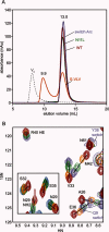
(A) Size exclusion chromatograms of Arc-S-VLV (orange), switch Arc (lavender), Arc-N11L (green), and Arc-WT (red), at 300 μM protein concentration in 50 mM MES (pH 5.5), 50 mM KCl. Switch Arc, Arc-N11L, and WT migrate as dimers with elution volumes of 13.0 ± 0.1 mL, while Arc-S-VLV migrates as a mixture of dimer (13.0 mL) and higher-order oligomer (9.9 mL). V0 (dashed line) indicates the void volume of the column, measured using blue dextran. B) Overlay of HSQC spectra for the four Arc variants at 25°C, colored as in panel A. Black lines track chemical shift changes for equivalent resonances. For all resonances shown, Arc-N11L and S-VLV have intermediate chemical shift values, indicative of two different Arc dimer folds in fast conformational exchange.
Figure 3
Figure 3
Size exclusion chromatograms of wild-type Arc (blue solid trace) and heated S-VLV (orange solid trace), both at 350 μM loading concentration, along with calibration standards (green dashed trace) labeled with molecular weights in kDa. The column void volume (V0) is measured as the elution volume of blue dextran. The inset shows a calibration plot for estimation of molecular weight of S-VLV (65 kDa based on the regression) and wild-type Arc (15 kDa based on the regression, with an actual dimeric size of 15.4 kDa).
Figure 4
Figure 4
Native nESI-MS of wild-type Arc and S-VLV, each at 50 μM protein in 0.1M ammonium acetate (pH 7.4), and CID of the S-VLV octamer. Under native MS conditions, wild-type Arc shows peaks for monomeric (M) and dimeric (D) states, while S-VLV contains peaks for monomer, dimer, and octamer (O); note that relative intensities do not directly reflect relative abundances of different oligomeric states because of instrument discrimination at higher m/z. CID of the S-VLV octamer (z = 17) produced highly charged monomer (consistent with the commonly expected oligomer CID pathway that involves unfolding and release of one subunit) and complementary heptamer (H). Charge states are indicated by superscripts.
Figure 5
Figure 5
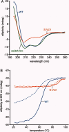
(A) Far UV circular dichroism spectra of wild-type Arc (blue), heated S-VLV (orange), and switch Arc (green) in SB250 (100 μM protein, 0.1 mm pathlength, 20°C), B) Thermal denaturation of wild-type Arc (blue) and heated S-VLV (orange) monitored at 222 nm (50 μM protein, 2.0 mm pathlength), with forward melts shown as solid lines and reverse melts shown as dashed lines.

References

    1. Chan HS, Bornberg-Bauer E. Perspectives on protein evolution from simple exact models. Appl Bioinformatics. 2002;1:121–144. - PubMed
    1. Murzin AG. Metamorphic proteins. Science. 2008;320:1725–1726. - PubMed
    1. Bryan PN, Orban J. Proteins that switch folds. Curr Opin Struct Biol. 2010;20:482–488. - PMC - PubMed
    1. Cordes MHJ, Stewart KL. The porous borders of the protein world. Structure. 2012;20:199–200. - PubMed
    1. Meier S, Ozbek S. A biological cosmos of parallel universes: does protein structural plasticity facilitate evolution? Bioessays. 2007;29:1095–1104. - PubMed

Publication types

MeSH terms

Substances

LinkOut - more resources