. 2013 Apr 19;288(16):11242-51.
doi: 10.1074/jbc.M113.457002. Epub 2013 Mar 7.

Chloroquine binding reveals flavin redox switch function of quinone reductase 2

Affiliations

Chloroquine binding reveals flavin redox switch function of quinone reductase 2

Kevin K K Leung et al. J Biol Chem. .

Abstract

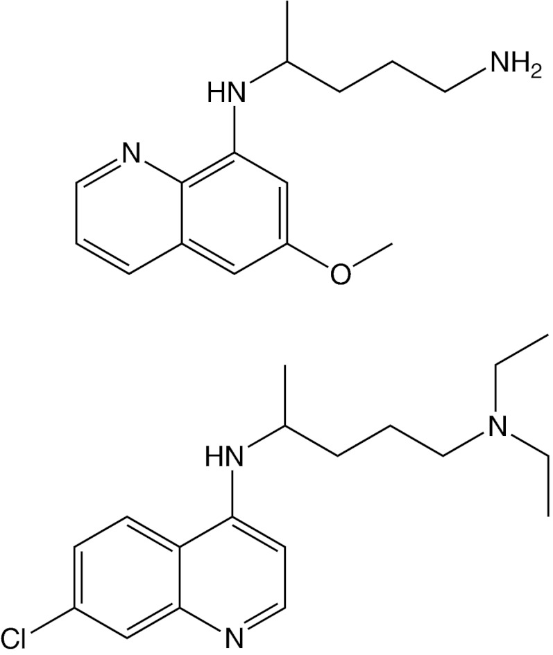
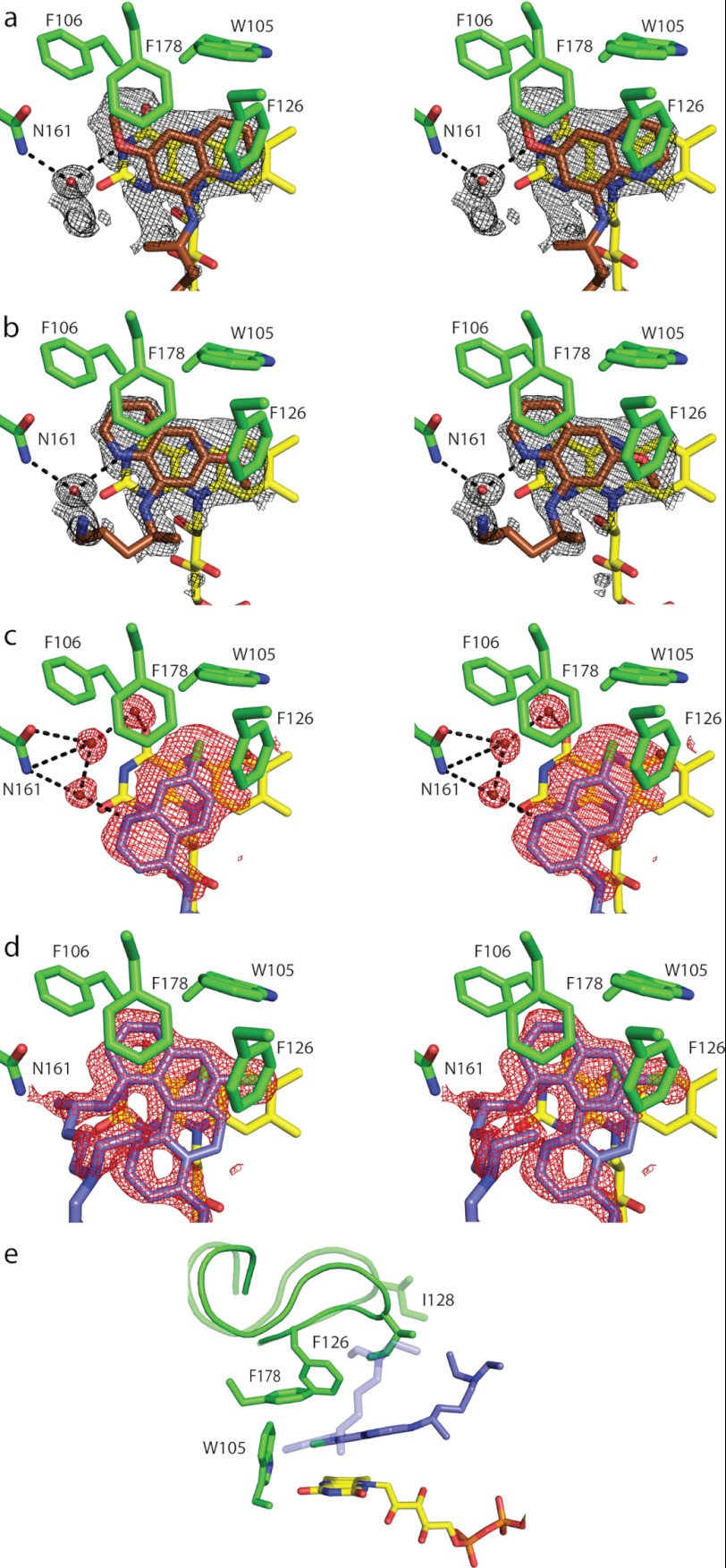
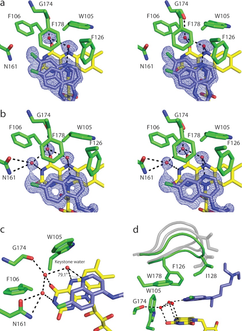
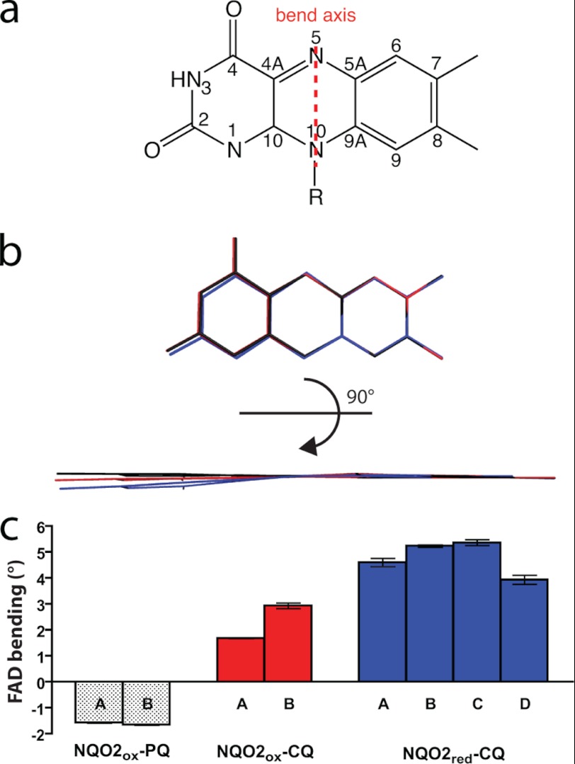
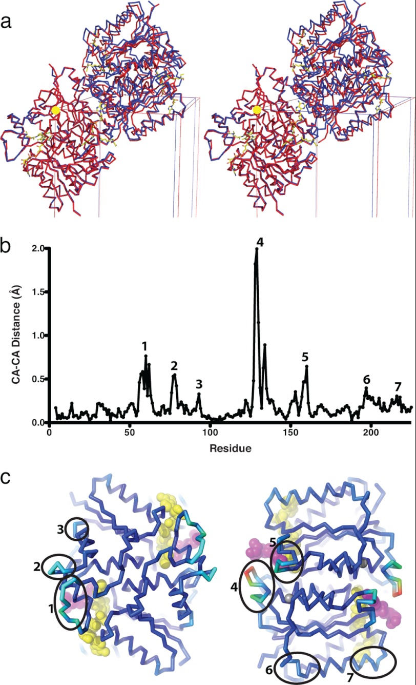
Quinone reductase 2 (NQO2) is an FAD-linked enzyme and the only known human target of two antimalarial drugs, primaquine (PQ) and chloroquine (CQ). The structural differences between oxidized and reduced NQO2 and the structural basis for inhibition by PQ and CQ were investigated by x-ray crystallography. Structures of oxidized NQO2 in complex with PQ and CQ were solved at 1.4 Å resolution. CQ binds preferentially to reduced NQO2, and upon reduction of NQO2-CQ crystals, the space group changed from P2(1)2(1)2(1) to P2(1), with 1-Å decreases in all three unit cell dimensions. The change in crystal packing originated in the negative charge and 4-5º bend in the reduced isoalloxazine ring of FAD, which resulted in a new mode of CQ binding and closure of a flexible loop (Phe(126)-Leu(136)) over the active site. This first structure of a reduced quinone reductase shows that reduction of the FAD cofactor and binding of a specific inhibitor lead to global changes in NQO2 structure and is consistent with a functional role for NQO2 as a flavin redox switch.


Figures

FIGURE 1.
FIGURE 1.
Structures of primaquine and chloroquine. Primaquine (top) and chloroquine (bottom) are two antimalarial drugs that act at different stages of the Plasmodium life cycle.
FIGURE 2.
FIGURE 2.
Binding of primaquine and chloroquine to oxidized NQO2. a–d, stereodiagrams illustrating the binding of PQ and CQ to NQO2ox. The electron density represents the final 2FoFc maps contoured at 1σ around the inhibitors. a and b show PQ bound to the A subunit in two alternate conformations. The B subunit of the NQO2ox-PQ homodimer contained similar electron density and was modeled in the same way as the A subunit. c and d show the active sites for the NQO2ox-CQ structure. Electron density in the NQO2ox-CQ subunit A (c) was different from that observed in the B subunit (d), and CQ was modeled in two alternate conformations in the B subunit. E, the two positions of CQ in the B subunit correspond to two different conformations of the active site loop, comprising residues 126–136. The active site loop in the “open” conformation (light shading) accommodates CQ when it is deeply buried in the active site, whereas the “closed” conformation (dark shading) is adopted when CQ is bound in a more peripheral location. One-letter amino acid codes are used.
FIGURE 3.
FIGURE 3.
Binding of chloroquine to reduced NQO2. a and b, stereodiagrams illustrating the binding of CQ to the NQO2red active sites in the two dimers of the asymmetric unit; the electron density corresponds to 2FoFc maps contoured at 1σ around CQ. a, the electron density for AB dimer. Shown is the active site of the A subunit, with CQ bound in two alternate positions, one of which corresponds to the position observed in the C and D subunits, whereas the second is shifted slightly. The electron density and positions of bound CQ were similar for the B subunit. b, the electron density and position of bound CQ in the CD dimer. Shown is the active site of the C subunit; the electron density in the D subunit was similar, and for both subunits, CQ was modeled in the same single position. c, the hydrogen bonding network, with the “keystone water” bridging N4 of CQ and N5 of the FAD isoalloxazine ring. d, the active site loop (residues 126–136) is shown in the fully closed conformation (green) observed in NQO2red-CQ; in this state, residue Ile128 makes van der Waals contact with CQ. For reference, the conformations of the loop observed in crystals of oxidized NQO2 are shown in gray. One-letter amino acid codes are used.
FIGURE 4.
FIGURE 4.
Structural changes in the FAD isoalloxazine ring. a, chemical structure and atom designations in the FAD isoalloxazine ring, and the axis for the “butterfly bend” that is brought about by reduction of FAD to FADH2. b, an edge-on view of the structure of the isoalloxazine ring for the following: NQO2ox with PQ (black), NQO2ox with CQ (red), and NQO2red with CQ (blue). c, binding of PQ induces a slight (−1.6°) concave bend in the isoalloxazine system, whereas binding of CQ induces a convex bend of ∼2.3°. Reduction of FAD further increases this bend to 4.8°. To determine the bend in the isoalloxazine rings, the crystal structures were subjected to simulated annealing without stereochemical restraints at the N5 and N10 positions. FAD bending along the N5–N10 axis (butterfly bend) was calculated using the atomic positions of the dimethylbenzene and pyrimidine “wings” and principal component analysis to find the angle between the two best fit planes. Error bars, S.D.
FIGURE 5.
FIGURE 5.
Conformational changes in NQO2 upon reduction and binding of CQ. a, stereodiagram illustrating the relationship between NQO2 in the oxidized (red) and reduced (blue) states. CA traces of two dimers are shown, with the origin of the unit cells indicated by a yellow sphere; the FAD molecules are drawn as stick representations. To show the changes that take place in the crystal, the dimer from the orthorhombic (P212121) NQO2ox-CQ crystal has been superimposed on one of the dimers from the monoclinic (P21) crystal of NQO2red-CQ. In orthorhombic crystals of NQO2ox, there is a single dimer in the asymmetric unit, and the second dimer shown is related by crystallographic symmetry. When NQO2-CQ is reduced (blue), the relationship between the two dimers changes, and the crystallographic symmetry is broken, resulting in a monoclinic space group with two dimers in the asymmetric unit. b, a plot of differences in average CA positions between protomers of oxidized NQO2 (NQO2ox-PQ and NQO2ox-CQ) and protomers of NQO2red-CQ. c, two views of the NQO2 dimer (CA trace) colored from blue to red according to the magnitude of the difference in average CA position between reduced and oxidized NQO2-CQ. The views are related by 180° rotation around a vertical axis; the FADH cofactor and bound CQ are represented as yellow and magenta CPK models. Regions of NQO2 (numbered 1–7) that demonstrate a significant shift upon reduction and binding of CQ (b) are indicated.

References

    1. Hill D. R., Baird J. K., Parise M. E., Lewis L. S., Ryan E. T., Magill A. J. (2006) Primaquine. Report from CDC expert meeting on malaria chemoprophylaxis I. Am. J. Trop. Med. Hyg. 75, 402–415 - PubMed
    1. Lalloo D. G., Shingadia D., Pasvol G., Chiodini P. L., Whitty C. J., Beeching N. J., Hill D. R., Warrell D. A., Bannister B. A., and HPA Advisory Committee on Malaria Prevention in UK Travellers (2007) UK malaria treatment guidelines. J. Infect. 54, 111–121 - PubMed
    1. Foley M., Tilley L. (1998) Quinoline antimalarials. Mechanisms of action and resistance and prospects for new agents. Pharmacol. Ther. 79, 55–87 - PubMed
    1. Fitch C. D. (2004) Ferriprotoporphyrin IX, phospholipids, and the antimalarial actions of quinoline drugs. Life Sci. 74, 1957–1972 - PubMed
    1. Vale N., Moreira R., Gomes P. (2009) Primaquine revisited six decades after its discovery. Eur. J. Med. Chem. 44, 937–953 - PubMed

Publication types