

Contribution of multiparameter flow cytometry immunophenotyping to the diagnostic screening and classification of pediatric cancer
- PMID: 23472067
- PMCID: PMC3589426
- DOI: 10.1371/journal.pone.0055534
Contribution of multiparameter flow cytometry immunophenotyping to the diagnostic screening and classification of pediatric cancer
Abstract
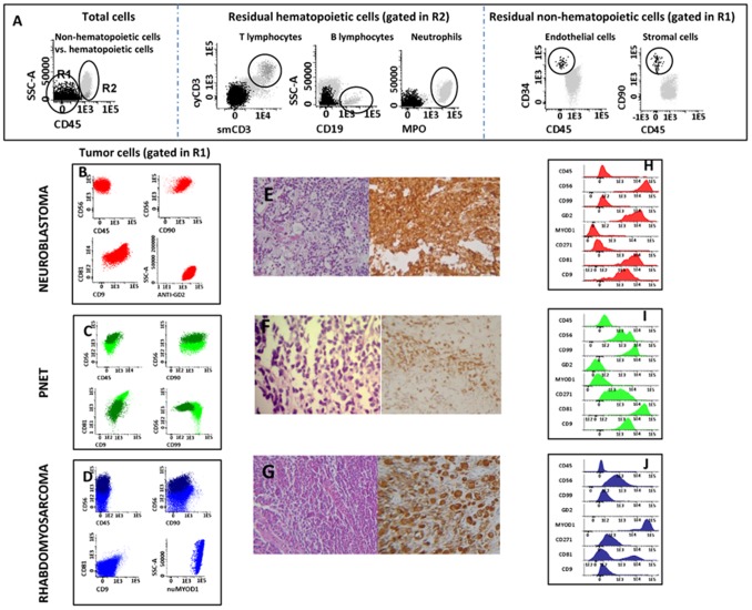
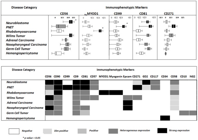
Pediatric cancer is a relatively rare and heterogeneous group of hematological and non-hematological malignancies which require multiple procedures for its diagnostic screening and classification. Until now, flow cytometry (FC) has not been systematically applied to the diagnostic work-up of such malignancies, particularly for solid tumors. Here we evaluated a FC panel of markers for the diagnostic screening of pediatric cancer and further classification of pediatric solid tumors. The proposed strategy aims at the differential diagnosis between tumoral vs. reactive samples, and hematological vs. non-hematological malignancies, and the subclassification of solid tumors. In total, 52 samples from 40 patients suspicious of containing tumor cells were analyzed by FC in parallel to conventional diagnostic procedures. The overall concordance rate between both approaches was of 96% (50/52 diagnostic samples), with 100% agreement for all reactive/inflammatory and non-infiltrated samples as well as for those corresponding to solid tumors (n = 35), with only two false negative cases diagnosed with Hodgkin lymphoma and anaplastic lymphoma, respectively. Moreover, clear discrimination between samples infiltrated by hematopoietic vs. non-hematopoietic tumor cells was systematically achieved. Distinct subtypes of solid tumors showed different protein expression profiles, allowing for the differential diagnosis of neuroblastoma (CD56(hi)/GD2(+)/CD81(hi)), primitive neuroectodermal tumors (CD271(hi)/CD99(+)), Wilms tumors (>1 cell population), rhabdomyosarcoma (nuMYOD1(+)/numyogenin(+)), carcinomas (CD45(-)/EpCAM(+)), germ cell tumors (CD56(+)/CD45(-)/NG2(+)/CD10(+)) and eventually also hemangiopericytomas (CD45(-)/CD34(+)). In summary, our results show that multiparameter FC provides fast and useful complementary data to routine histopathology for the diagnostic screening and classification of pediatric cancer.
Conflict of interest statement
Figures
References
-
- Dang-Tan T, Franco EL (2007) Diagnosis delays in childhood cancer. Cancer 110: 703–713. - PubMed
-
- Triche TJ, Hicks J, Sorensen PHB (2010) Diagnostic pathology of pediatric malignancies. Principles and Practices of Pediatric Oncology (6th ed), Pizzo PA, Poplack DG (ed). Lippincott Willians & Wilkins, Philadelphia 165–215.
-
- Wick MR (2012) Histochemistry as a tool in morphological analysis: a historical review. Ann Diagn Pathol 16: 71–78. - PubMed
-
- Rushton J, López-Terrada D (2011) Molecular and genetic basis of childhood cancer. Cancer Biomark 9: 211–234. - PubMed
Publication types
MeSH terms
LinkOut - more resources
Full Text Sources
Other Literature Sources
Research Materials
Miscellaneous
