. 2013;8(3):e57631.
doi: 10.1371/journal.pone.0057631. Epub 2013 Mar 5.

Migalastat HCl reduces globotriaosylsphingosine (lyso-Gb3) in Fabry transgenic mice and in the plasma of Fabry patients

Affiliations

Migalastat HCl reduces globotriaosylsphingosine (lyso-Gb3) in Fabry transgenic mice and in the plasma of Fabry patients

Brandy Young-Gqamana et al. PLoS One. 2013.

Abstract

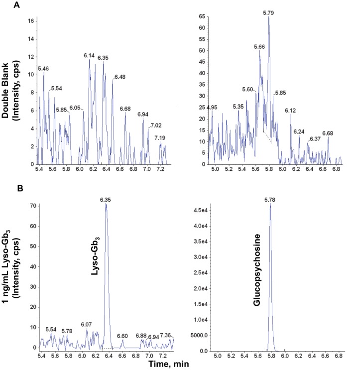
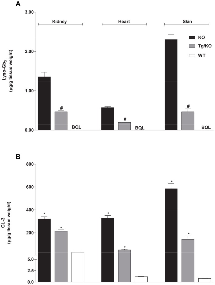
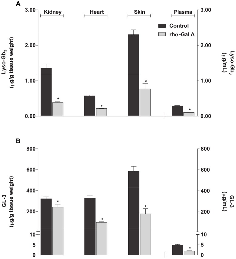
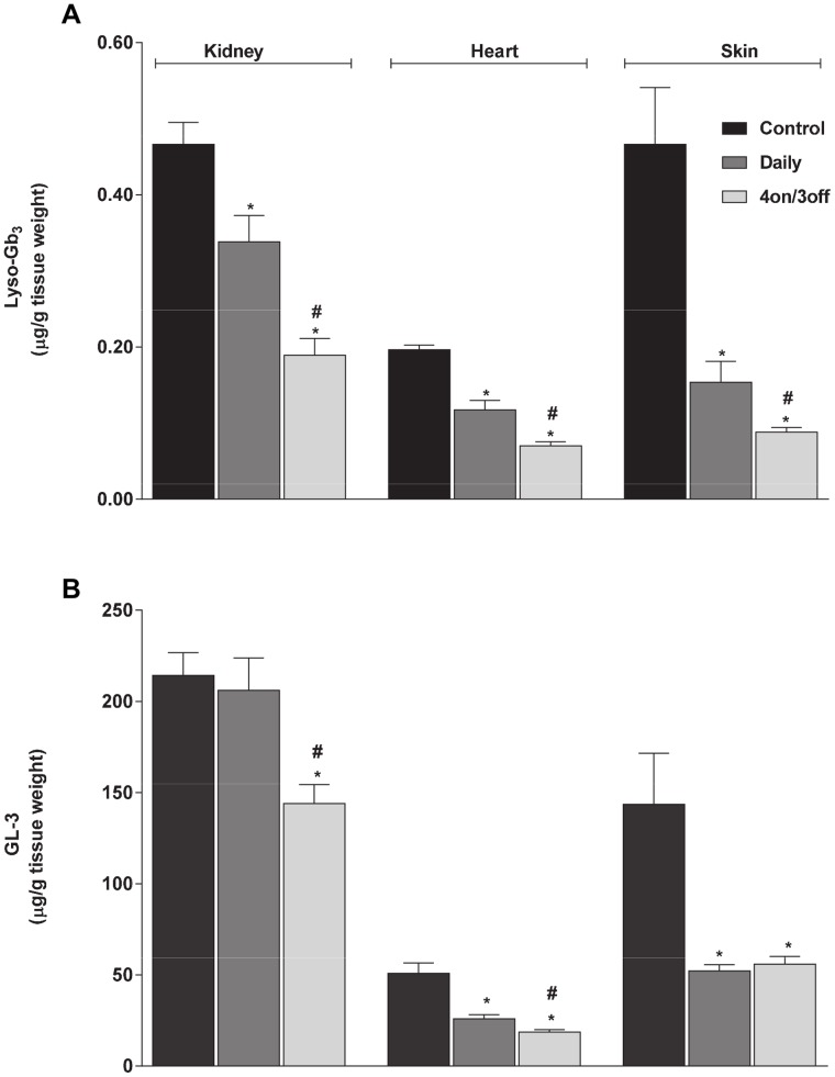
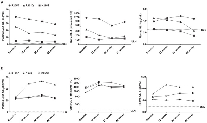
Fabry disease (FD) results from mutations in the gene (GLA) that encodes the lysosomal enzyme α-galactosidase A (α-Gal A), and involves pathological accumulation of globotriaosylceramide (GL-3) and globotriaosylsphingosine (lyso-Gb3). Migalastat hydrochloride (GR181413A) is a pharmacological chaperone that selectively binds, stabilizes, and increases cellular levels of α-Gal A. Oral administration of migalastat HCl reduces tissue GL-3 in Fabry transgenic mice, and in urine and kidneys of some FD patients. A liquid chromatography-tandem mass spectrometry method was developed to measure lyso-Gb3 in mouse tissues and human plasma. Oral administration of migalastat HCl to transgenic mice reduced elevated lyso-Gb3 levels up to 64%, 59%, and 81% in kidney, heart, and skin, respectively, generally equal to or greater than observed for GL-3. Furthermore, baseline plasma lyso-Gb3 levels were markedly elevated in six male FD patients enrolled in Phase 2 studies. Oral administration of migalastat HCl (150 mg QOD) reduced urine GL-3 and plasma lyso-Gb3 in three subjects (range: 15% to 46% within 48 weeks of treatment). In contrast, three showed no reductions in either substrate. These results suggest that measurement of tissue and/or plasma lyso-Gb3 is feasible and may be warranted in future studies of migalastat HCl or other new potential therapies for FD.


Conflict of interest statement

Competing Interests: Brandy Young-Gqamana is a former employee of Amicus Therapeutics. Nastry Brignol, Hui-Hwa Chang, Richie Khanna, Rebecca Soska, Sheela A. Sitaraman, Pol Boudes, David J. Lockhart, Kenneth J. Valenzano, and Elfrida R. Benjamin are employed by Amicus Therapeutics and are shareholders in the company. Maria Fuller was a collaborator with Amicus Therapeutics. All other authors were investigators on the Phase 2 clinical studies of migalastat HCl. Dominique P. Germain has received research funding, consultancy fees, and travel expenses from Genzyme and Shire HGT. Roberto Giugliani is a consultant and investigator for Actelion, Amicus, BioMarin, Genzyme and Shire HGT. Derralynn A. Hughes is a consultant for Amicus, Shire HGT, Genzyme, Actelion, has been on a Speaker's Bureau for Amicus, Shire HGT, Genzyme, and Actelion, and has received grants from Amicus, Shire HGT, and Genzyme. Atul Mehta has received honoraria, research funding, consultancy fees, and travel expenses from Shire HGT, Genzyme, Actelion, Protalix, and Amicus. Kathy Nicholls has received travel and research support and speaker's honoraria from Amicus, Shire HGT and Genzyme. Amicus Therapeutics funded the research and any publication fees. The α-Gal A (Fabrazyme) used in this study is a product produced by Genzyme. Maria Fuller is employed by SA Pathology, Adelaide. There are no further patents, products in development or marketed products to declare. This does not alter the authors' adherence to all of the PLOS ONE policies on sharing data and materials.

Figures

Figure 1
Figure 1. Lyso-Gb3: structure, MS, and product ion spectra.
The lyso-Gb3 molecule (A) was detected and confirmed in positive electrospray ionization mode (ESI+), where (B) shows the lyso-Gb3 [M+H]+ ion at m/z of 787, and (C) shows the most intense product ion at m/z 282. The data were collected in product ion scan mode.
Figure 2
Figure 2. Glucopsychosine (IS): structure, MS, and product ion spectra.
The glucopsychosine molecule (A) was detected and confirmed in positive electrospray ionization mode (ESI+), where (B) shows the glucopsychosine [M+H]+ ion at m/z of 460, and (C) shows the most intense product ion at m/z 280. The data were collected in product ion scan mode.
Figure 3
Figure 3. Representative LC-MS/MS chromatograms of lyso-Gb3 and glucopsychosine extracted from human plasma.
(A) Blank human plasma without spiked lyso-Gb3 or glucopsychosine; (B) lyso-Gb3 calibration standard at LLOQ = 1 ng/mL; glucopsychosine (IS) in control human plasma.
Figure 4
Figure 4. Baseline levels of lyso-Gb3 and GL-3 in normal and Fabry mouse tissues.
Baseline levels of (A) lyso-Gb3 and (B) GL-3 were measured in kidney, heart, and skin tissues of twelve-week-old male wild-type (C57BL/6; WT), hR301Q α-Gal A Tg/KO (Tg/KO), and GLA KO (KO) mice. *p<0.05 compared to WT, t-test; #p<0.05 compared to KO, t-test; WT contained non-detectable levels of lyso-Gb3. BQL = Below Quantitation Limit <0.034 ug/g tissue weight; the lyso-Gb3 and GL-3 data represent the mean ± SEM of 5–10 mice/group.
Figure 5
Figure 5. Lyso-Gb3 and GL-3 reductions in GLA KO mice administered rhα-Gal A.
Twelve-week old male GLA KO mice were used as control or administered 1 mg/kg rhα-Gal A via bolus tail vein injection. Kidney, heart, skin, and plasma were harvested 7 days post-administration for the determination of (A) lyso-Gb3 and (B) GL-3 levels. The lyso-Gb3 and GL-3 data represent the mean ± SEM of 5 mice/group. *p<0.05 compared to untreated; t-test.
Figure 6
Figure 6. Lyso-Gb3 and GL-3 reduction in hR301Q α-Gal A Tg/KO mice administered migalastat HCl.
Eight-week old male Fabry hR301Q α-Gal A Tg/KO mice were administered either water or migalastat HCl (100 mg/kg ad libitum in drinking water) daily or less frequently (4 on/3 off) for 28 days. Kidney, heart, and skin were subsequently harvested and analyzed for (A) lyso-Gb3 and (B) GL-3 levels. The lyso-Gb3 and GL-3 data represent the mean ± SEM of 10 mice/group. *p<0.05 compared to control; #p<0.05 compared to daily; t-test.
Figure 7
Figure 7. Plasma lyso-Gb3, as well as urine and plasma GL-3 levels in male FD patients after oral administration of migalastat HCl.
(A) Male FD patients who showed urine GL-3 reductions after oral administration of migalastat HCl; (B) male FD patients who did not show urine GL-3 reductions after oral administration of migalastat HCl; ULN, upper limit of normal. The ULN was determined for urine GL-3, and the value is 74.6 pmol/nmol PC. The same acronym denotes the upper range of normal for plasma lyso-Gb3 (value is 1.12 ng/mL; [36]) and plasma GL-3 (value is 2.50 µg/mL).

References

    1. Desnick R, Ioannou Y, Eng C (2001) α-Galactosidase A deficiency: Fabry disease. In: Scriver C, Beaudet A, Sly W, Valle D, editors. The Metabolic and Molecular Bases of Inherited Disease. New York, New York, USA: McGraw-Hill. 3733–3774.
    1. Askari H, Kaneski CR, Semino-Mora C, Desai P, Ang A, et al. (2007) Cellular and tissue localization of globotriaosylceramide in Fabry disease. Virchows Arch 451: 823–834. - PubMed
    1. Brady OR, Gal AE, Bradley RM, Martensson E, Warshaw AL, et al. (1967) Enzymatic defect in Fabry's disease: ceramide trihexosidase deficiency. N Engl J Med 276: 1163–1167. - PubMed
    1. Sweeley CC, Klionsky B (1963) Fabry's disease: classification as a sphingolipidosis and partial characterization of a novel glycolipid. J Biol Chem 238: PC3148–PC3150. - PubMed
    1. Germain DP (2010) Fabry disease. Orphanet J Rare Dis doi:10.1186/1750-1172-5-30. - PMC - PubMed

Publication types

MeSH terms

LinkOut - more resources