. 2013;8(3):e58283.
doi: 10.1371/journal.pone.0058283. Epub 2013 Mar 5.

The AMPK family member Snf1 protects Saccharomyces cerevisiae cells upon glutathione oxidation

Affiliations

The AMPK family member Snf1 protects Saccharomyces cerevisiae cells upon glutathione oxidation

Maria Pérez-Sampietro et al. PLoS One. 2013.

Abstract

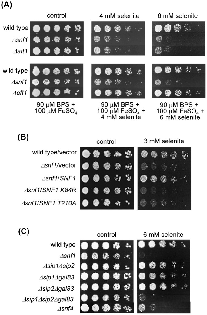
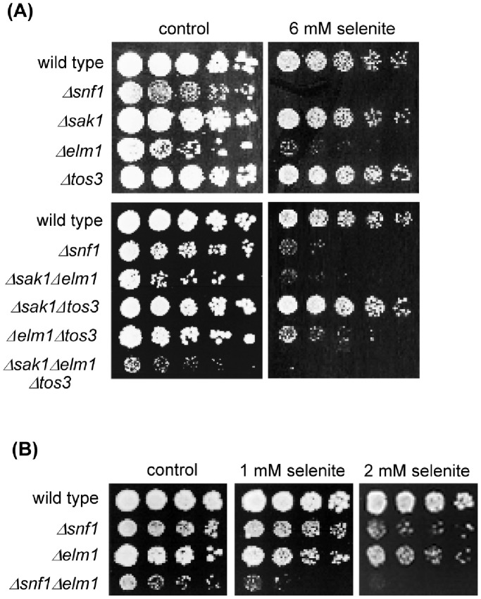
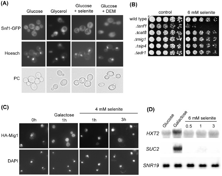
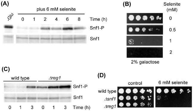
The AMPK/Snf1 kinase has a central role in carbon metabolism homeostasis in Saccharomyces cerevisiae. In this study, we show that Snf1 activity, which requires phosphorylation of the Thr210 residue, is needed for protection against selenite toxicity. Such protection involves the Elm1 kinase, which acts upstream of Snf1 to activate it. Basal Snf1 activity is sufficient for the defense against selenite, although Snf1 Thr210 phosphorylation levels become increased at advanced treatment times, probably by inhibition of the Snf1 dephosphorylation function of the Reg1 phosphatase. Contrary to glucose deprivation, Snf1 remains cytosolic during selenite treatment, and the protective function of the kinase does not require its known nuclear effectors. Upon selenite treatment, a null snf1 mutant displays higher levels of oxidized versus reduced glutathione compared to wild type cells, and its hypersensitivity to the agent is rescued by overexpression of the glutathione reductase gene GLR1. In the presence of agents such as diethyl maleate or diamide, which cause alterations in glutathione redox homeostasis by increasing the levels of oxidized glutathione, yeast cells also require Snf1 in an Elm1-dependent manner for growth. These observations demonstrate a role of Snf1 to protect yeast cells in situations where glutathione-dependent redox homeostasis is altered to a more oxidant intracellular environment and associates AMPK to responses against oxidative stress.


Conflict of interest statement

Competing Interests: The authors have declared that no competing interests exist.

Figures

Figure 1
Figure 1. Snf1 activity is required for protection against selenite.
(A) Growth assays of serial dilutions of the respective strains on YPD medium with the indicated additions. Growth was recorded after 48 hours at 30°C. Strains employed: wild type (W303-1A), Δsnf1 (Wsnf1) and Δaft1 (MML348). (B) Growth assays of serial dilutions of the following strains, plated on SC medium with sodium selenite: wild type (W303-1A) and Δsnf1 (Wsnf1) cells transformed with vector pWS93, and Δsnf1 cells transformed with pWS-Snf1, pWS-Snf1-T210A and pWS-Snf1-K84R. Growth was recorded after 3 days at 30°C. (C) As in (A), with the following strains in addition to wild type and Δsnf1: Δsip1Δsip2 (MML1445), Δsip1Δgal83 (MML1452), Δsip2Δgal83 (MML1454), Δsip1Δsip2Δgal83 (MML1459) and Δsnf4 (MML1407).
Figure 2
Figure 2. Protection against selenite preferentially requires the Elm1 kinase.
(A) Growth assays of serial dilutions of the following strains on YPD medium with sodium selenite: wild type (W303-1A), Δsnf1 (Wsnf1), Δsak1 (MML1370), Δelm1 (YPDahl21), Δtos3 (YPDahl19), Δsak1Δelm1 (MML1387), Δsak1Δtos3 (MML1389), Δelm1Δtos3 (MML1390) and Δsak1Δelm1Δtos3 (MML1392). (B) As in (A) with the strains: wild type, Δsnf1, Δelm1 and Δsnf1Δelm1 (MML1724).
Figure 3
Figure 3. Protection against selenite toxicity does not require activity of the known nuclear effectors of Snf1.
(A) Localization of Snf1 upon different treatments. The Snf1-GFP protein expressed in pOV84-transformed wild type cells was visualized by fluorescence microscopy in cells growing in SC medium without treatment (Glucose) or after 2 hours treatment with 4 mM sodium selenite or 1 mM DEM. In parallel, cell samples were shifted to YPGly and observed one hour later (Glycerol). Prior to observations, samples were stained with Hoesch for nuclei localization. The corresponding phase contrast fields (PC) are shown. (B) Growth assays of serial dilutions of the following strains on YPD medium with sodium selenite: wild type (W303-1A), Δsnf1 (Wsnf1), Δcat8 (MML1417), Δmig1 (MML1408), Δsip4 (MML1396) and Δadr1 (MML1419). (C) Localization of HA-Mig1 upon different treatments. Cells transformed with pHA-Mig1 were grown in SC medium and treated with selenite for the indicated times or shifted to medium with 2% galactose instead of glucose. Cells were observed by immunofluorescence experiments with anti-HA antibodies, with parallel nuclear staining with DAPI. (D) Northern blot expression analysis of the indicated genes in wild type (W303-1A) cells growing in YPD medium without (Glucose) or with selenite for the indicated times (hours), or in YPGal medium (Galactose) for 1 hour. SNR19 was employed as loading control. The same blotted membrane was successively hybridized with the three probes after extensive washings.
Figure 4
Figure 4. Phosphorylation levels of Snf1 at Thr210 do not correlate with protection against selenite treatment.
(A) Western blot analysis of phosphorylated Snf1 at Thr210 with anti-phospho-Thr172-AMPK (upper panel). Blots were rehybridized with anti-Snf1 antibodies for total Snf1 (lower panel). Samples were obtained from wild type (W303-1A) exponential cultures in YPD treated with sodium selenite for the indicated times. Control samples were run from YPD-grown wild type cells that were shifted for 1 hour to YPGly (-Glu). (B) Growth assays of serial dilutions of wild type (W303-1A) cells in YPGal medium with the indicated concentrations of selenite. (C) As in (A) with samples from wild type and Δreg1 (MML1442) cells. (D) Growth assays of serial culture dilutions of wild type (W303-1A), Δsnf1 (Wsnf1) and Δreg1 (MML1442) strains on YPD medium with selenite.
Figure 5
Figure 5. Effect of selenite on growth in low or normal phosphate conditions.
Relative growth of wild type (W303-1A), Δsnf1 (Wsnf1), Δpho84 (MML1304) and Δsnf1Δpho84 (MML1401) cells in low or normal phosphate medium without or with 3 mM selenite. Growth in shaken microtiter plates was automatically recorded and the growth values reached by each strain after 24 hours were made relative to the growth of wild type cells, which was given the unit value for each growth condition considered. The mean of three independent experiments (± s.d.) is represented. Note that different scales of the y-axis are employed in both panels.
Figure 6
Figure 6. The ratio of reduced vs oxidized glutathione is altered upon selenite treatment.
(A) Intracellular concentration of GSH (left) and GSSG (right) in cells treated with 2 mM sodium selenite for the indicated times. Wild type (W303-1A, continuous lines) and mutant Δsnf1 (Wsnf1, dashed lines) cells were grown in SC medium. Values (± s.d.) are the mean of three independent experiments. (B) GSH/GSSG ratio as determined from the concentration values shown in part (A). (C) GSH redox potential (EGSH) in wild type (continuous lines) and mutant Δsnf1 (dashed lines) cells treated with selenite. EGSH was calculated from the GSH and GSSG concentration values in each of the three experiments indicated in part (A), using the Nernst equation for the GSH/GSSG pair. The mean (± s.d.) is represented. (D) Growth assays in SC medium of serial dilutions of the strains indicated in part (A) transformed with vector pCM189 or its derivative pMM1039 (tetO-GLR1), or of the same strains transformed with the multicopy vector YEplac195 or its derivative P1116 overexpressing GLR1 (right panels).
Figure 7
Figure 7. Snf1 is required for protection against glutathione-oxidizing agents.
(A) Growth assays of serial dilutions of the following strains on YPD medium with DEM, diamide or t-BOOH: wild type (W303-1A), Δsnf1 (Wsnf1) and Δsip1Δsip2Δgal83 (MML1459). (B) Growth assays of serial dilutions of the following strains, plated on SC medium with DEM: wild type (W303-1A) and Δsnf1 (Wsnf1) cells transformed with vector pWS93, and Δsnf1 cells transformed with pWS-Snf1, pWS-Snf1-T210A and pWS-Snf1-K84R. (C) Growth assays of serial dilutions of the following strains on YPD medium with DEM: wild type (W303-1A), Δsak1Δelm1 (MML1387), Δsak1Δtos3 (MML1389), Δelm1Δtos3 (MML1390) and Δsak1Δelm1Δtos3 (MML1392). (D) Western blot analysis of Thr210-phosphorylated Snf1 (Snf1-P) and of total Snf1. The same membrane was successively hybridized with the corresponding antibodies. Samples were from wild type (W303-1A) exponential cultures in YPD treated with DEM or diamide. Control samples were from YPD-grown wild type cells shifted for 1 hour to YPGly (-Glu). (E) Growth assays of wild type (W303-1A) and Δsnf1 (Wsnf1) cells transformed with multicopy vector YEplac195 or its derivative P1116 overexpressing GLR1, in SC medium with DEM.

Similar articles

Cited by

References

    1. Hardie DG (2011) AMP-activated protein kinase- an energy sensor that regulates all aspects of cell function. Genes Dev 25: 1895–1908. - PMC - PubMed
    1. Sanz P (2003) Snf1 protein kinase: a key player in the response to cellular stress in yeast. Biochem Soc Trans 31: 178–181. - PubMed
    1. Hedbacker K, Carlson M (2008) SNF1/AMPK pathways in yeast. Front Biosci 13: 2408–2420. - PMC - PubMed
    1. McCartney RR, Schmidt MC (2001) Regulation of Snf1 kinase. Activation requires phosphorylation of threonine 210 by an upstream kinase as well as a distinct step mediated by the Snf4 subunit. J Biol Chem 276: 36460–36466. - PubMed
    1. Hong SP, Leiper FC, Woods A, Carling D, Carlson M (2003) Activation of yeast Snf1 and mammalian AMP-activated protein kinase by upstream kinases. Proc Natl Acad Sci USA 100: 8839–8843. - PMC - PubMed

Publication types

MeSH terms

LinkOut - more resources