. 2013;8(3):e58519.
doi: 10.1371/journal.pone.0058519. Epub 2013 Mar 5.

Eye-size variability in deep-sea lanternfishes (Myctophidae): an ecological and phylogenetic study

Affiliations

Eye-size variability in deep-sea lanternfishes (Myctophidae): an ecological and phylogenetic study

Fanny de Busserolles et al. PLoS One. 2013.

Abstract

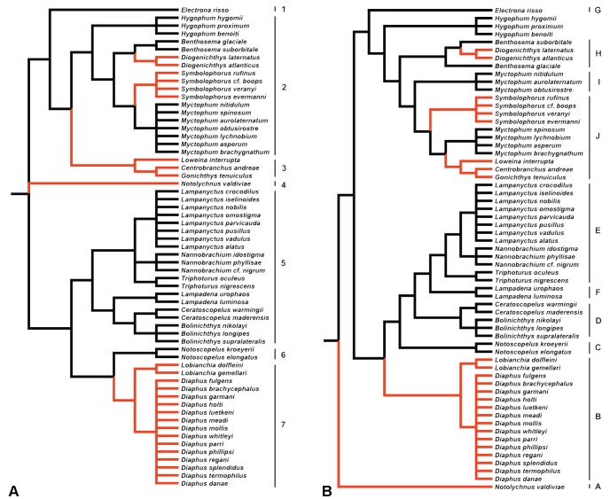
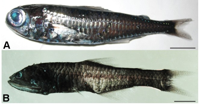
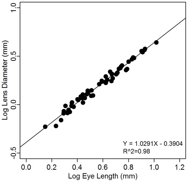
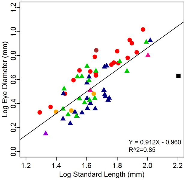
One of the most common visual adaptations seen in the mesopelagic zone (200-1000 m), where the amount of light diminishes exponentially with depth and where bioluminescent organisms predominate, is the enlargement of the eye and pupil area. However, it remains unclear how eye size is influenced by depth, other environmental conditions and phylogeny. In this study, we determine the factors influencing variability in eye size and assess whether this variability is explained by ecological differences in habitat and lifestyle within a family of mesopelagic fishes characterized by broad intra- and interspecific variance in depth range and luminous patterns. We focus our study on the lanternfish family (Myctophidae) and hypothesise that lanternfishes with a deeper distribution and/or a reduction of bioluminescent emissions have smaller eyes and that ecological factors rather than phylogenetic relationships will drive the evolution of the visual system. Eye diameter and standard length were measured in 237 individuals from 61 species of lanternfishes representing all the recognised tribes within the family in addition to compiling an ecological dataset including depth distribution during night and day and the location and sexual dimorphism of luminous organs. Hypotheses were tested by investigating the relationship between the relative size of the eye (corrected for body size) and variations in depth and/or patterns of luminous-organs using phylogenetic comparative analyses. Results show a great variability in relative eye size within the Myctophidae at all taxonomic levels (from subfamily to genus), suggesting that this character may have evolved several times. However, variability in eye size within the family could not be explained by any of our ecological variables (bioluminescence and depth patterns), and appears to be driven solely by phylogenetic relationships.


Conflict of interest statement

Competing Interests: The authors have declared that no competing interest exist.

Figures

Figure 1
Figure 1. Phylogenies of Myctophidae reconstructed from (A) Paxton et al.
, (B) Poulsen et al. . The red branches indicate the main differences between the two phylogenies. Branch lengths are arbitrarily ultrametricized on the figure. In A, the numbers identify the different tribes of Paxton et al. , 1. Electronini, 2.Myctophini, 3.Gonichthyini, 4.Notolychnini, 5.Lampanyctini, 6.Gymnoscopelini, 7.Diaphini. In B, the letters identify the different clades of Poulsen et al. , A. Notolychnini, B. Diaphini, C. Notoscopelini, D-E-F. Lampanyctini, G. Electronini, H. Myctophini, I. Myctophini (cycloid-species-group), J. Myctophini (ctenoid-species group)+Gonichthyini.
Figure 2
Figure 2. Difference in eye size compared to body size in two species of lanternfish.
(A) Myctophum brachygnathum,(B) Nannobrachium phyllisae. Scale bar, 10 mm.
Figure 3
Figure 3. Relationship between lens diameter and eye diameter after correcting for phylogeny (PGLS).
The fitted line is the linear regression corrected for phylogeny (PGLS) using the phylogeny of Paxton et al. .
Figure 4
Figure 4. Relationship between eye diameter and standard length in 61 species of Myctophidae.
Each point represents the mean for the species; individual details are in Table 2. Shapes represent the subfamilies, circles = Myctophinae, triangles = Lampanyctinae. Colors represent the tribes of Paxton et al. , brown = Electronini, red = Myctophini, blue = Lampanyctini, green = Diaphini, yellow = Gonichthyini, pink = Gymnoscopelini, purple = Notolychnini. The fitted line is the linear regression corrected for phylogeny (PGLS) using the phylogeny of Paxton et al. . The black square represents one individual, Scopelengys tristis, from the sister family Neoscopelidae.
Figure 5
Figure 5. Residuals eye size corrected for body size of Myctophidae after correcting for phylogeny: (A) by tribes (phylogeny A, [43]) and (B) by clades (phylogeny B, [44]).
Colours represent the tribes of Paxton et al. for comparison, as in Fig. 4. Groups sharing the same superscript letter are not significantly different from one another based on the post-hoc analyses. Groups with no superscript letters were not included in the analysis due to the low samples size (n<3). #, $ indicates genera that were significantly different for one of the ten randomly resolved polytomy phylogenies (P# = 0.04, P$ = 0.03).
Figure 6
Figure 6. Residuals eye size corrected for body size by genera of Myctophidae, corrected for phylogeny (phylogeny A, [43]).
Colors represent the tribes of Paxton et al. , as in Figure 4. The genus Diaphus was divided into two groups based on the presence/absence of the So. Groups with the same superscript letters are not significantly different from one another, independent of the phylogeny used based on the post-hoc analyses. Groups with no superscript letters were not included in the analyses due to the low samples size (n<3). # indicates genera that were not significantly different when analysed with phylogeny B (P = 1) and for four of the ten randomly resolved polytomy phylogenies of phylogeny A (P = 0.06–0.08).
Figure 7
Figure 7. Variation in the size and location of the Dn, Vn and So luminous organs within the genus Diaphus.
A = D. luetkeni, B = D. brachycephalus, C = D. danae, D = D. mollis, E = D. phillipsi, F = D. parri, G = D. termophilus, H = D. holti. A, C, E, G = Diaphus group 1 (So absent); B, D, F, H = Diaphus group 2 (So present). The yellow arrows indicate the position of the So photophore.

References

    1. Land MF (1990) Optics of the eyes of marine animals. In: Herring PJ, Campbell AK, Whitfield M, Maddock L, editors. Light and life in the sea: Cambridge University Press. pp. 149–166.
    1. Matthiessen L (1882) Über die Beziehungen, welche zwischen dem Brechungsindex des Kernzentrums der Krystalllinse und den Dimensionen des Auges bestehen. Pflügers Archiv für die gesamate Physiologie des Menschen und der Tiere 27: 510–523.
    1. Matthiessen L (1886) Über den physikalisch-optischen Bau des Auges der Cetacean und der Fische. Pflügers Archiv für die gesamate Physiologie des Menschen und der Tiere 38: 521–528.
    1. Warrant E (2004) Vision in the dimmest habitats on Earth. Journal of Comparative Physiology A 190: 765–789. - PubMed
    1. Denton EJ (1990) Light and vision at depths greater than 200 metres. In: Herring PJ, Campbell AK, Whitfield M, Maddock L, editors. Light and life in the sea: Cambridge University Press. pp. 127–148.

Publication types

LinkOut - more resources