Review
. 2013 Jun 19;4(6):909-17.
doi: 10.1021/cn400027z. Epub 2013 Mar 21.

A calcium-relay mechanism in vertebrate phototransduction

Affiliations
Review

A calcium-relay mechanism in vertebrate phototransduction

Karl-Wilhelm Koch et al. ACS Chem Neurosci. .

Abstract

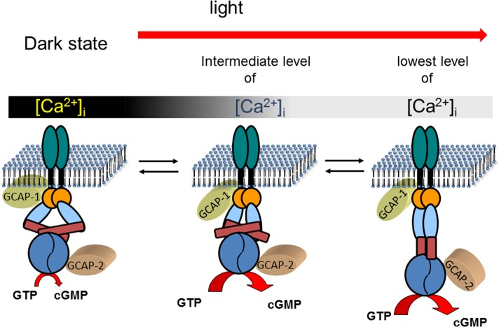
Calcium-signaling in cells requires a fine-tuned system of calcium-transport proteins involving ion channels, exchangers, and ion-pumps but also calcium-sensor proteins and their targets. Thus, control of physiological responses very often depends on incremental changes of the cytoplasmic calcium concentration, which are sensed by calcium-binding proteins and are further transmitted to specific target proteins. This Review will focus on calcium-signaling in vertebrate photoreceptor cells, where recent physiological and biochemical data indicate that a subset of neuronal calcium sensor proteins named guanylate cyclase-activating proteins (GCAPs) operate in a calcium-relay system, namely, to make gradual responses to small changes in calcium. We will further integrate this mechanism in an existing computational model of phototransduction showing that it is consistent and compatible with the dynamics that are characteristic for the precise operation of the phototransduction pathways.


Figures

Figure 1
Figure 1
Idealized single photon responses of mice rods. Responses of rods from wild-type mice (black trace), from GCAPs–/– mice (green trace), and from mice expressing GCAP2 on a GCAPs–/– genetic background (blue trace) were superimposed. Red arrows indicate the time at which maximum amplitude is reached (time-to-peak). Traces represent typical idealized responses and were drawn according to published results in ref (15). The lower part of the figure shows the change of intracellular Ca2+-concentration during a light response.
Figure 2
Figure 2
Heterogeneity of light responses from WT and transgenic mice. Original recordings were adapted from Figure 6 of ref (21) (Makino et al., PLoS One 7, e47637) in accordance with the Creative Commons Attribution License. (A) Comparison of averaged dim flash recordings from WT, GCAP1–/–, and GCAPs1,2–/– as indicated. (B–D) Variability of integration times of single photon responses as observed by Makino et al.
Figure 3
Figure 3
Ca2+-dependent activation profile of the photoreceptor guanylate cyclase is regulated by GCAP1 and GCAP2. The GC activity is normalized to 100%. The curves are typical for mammalian GCAPs and summarize the principal finding of many references (see main text). The gray bar symbolizes the physiological range of cytoplasmic Ca2+ that changes from a high (dark gray) to a low value (light gray).
Figure 4
Figure 4
Activity profiles of cone specific GCAPs from zebrafish. The GC activity is normalized as in Figure 3. The curves were obtained by measuring the GC activity in the presence of zGCAPs as indicated. The figure is based on the original data published in ref (53), where a bovine ROS membrane preparation containing ROS-GCs was used for assaying zebrafish GCAPs.
Figure 5
Figure 5
Ca2+-relay model of sequential GCAP action. Photoreceptor guanylate cyclases are dimers with one transmembrane domain in each monomer. In addition, each monomer consists of one extracelllar domain (in rods, this domain is in the lumen of the disks), one juxtamembrane domain (orange), and one kinase homology domain (light blue). Further, the dimerization domain (dark red rectangle) is important for the formation of an active enzyme controlling the correct positioning of the two catalytic domains (blue). In the dark state of the cell, GCAPs are fully or partially saturated with Ca2+ which keeps the guanylate cyclase activity at a very low level that is sufficient to keep a fraction of the CNG-channels in the plasma membrane open. GCAPs form with the target guanylate cyclase a complex in the presence and absence of Ca2+, which enables a rapid response to changing Ca2+-concentration after illumination. When the intracellular Ca2+-concentration falls to an intermediate level, Ca2+ dissociates form GCAP1. This process triggers a conformational change in GCAP1, leading to the activation of guanylate cyclase. When Ca2+ reaches its final lower intracellular level, GCAP2 turns into an activator. The different conformations of the guanylate cyclase are hypothetical and need to be verified in future experiments. It is further suggested that GCAPs stabilize the transition states of the cGMP catalytic step, but experimental proof is also lacking so far. The stepwise and reversible action of GCAPs would allow the cell to react on small incremental changes in Ca2+ with a fine-tuned response system.
Figure 6
Figure 6
Simulated photocurrents of an amphibian rod stimulated by flashes of light of increasing intensity. The duration of each flash is 24 ms, and each stimulus leads to 1.54, 46, 280, 985, 30 400, and 115 000 photoisomerizations of rhodopsin. The mathematical model used for the simulation is the one reported in ref (57) in which the Ca2+-relay mechanism has been implemented as described here.
Figure 7
Figure 7
Simulated time course of the synthesis of cGMP by GC1 under the Ca2+-mediated regulation by GCAP1 and GCAP2 according to the Ca2+-relay mechanism. Panels A–F refer to the same flashes of increasing intensity reported in Figure 6 (i.e., panel A refers to 1.54 photoisomerization, panel B to 46 photoisomerizations, and so forth). Blue lines report on the contribution of GCAP1 to the rate of synthesis, while red lines to that of GCAP2. The two contributions add up to form the overall rate (black line).

References

    1. Pugh E. N. Jr., and Lamb T. D. (2000) Phototransduction in vertebrate rods and cones: molecular mechanisms of amplification, recovery and light adaptation. In Handbook of Biological Physics (Stavenga D. G., DeGrip W. J., and Pugh E. N. Jr., Eds.), Vol. 4, pp 183–255, Elsevier Science BV: Amsterdam, The Netherlands.
    1. Luo D. G.; Xue T.; Yau K.-W. (2008) How vision begins: an odyssey. Proc. Natl. Acad. Sci. U.S.A. 105, 9855–9862. - PMC - PubMed
    1. Stephen R.; Filipek S.; Palczewski K.; Sousa M. C. (2008) Ca2+-dependent regulation of phototransduction. Photochem. Photobiol. 84, 903–910. - PMC - PubMed
    1. Koch K.-W.; Duda T.; Sharma R. K. (2010) Ca2+-modulated vision-linked ROS-GC guanylate cyclase transduction machinery. Mol. Cell. Biochem. 334, 105–115. - PubMed
    1. Kaupp U. B.; Seifert R. (2002) Cyclic nucleotide-gated ion channels. Physiol. Rev. 82, 769–824. - PubMed

MeSH terms

Substances

LinkOut - more resources