

Liver angulometry: a simple method to estimate liver volume and ratios
- PMID: 23472855
- PMCID: PMC3843616
- DOI: 10.1111/hpb.12079
Liver angulometry: a simple method to estimate liver volume and ratios
Abstract
Objectives: Volumetry is standard method for evaluating the volumes of the right liver (RL), left liver (LL), left lateral segments (LLS), total liver (TL) and future liver remnant (FLR). The aim of this study was to report a simple technique based on measurements of liver angles (angulometry) that can be used to predict liver ratios.
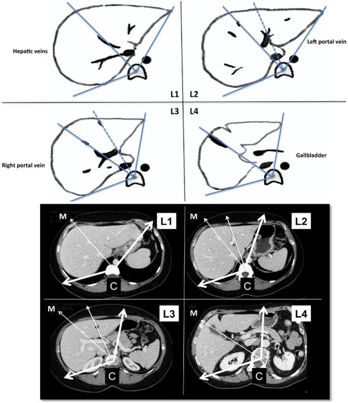
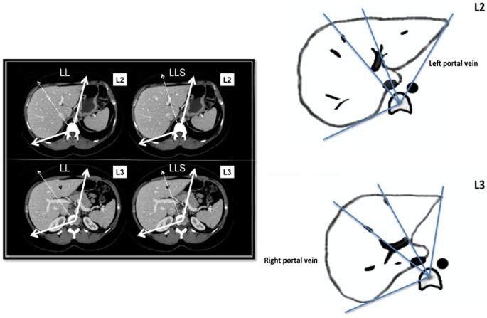
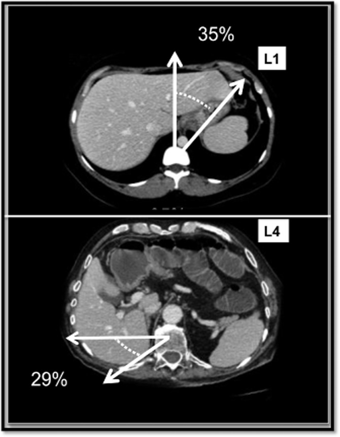
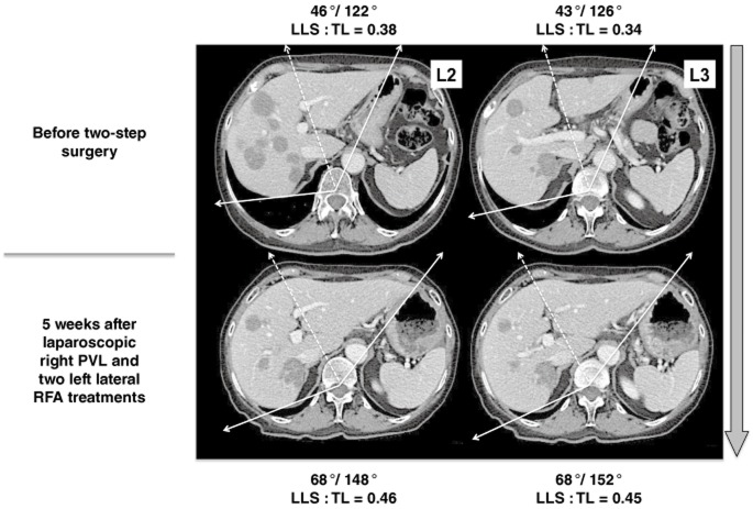
Methods: Fifty computed tomography (CT) scans obtained in subjects with normal liver were studied. Four CT scan levels were preselected: level 1 passed by the upper part of the hepatic veins; level 2 passed by the left portal vein branch division; level 3 passed by the right portal vein branch division, and level 4 passed by the gallbladder bed. Left and right tangent lines passing the liver edges were drawn and joined to the centre of the vertebra defining the TL angle. Two lines through, respectively, the plane of the middle hepatic vein and the left portal branches determined the angles of the RL, LL and LLS. Volumetric and angulometric data obtained on levels 2 and 3 in 50 different subjects were compared.
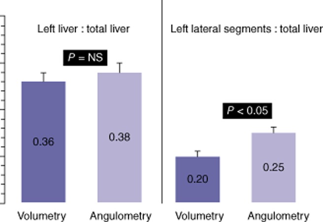
Results: Level 2 CT scans represented the most accurate way of obtaining angulometric measurements. The mean ± standard deviation (SD) angles of the TL and LL were 134 ± 12 ° and 55 ± 12 °, respectively. The mean ± SD percentages of the TL represented by the LL in angulometry and volumetry were 38 ± 7% and 36 ± 6%, respectively (non-significant difference). The mean ± SD percentages of the TL represented by the LLS in angulometry and volumetry were 25 ± 4% and 20 ± 3%, respectively (P < 0.05). The mean ± SD overestimation of the percentage of the TL represented by the LLS in angulometry was 2.7 ± 7.0%.
Conclusions: Angulometry is a simple and accurate technique that can be used to estimate the ratio of the FLR to TL volume on one or two CT (or magnetic resonance imaging) slices. It can be helpful for clinicians, especially before right or extended right hepatectomy and after right portal vein occlusion techniques.
© 2013 International Hepato-Pancreato-Biliary Association.
Figures
References
-
- Abdalla EK. Portal vein embolization (prior to major hepatectomy) effects on regeneration, resectability, and outcome. J Surg Oncol. 2010;102:960–967. - PubMed
-
- Tongyoo A, Pomfret EA, Pomposelli JJ. Accurate estimation of living donor right hemi-liver volume from portal vein diameter measurement and standard liver volume calculation. Am J Transplant. 2012;12:1229–1239. - PubMed
-
- Nakayama Y, Li Q, Katsuragawa S, Ikeda R, Hiai Y, Awai K, et al. Automated hepatic volumetry for living related liver transplantation at multisection CT. Radiology. 2006;240:743–748. - PubMed
-
- Yoshizumi T, Taketomi A, Kayashima H, Yonemura Y, Harada N, Ijichi H, et al. Estimation of standard liver volume for Japanese adults. Transplant Proc. 2008;40:1456–1460. - PubMed
Publication types
MeSH terms
Substances
LinkOut - more resources
Full Text Sources
Other Literature Sources
Medical
