Case Reports
. 2013 Mar 5;17(3):448-455.
doi: 10.1016/j.cmet.2013.02.001.

Identification of a SIRT1 mutation in a family with type 1 diabetes

Affiliations
Case Reports

Identification of a SIRT1 mutation in a family with type 1 diabetes

Anna Biason-Lauber et al. Cell Metab. .

Abstract

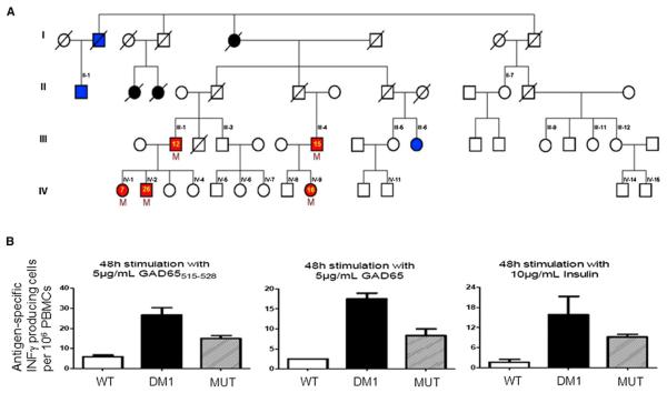
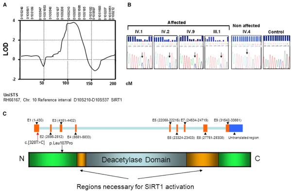
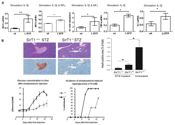
Type 1 diabetes is caused by autoimmune-mediated β cell destruction leading to insulin deficiency. The histone deacetylase SIRT1 plays an essential role in modulating several age-related diseases. Here we describe a family carrying a mutation in the SIRT1 gene, in which all five affected members developed an autoimmune disorder: four developed type 1 diabetes, and one developed ulcerative colitis. Initially, a 26-year-old man was diagnosed with the typical features of type 1 diabetes, including lean body mass, autoantibodies, T cell reactivity to β cell antigens, and a rapid dependence on insulin. Direct and exome sequencing identified the presence of a T-to-C exchange in exon 1 of SIRT1, corresponding to a leucine-to-proline mutation at residue 107. Expression of SIRT1-L107P in insulin-producing cells resulted in overproduction of nitric oxide, cytokines, and chemokines. These observations identify a role for SIRT1 in human autoimmunity and unveil a monogenic form of type 1 diabetes.


Figures

Figure 1
Figure 1. The Patients' Family Tree and Functional Analysis of Peripheral Blood Mononuclear Cells
(A) Red symbols indicate family members who developed an autoimmune disease: type 1 diabetes (III-1, III-4, IV-1, IV-2) or colitis (IV-9). The yellow numbered symbols denote the age of onset of the disease. Blue symbols indicate type 2 diabetes, while black symbols indicate an unclear diabetes phenotype. Black numbered symbols identify all family members who were tested for the presence of the mutation, and the letter “M” identifies the ones who were positive. A slash denotes deceased family members. Individuals IV-1 to IV 15, III-1, III-4, III-5, III-9, III-11, and III-12 are lean (body mass index <25 Kg/m2), and II-1 and III-6 have body mass indices of 27 and >40 Kg/m2, respectively. (B) Peripheral blood mononuclear cells were obtained from healthy control subjects (WT) and from patients with type 1 diabetes carrying wild-type SIRT1 (DM1) or the L107P mutation (MUT). GAD65515–528 peptide, whole GAD65 protein, and insulin elicited antigen-specific IFN-γ production. n = 3 for each of the three groups. All data are presented as mean ± SEM.
Figure 2
Figure 2. Impaired Insulin Secretion and Sensitivity in a Patient Carrying a Mutation in the SIRT1 Gene
(A) Plasma glucose and insulin levels during oral glucose tolerance tests of the index patient with a SIRT1 mutation (red lines) and a control 20-year-old non-affected male family member (green lines). (B) Impaired insulin stimulated glucose uptake of cultured human skeletal myotubes obtained from biopsies of the vastus lateralis muscle of the index patient with a SIRT1 mutation (diabetes) compared to the control 20-year-old nonaffected male family member (control). (C) The cell lysates of the myotubes were analyzed by immunoblotting with antibodies to phosphorylated and nonphosphorylated AKT, ERK, NF-κB (p65), and AMP-K. n = 3 myotube subpopulations. All data are presented as mean ± SEM.
Figure 3
Figure 3. Description of the Mutation
(A) Whole-genome microsatellite linkage analysis. Details of chromosome 10, for which the LOD reached significance (LOD 4.1; whole-genome significance, LOD > 2.2). The length of the chromosome is given in centimorgans (cM). According to the UniSTS database, the region between microsatellite D10S210 and D10S537 contains the SIRT1 gene. (B) Sanger sequence analysis of the mutation. DNA sequencing chromatograms obtained by direct sequencing of PCR products showing the presence of the heterozygous c.[320T > C] substitution in exon 1, not present in nonaffected members of the family and normal individuals (control, representative example out of 200 alleles). The roman numerals correspond to those in Figure 1A. (C) SIRT1 gene structure and predicted protein structure.
Figure 4
Figure 4. Decreased Anti-Inflammatory Activity of SIRT1-L107P
(A) MIN6 cells were stably transduced with empty, wild-type (WT), or mutant SIRT1 L107P virus and stimulated with IL-1β or a combination of IL-1β and IFN-γ. iNOS mRNA expression relative to empty virus-transduced cells MIN6 cells, and NO formation following cytokine stimulation expressed as fold of unstimulated cells. Findings were verified with five separately produced sets of sirt1 WT and L107P-overexpressing cell lines each time at least in triplicate (mean ± SD, *p < 0.05; **p < 0.01). (B) Wild-type and SIRT1-deficient mice were injected with five daily injections of streptozotocin (40 mg/kg). Glycemia and onset of diabetes (serum glucose >10 mM) were monitored for 16 days. Islet histology was visualized by HE and immunostaining for insulin (brown). The proportion of surface area that stained positive for insulin is shown. All data are presented as mean ± SEM. *p < 0.05; n = 6 for +/+ and +/−, and n = 5 for −/−.

Comment in

References

    1. American Diabetes Association Diagnosis and classification of diabetes mellitus. Diabetes Care. 2010;33(Suppl 1):S62–S69. - PMC - PubMed
    1. Autiero I, Costantini S, Colonna G. Human sirt-1: molecular modeling and structure-function relationships of an unordered protein. PLoS ONE. 2009a;4:e7350. http://dx.doi.org/10.1371/journal.pone.0007350. - DOI - PMC - PubMed
    1. Autiero I, Costantini S, Colonna G. Modeling of the bacterial mechanism of methicillin-resistance by a systems biology approach. PLoS ONE. 2009b;4:e6226. http://dx.doi.org/10.1371/journal.pone.0006226. - DOI - PMC - PubMed
    1. Banks AS, Kon N, Knight C, Matsumoto M, Gutiérrez-Juárez R, Rossetti L, Gu W, Accili D. SirT1 gain of function increases energy efficiency and prevents diabetes in mice. Cell Metab. 2008;8:333–341. - PMC - PubMed
    1. Barrett JC, Clayton DG, Concannon P, Akolkar B, Cooper JD, Erlich HA, Julier C, Morahan G, Nerup J, Nierras C, et al. Type 1 Diabetes Genetics Consortium. (2009). Genome-wide association study and meta-analysis find that over 40 loci affect risk of type 1 diabetes. Nat. Genet. 41:703–707. - PMC - PubMed

Publication types

LinkOut - more resources