Review
. 2013 Mar 6;77(5):810-24.
doi: 10.1016/j.neuron.2013.02.018.

Hypothalamic survival circuits: blueprints for purposive behaviors

Affiliations
Review

Hypothalamic survival circuits: blueprints for purposive behaviors

Scott M Sternson. Neuron. .

Abstract

Neural processes that direct an animal's actions toward environmental goals are critical elements for understanding behavior. The hypothalamus is closely associated with motivated behaviors required for survival and reproduction. Intense feeding, drinking, aggressive, and sexual behaviors can be produced by a simple neuronal stimulus applied to discrete hypothalamic regions. What can these "evoked behaviors" teach us about the neural processes that determine behavioral intent and intensity? Small populations of neurons sufficient to evoke a complex motivated behavior may be used as entry points to identify circuits that energize and direct behavior to specific goals. Here, I review recent applications of molecular genetic, optogenetic, and pharmacogenetic approaches that overcome previous limitations for analyzing anatomically complex hypothalamic circuits and their interactions with the rest of the brain. These new tools have the potential to bridge the gaps between neurobiological and psychological thinking about the mechanisms of complex motivated behavior.


Figures

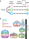
Figure 1
Figure 1. Physiological Needs Influence Behavior through Discrete Hypothalamic Circuits
(A) Circulating and neural signals of physiological needs are sensed by interoceptive cell types, which influence action selection to achieve goals that alleviate the needs. (B) Discrete hypothalamic regions contain interoceptors for a variety of substances and also have been shown to evoke behavior with localized electrical or chemical perturbations. E, estrogen; T, testosterone; Agt, angiotensin; Osmol, osmolarity; Temp, temperature; ARC, arcuate nucleus; VMH, ventromedial hypothalamus; LH, lateral hypothalamus; PVH, paraventricular hypothalamus; SFO, subfornical organ; OVLT, organosum vaculosum of lamina terminalis; MPOA, medial preoptic area.
Figure 2
Figure 2. Functional Circuit Mapping and Behavioral Evaluation of Cell Type-Specific Circuit Connections
(A) For molecularly defined neurons, channelrhodopsin-assisted circuit mapping is used to determine postsynaptic targets as well as their functional synaptic properties. Upper right: schematic postsynaptic current. (B) For an example inhibitory circuit, cell type-specific neuron activation and silencing techniques are used to test whether activation of the presynaptic population or inhibition of the postsynaptic population are each sufficient for the behavioral response. (C) Photostimulation of axon projections from a cell type (inhibitory) over a specific target region is performed while monitoring the behavioral response. The axon anatomy of a given molecularly defined cell type is often not known and may involve each cell within a molecularly defined type projecting to multiple target regions (collateral axon connectivity) or each projection target arising from a single subpopulation of the molecularly defined cell class (“one-to-one” connectivity). For axons with collateral connectivity, a back propagating action potential can activate axon collaterals projecting to other regions; thus, additional experiments are required to prove the behavioral consequence of activating a specific projection. (D) Test of the functional necessity of specific circuit connections. Left: the behavioral role of identified inhibitory connections has typically been tested by blocking inhibition regionally with reagents that are not cell type-specific to the identified postsynaptic neuron. Thus, all neurons in the region, regardless of connectivity, are released from inhibition. Right: cell type-specific occlusion of synaptic inhibition by simultaneously activating presynaptic neurons (or their axons) and a molecularly defined postsynaptic neuron population. Because the presynaptic neuron also projects to other populations, this is a test for the behavioral necessity of this specific circuit connection while all other targets are receiving evoked synaptic input. Figure modified from Atasoy et al. (2012).
Figure 3
Figure 3. AGRP Neurons Are Necessary and Sufficient for Feeding
(A) Configuration for optogenetic activation of AGRP neurons in a behaving mouse. ARC, arcuate nucleus. Photo from Igor Siwanowicz. (B) Photostimulation-evoked feeding from mice engineered to express channelrhodopsin in AGRP neurons (Aponte et al., 2011). (C) Food intake from AGRP neuron photo-stimulation in ad libitum fed mice is similar to re-feeding 24 hr food-deprived mice (Aponte et al., 2011). (D) Scheme for diphtheria toxin receptor-mediated acute AGRP neuron ablation. (E) AGRP neuron ablation leads to aphagia (Luquet et al., 2005).
Figure 4
Figure 4. AGRP Neuron Axon Projections Regulate a Hindbrain Circuit that Mediates Aphagia
(A) Summary of neuron and pharmacological manipulations individually and together that probe epistatic relationships for their effect on survival (resulting from rescue of feeding). Abl, ablation; Bz, bretazanil; Ond, ondansetron; icv, intracerebroventricular; LS, lateral septum. (B) AGRP neurons project local and long-range axons to inhibit POMC, PVH, LS, and PBN neurons along with other targets. ARCAGRP/PBN connections are required to suppress visceral malaise, which is mediated in the PBN by glutamatergic excitatory drive from the nucleus of the solitary tract (NTS) that is regulated by serotonergic inputs.
Figure 5
Figure 5. Distinct AGRP Neuron Axon Projections Regulate Different Aspects of Feeding Behavior
(A) Summary of cell type-specific activity manipulations to probe epistatic relationships for their effect on feeding behavior. Act, activation; Sup, suppression; bl, pharmacological blockade. (B) AGRP neurons project local and long-range axons to inhibit POMC, PVH, and PBN neurons along with other targets. ARCAGRP→ARCPOMC connections influence long-term regulation of food intake and other aspects of energy homeostasis. ARCAGRP→PVH and ARCAGRP→PVHOXT connections regulate acute food intake. PVH neurons in turn communicate with brainstem satiety centers (NTS, nucleus of the solitary tract; DVC, dorsal vagal complex; SC, spinal cord). Figure modified from Atasoy et al. (2012).
Figure 6
Figure 6. An Aggression Locus in the Ventrolateral Subdivision of the Ventromedial Hypothalamus
(A) In vivo electophysiological recordings in mice show electrical activity from neurons in the VMHvl during male-male social interactions (Lin et al., 2011). High levels of activity are observed during attack behaviors. (B) Summary of mean firing rate changes across different social interactions between male and female mice (Lin et al., 2011). (C) Configuration of optogenetic experiments for VMHvl photostimulation (Lin et al., 2011). (D) Ethogram showing VMHvl photostimulation-evoked attack behavior (Lin et al., 2011). (E) Schematic of functional interactions between antagonistic circuits that mediate fighting and mating. Circuits to support the proposed inhibitory interactions have not been identified. Inhibitory interactions may be at the level of inputs to circuits mediating fighting and mating or may result from antagonism between these circuits.

Similar articles

Cited by

References

    1. Anderson DJ. Optogenetics, sex, and violence in the brain: implications for psychiatry. Biol. Psychiatry. 2012;71:1081–1089. - PMC - PubMed
    1. Aponte Y, Atasoy D, Sternson SM. AGRP neurons are sufficient to orchestrate feeding behavior rapidly and without training. Nat. Neurosci. 2011;14:351–355. - PMC - PubMed
    1. Arenkiel BR, Peca J, Davison IG, Feliciano C, Deisseroth K, Augustine GJ, Ehlers MD, Feng G. In vivo light-induced activation of neural circuitry in transgenic mice expressing channelrhodopsin-2. Neuron. 2007;54:205–218. - PMC - PubMed
    1. Atasoy D, Aponte Y, Su HH, Sternson SM. A FLEX switch targets Channelrhodopsin-2 to multiple cell types for imaging and long-range circuit mapping. J. Neurosci. 2008;28:7025–7030. - PMC - PubMed
    1. Atasoy D, Betley JN, Su HH, Sternson SM. Deconstruction of a neural circuit for hunger. Nature. 2012;488:172–177. - PMC - PubMed

Publication types

Substances