. 2013 Mar 6;77(5):859-66.
doi: 10.1016/j.neuron.2012.12.027.

Anatomical plasticity of adult brain is titrated by Nogo Receptor 1

Affiliations

Anatomical plasticity of adult brain is titrated by Nogo Receptor 1

Feras V Akbik et al. Neuron. .

Erratum in

  • Neuron. 2014 Jun 4;82(5):1184-5

Abstract

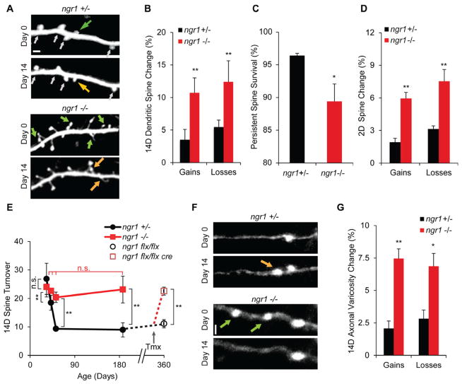
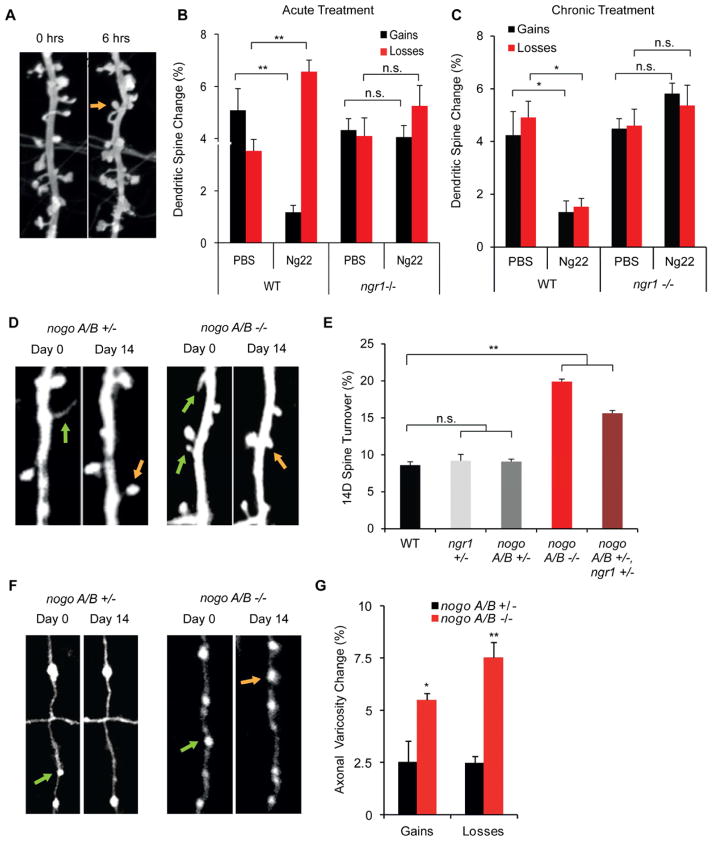
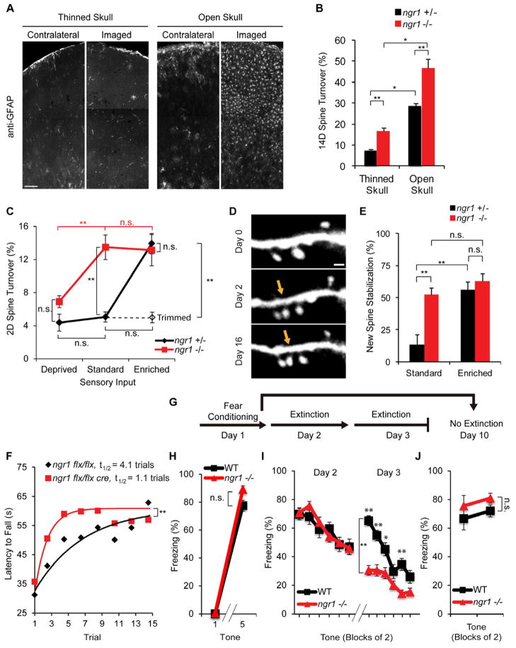
Experience rearranges anatomical connectivity in the brain, but such plasticity is suppressed in adulthood. We examined the turnover of dendritic spines and axonal varicosities in the somatosensory cortex of mice lacking Nogo Receptor 1 (NgR1). Through adolescence, the anatomy and plasticity of ngr1 null mice are indistinguishable from control, but suppression of turnover after age 26 days fails to occur in ngr1-/- mice. Adolescent anatomical plasticity can be restored to 1-year-old mice by conditional deletion of ngr1. Suppression of anatomical dynamics by NgR1 is cell autonomous and is phenocopied by deletion of Nogo-A ligand. Whisker removal deprives the somatosensory cortex of experience-dependent input and reduces dendritic spine turnover in adult ngr1-/- mice to control levels, while an acutely enriched environment increases dendritic spine dynamics in control mice to the level of ngr1-/- mice in a standard environment. Thus, NgR1 determines the low set point for synaptic turnover in adult cerebral cortex.


Figures

Figure 1
Figure 1. NgR1 Restricts Dendritic Spine and Axonal Varicosity Turnover In Adult Brain
(A) Repeated transcranial two-photon imaging of YFP-H transgenic mice reveals anatomical plasticity of layer 5 dendritic spines in layer 1 in vivo over two weeks. Orange and green arrowheads indicate spine gain and loss, respectively, over two weeks. White arrowheads indicate stable spines. (B) The percentage of spines gained and lost over two weeks in ngr1+/− (n=7) and ngr1−/− mice (n=8) at P180–P220. (C) The survival of persistent dendritic spines observed on both day 0 and 2 was monitored from day 2 to 16. (D) Dendritic spine gain and loss during a 2 day period is plotted for ngr1+/− (n=7) and ngr1−/− mice (n=8) at P180–P220. (E) Turnover of dendritic spines as a function of age. The two week turnover for ngr1+/− and ngr1−/− mice was measured starting at P26 (n=5, n=6, respectively), P35 (n=5 per genotype), P45 (n=5 per genotype), and P180 (n=7, n=8, respectively). Turnover is unchanged during maturation in ngr1 −/− mice, but decreases significantly in ngr1+/− mice from P26 to P45. ngr1 flx/flx mice with and without Cre (n=4, n=6, respectively) were treated with tamoxifen at P330. The 2-week turnover of dendritic spines was measured starting at P360. (F) Repeated transcranial two-photon imaging of en passant axonal varicosities in S1 in vivo. Green arrowheads indicate varicosity loss over two weeks. (G) The percentage of axonal varicosities gained and lost over two weeks in ngr1+/− (n=5) and ngr1−/− mice (n=6) at P180. Data are presented as mean ± s.e.m. **P<0.01. Scale bars, 1 μm.
Figure 2
Figure 2. Nogo Ligand Regulates Dendritic and Axonal Turnover In Adult Brain
(A) Time-lapse spinning-disc-confocal microscopy of 19–22DIV primary cortical neurons electroporated with myristoylated-GFP reveals dendritic spine turnover in vitro over six hours. WT or ngr1−/− cultures were imaged every hour for six hours. After the first hour, cultures were treated with 100 nM Nogo-22 or PBS. The orange arrowhead indicates spine gain. (B) The percentage of spines gained and lost over six hours (n=6 plates, 5806 spines) in response to PBS or 100 nM Nogo-22 treatment at 1 hour. (C) The percentage of spines gained and lost over six hours (n=4 plates, 3494 spines) in response to 6 day pre-treatment with 100 nM Nogo-22. (D) Repeated transcranial two-photon imaging of dendritic spines in vivo in Nogo-A/B mutants transgenic for YFP-H. Orange and green arrowheads indicate spine gain and loss, respectively, over two weeks. (E) The percentage of spines gained and lost over two weeks in WT (n=3), ngr1+/−(n=7), nogo-A/B+/− (n=6), nogo-A/B−/− mice (n=4), and nogo-A/B+/− ngr1+/− adult mice. (F) Repeated transcranial two-photon imaging of en passant boutons (EPBs) in vivo. Orange and green arrowheads indicate varicosity gain and loss, respectively, over two weeks. (G) The percentage of EPBs gained and lost over two weeks in nogo-A/B+/− (n=6) and nogo-A/B−/− mice (n=4) measured from P120-P134. Data are presented as mean ± s.e.m. *P<0.05, **P<0.01. Scale bars, 1 μm.
Figure 3
Figure 3. NgR1 Cell Autonomously Restricts Anatomical Plasticity
(A) Subcellular fractionation of somatosensory cortex in P180 mice. (B,C) WT or ngr1−/− cortical neurons were electroporated with myristoylated-GFP and then co-cultured with unlabeled neurons to assess cell autonomy in vitro at 19–22 DIV. Neurons were imaged over six hours. Cultures were treated with 100 nM Nogo-22 or PBS after the first hour. (B) Labeled WT neurons were imaged in an excess of unlabeled ngr1−/− neurons (n=3 plates, 2802 spines). (C) Labeled ngr1−/− neurons were imaged in an excess of unlabeled WT neurons (n=3 plates, 2670 spines). (D) Schematic of assessment of cell autonomy in vivo. Labeled (green) neurons are transgenic for both YFP and Cre-ERT2, while unlabeled neurons (black) have neither. Tamoxifen induced recombination creates a chimeric population of labeled ngr1−/− in an ngr1 flx/flx background. (E) Repeated transcranial two-photon imaging of dendritic spines in vivo of SLICK-V mice over two weeks. Orange arrowheads represent spine gain over two weeks. (F,G) The percentage of spines (F) and EPBs (G) gained and lost over two weeks in ngr1 flx/+ (n=4) and ngr1 flx/flx littermates (n=6) measured at P120. Data are presented as mean ± s.e.m. *P<0.05, **P<0.01. Scale bars, 1 μm.
Figure 4
Figure 4. NgR1 regulates inflammation-induced and experience-dependent anatomical plasticity
(A) Photomicrographs illustrate glial fibrillary acidic protein (GFAP) staining one month after craniotomy for the open-skull method, or two weeks after an initial thinned-skull surgery. Pictures are from barrel cortex contralateral or ipsilateral to the imaging site. Scale bars, 200 μm. (B) The two week turnover of dendritic spines in EGFP-M transgenics measured using the open (n=5 per genotype) or thinned skull preparations (n=4 per genotype) in primary motor cortex. (C) Turnover of dendritic spines over two days was assessed for mice of different genotypes and varied vibrissal sensory input levels. P180 ngr1+/− and ngr1−/− mutants are subjected to one of four sensory paradigms: 12 days of whisker trimming (n=5 per genotype), standard environment (n=6 per genotype), enriched environment (n=7–8 per genotype), or enriched environment with whisker trimming (n=5–6 per genotype). (D) In P180, YFP-H transgenic NgR1 mutants, repeated transcranial imaging of dendritic spines in barrel cortex at Days 0, 2, and 16 reveals a new persistent new spine (indicated by the orange arrow). Scale bars, 1 μm. (E) The fraction of new spines at Day 2 that are stabilized into persistent spines at Day 16. (F) Conditionally mutant 4–5 month old adult mice with and without Cre recombinase (n= 9 and 10, respectively) were treated with tamoxifen 11 days prior to training. Mice were trained three trials per day on an accelerating, rotating drum (Rotarod). Latency to fall off of the rod was recorded and used to generate a learning curve under one-phase decay assumptions and compared using an extra sum of squares F-test, P=0.0072. The half-time to saturation for Cre-positive and Cre-negative mice is 1.1 and 4.1 trials, respectively. Except for the first trial, all points represent the average of two trials. (G) Schematic for fear conditioning and extinction protocol. Adult mice were conditioned to an acoustic tone that co-terminated with a foot shock on Day 1. On Days 2 and 3 a subset of mice were presented with 12 unpaired tones during a 30-minute observation period. A separate cohort was presented with 4 unpaired tones on Day 10 to test memory of conditioning without extinction. (H) WT and ngr1−/− mice (4–5 month old, n=20 and 17, respectively) were conditioned to an auditory cue paired with a foot shock. Percentage of freezing time during the first and last tone of acquisition are shown. (I) Extinction of conditioned fear on Days 2 and 3 during exposure to 12 unpaired tones per day is presented as percentage of time freezing (n=15 WT, n=13 ngr1−/−). The two genotypes differed across trials during Day 3 by RM-ANOVA, bracket **, P<0.001, and differed on specific trial blocks by one-way ANOVA. (J) Without prior extinction, freezing to the unpaired conditioned tone was assessed on Day 10 (n=4 per genotype). All data are presented as mean ± s.e.m. *P<0.05, **P<0.01.

References

    1. Akbik F, Cafferty WB, Strittmatter SM. Myelin associated inhibitors: a link between injury-induced and experience-dependent plasticity. Exp Neurol. 2012;235:43–52. - PMC - PubMed
    1. Atwal JK, Pinkston-Gosse J, Syken J, Stawicki S, Wu Y, Shatz C, Tessier-Lavigne M. PirB is a functional receptor for myelin inhibitors of axonal regeneration. Science. 2008;322:967–970. - PubMed
    1. Bisler S, Schleicher A, Gass P, Stehle JH, Zilles K, Staiger JF. Expression of c-Fos, ICER, Krox-24 and JunB in the whisker-to-barrel pathway of rats: time course of induction upon whisker stimulation by tactile exploration of an enriched environment. Journal of Chemical Neuroanatomy. 2002;23:187–198. - PubMed
    1. De Paola V, Holtmaat A, Knott G, Song S, Wilbrecht L, Caroni P, Svoboda K. Cell type-specific structural plasticity of axonal branches and boutons in the adult neocortex. Neuron. 2006;49:861–875. - PubMed
    1. Delekate A, Zagrebelsky M, Kramer S, Schwab ME, Korte M. NogoA restricts synaptic plasticity in the adult hippocampus on a fast time scale. Proceedings of the National Academy of Sciences of the United States of America. 2011;108:2569–2574. - PMC - PubMed

Publication types

MeSH terms

LinkOut - more resources