. 2013 Mar 6;77(5):955-68.
doi: 10.1016/j.neuron.2012.12.038.

Differential control of learning and anxiety along the dorsoventral axis of the dentate gyrus

Affiliations

Differential control of learning and anxiety along the dorsoventral axis of the dentate gyrus

Mazen A Kheirbek et al. Neuron. .

Abstract

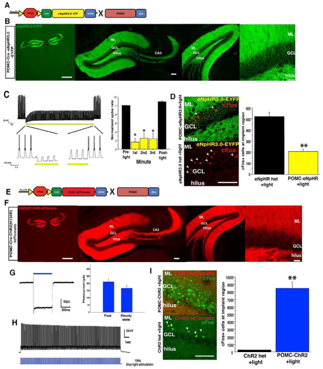
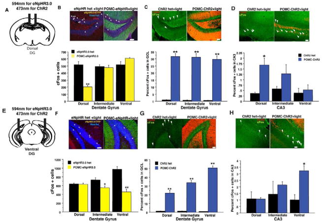
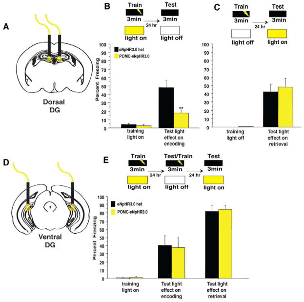
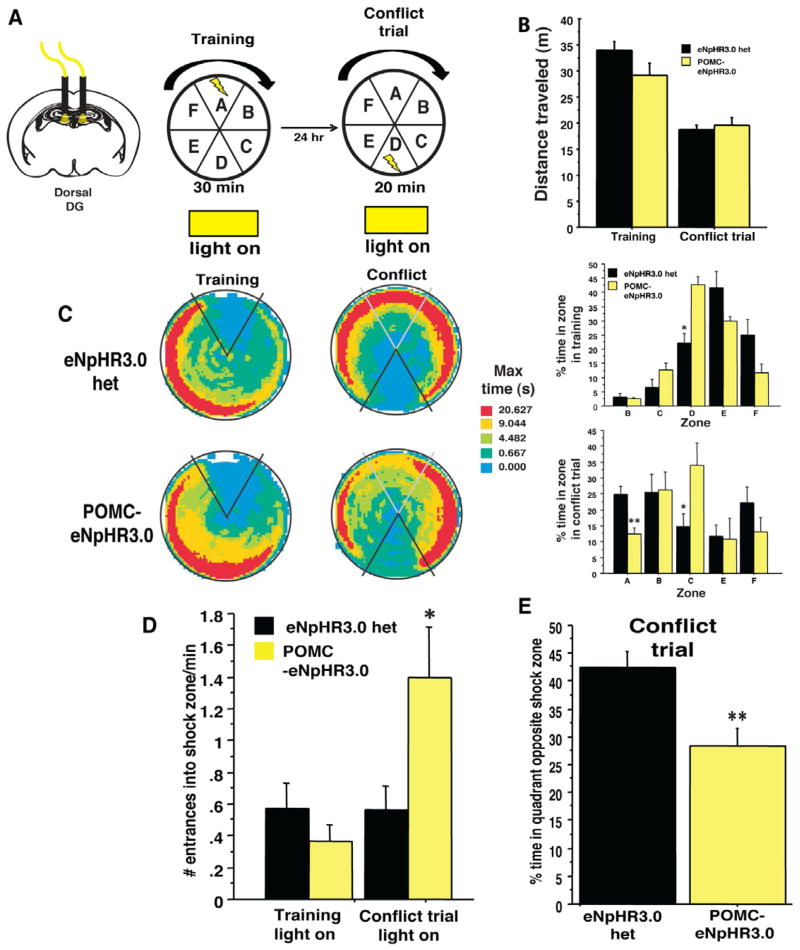
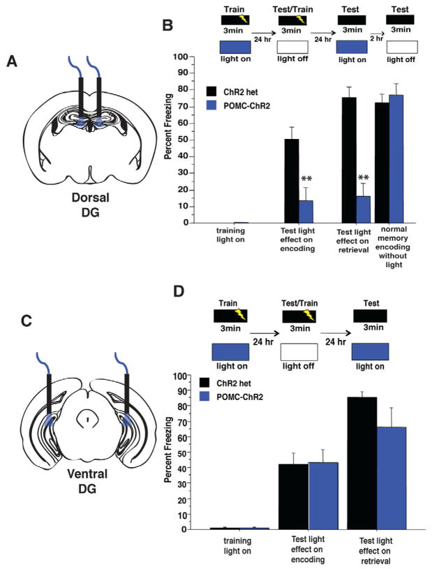
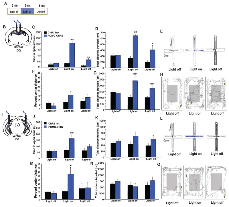
The dentate gyrus (DG), in addition to its role in learning and memory, is increasingly implicated in the pathophysiology of anxiety disorders. Here, we show that, dependent on their position along the dorsoventral axis of the hippocampus, DG granule cells (GCs) control specific features of anxiety and contextual learning. Using optogenetic techniques to either elevate or decrease GC activity, we demonstrate that GCs in the dorsal DG control exploratory drive and encoding, not retrieval, of contextual fear memories. In contrast, elevating the activity of GCs in the ventral DG has no effect on contextual learning but powerfully suppresses innate anxiety. These results suggest that strategies aimed at modulating the excitability of the ventral DG may be beneficial for the treatment of anxiety disorders.


Figures

Figure 1
Figure 1. Optogenetic control of dentate gyrus granule cells
(A) Genetic design for eNpHR3.0-YFP expression in DG GCs. (B) Expression of eNpHR3.0 in DG GCs, scale bar left, 1mm, middle 100um, right 30um. (C) Extended 3-minute illumination blocks evoked spiking in GCs, quantified on the right (n = 6, Mann-Whitney tests, planned comparisons, p < 0.05 for each minute). (D) Yellow light illumination of the dorsal DG in vivo reduces the number of cells positive for the immediate early gene cFos in the GCL region below the implanted fiber optic (scale bar, 100um), quantified on right (n=4/geno, t6= 6.7, p<0.001) (E) Genetic design for ChR2-tdTomato expression in DG GCs. (F) Expression of ChR2-tdTomato in DG GCs, scale bar left, 1mm, middle 100um, right 30um. (G) Voltage-clamp trace showing cationic current evoked by 1s pulse of 473nm light, average peak and steady state current amplitudes are quantified on the right. (H) Example current clamp record of spiking GC following a 10Hz train of 20 msec pulses. (I) Blue light illumination of the dorsal DG (10Hz, 20ms pulses) led to induction of cFos in a cohort of DG GCs (scale bar, 100um), quantified on the right (n=3–4/geno, t5=8.2, p<0.001). **p<0.01.
Figure 2
Figure 2. Illumination of the DG in POMC-eNpHR3.0 and POMC-ChR2 mice in vivo modulates cFos levels in the hippocampus in a region specific manner
Yellow light Illumination of the dorsal (B) or ventral (F) DG of POMC-eNpHR3.0 mice during exploration of a novel environment (20min constant illumination) reduces total number of cFos immunoreactive cells in the GCL in the region below the implanted fiber optic. (dorsal implants, n=4/geno, repeated measures ANOVA, genotypeXregion interaction F(2,12)=45.1, p<0.01, ventral implants, n-4–5/geno, F(2,14)=23.8, p<0.01. (C, G) Blue light illumination of the (5min, 10 Hz illumination in a novel environment) of the dorsal (C), or ventral DG (G) in POMC-ChR2 mice can increase percent of cFos+ cells (% Hoechst 33342) throughout the GCL, but leads to modest induction in CA3 in a region-specific manner (D, H), in the region of the implanted fiber optic, most likely via direct stimulation of mossy fiber axons projecting towards CA3 (dorsal implants DG: n=3–4/geno, repeated measures ANOVA, genotype effect, F(1,5)= 148.4, p<0.0001, geno X region F(2,10)=0.5, p=0.6, CA3:, repeated measures ANOVA, geno X region F(2,10)=6.8, p=0.01. Ventral implants DG: repeated measures ANOVA, genotype effect, F(1,3)= 297.5, p<0.01, geno X region, F(2,6)=1321, p<0.001, CA3:, repeated measures ANOVA, geno X region F(2,6)=5.2, p<0.05. Scale bars represent 50um, *p<0.05, **p<0.01. All error bars are +/− SEM.
Figure 3
Figure 3. GCs in the dorsal but not ventral DG selectively control the encoding of contextual fear memories
(A–B) Yellow light illumination of the dorsal DG during training blocked the encoding of context fear as POMC-eNpHR3.0 mice exhibited reduced freezing when tested 24hr later in the absence of light (n=6–7/geno, repeated measures ANOVA, genotype effect F(1,11)=13.2, p=0.004, genotypeXtraining interaction F(1,11)=9.4, p=0.01 (t11=3.4, p=0.006) (C) Optogenetic inhibition of the dorsal DG selectively impairs the encoding of contextual fear memories, as yellow light illumination of the dorsal DG during a recall session did not impair the retrieval of contextual fear memories. (n=7–8/geno, repeated measures ANOVA, geno effect F(1,13)= 0.19, p=0.7, training effect F(1,13)= 42.7, p=<0.0001, genotype X training interaction F(1,13)= 0.19, p=0.7 (t13=−.4, p=0.7) (D–E) POMC-eNpHR3.0 mice received yellow light stimulation of the ventral DG during encoding, were tested and retrained in the absence of light, then tested for light effects on retrieval. (E) Optogenetic inhibition of the ventral DG had no impact in acquisition or retrieval of contextual fear memories. (n=6/geno light on during encoding, repeated measures ANOVA, geno effect F(1,10)= 0.01, p=0.9, training effect F(1,10)= 19.5, p<0.01, genotype X training interaction F(1,10)= 0.03, p=0.9, light on during retrieval, geno effect F(1,10)= 0.1, p=0.8, genotype X training interaction F(1,10)= 0.05, p=0.8, (t10=−.3, p=0.8). **p<0.01. All error bars are +/− SEM.
Figure 4
Figure 4. Optogenetic inhibition of the dorsal DG impairs discrimination in an active place avoidance task
(A) Experimental design. Shown are the last training trial and the conflict trial. (B) Optogenetic inhibition of the dorsal DG did not impact exploration of the apparatus in either training or conflict trial phases (n=5–6/geno, repeated measures ANOVA, geno effect F(1,9)= 1.1, p=0.3, training effect F(1,9)= 1.3, p=0.3, genotype X training interaction F(1,9)= 2.7, p=0.1 (C) Time-in-location heat maps for POMC-eNpHR3.0 and control mice during the first 20 min of the last training trial and the 20min conflict trial. (D) Percent time in each of the zones during the training trial and the conflict trial. In training, a slightly different strategy was used, but both groups effectively avoided the shock zone (genotype X training interaction F(5,45)= 5.4, p<0.01, t-test on zone D, t9=−3.4, p<0.05). In the conflict trial, POMC-eNpHR3.0 mice spent significantly less time in the zone opposite the shock zone and more time adjacent to the shock zone (genotype X training interaction F(5,45)= 2.5, p<0.05, t-test on zone D, t9=4.1, p<0.05, zone C, t9=−2.3, p<0.05) (D–E) As a consequence, POMC-eNpHR3.0 mice exhibited an increased number of entrances into the shock zone after switching its location (unpaired t test, t9=−2.3, p<0.05, and the percent time in the quadrant opposite the new shock zone (t9=3.4, p<0.01). **p<0.01, *p<0.05. All error bars are +/− SEM.
Figure 5
Figure 5. Optogenetic stimulation of GCs in the dorsal, but not ventral, DG impairs the encoding and retrieval of contextual fear memories
(A–B) Blue light illumination of the dorsal DG during training blocked the context-shock association as POMC-ChR2 mice froze less to the training context when tested in the absence of light 24hr after training n=7–9/geno, repeated measures ANOVA, genotype effect F(1,14)=11.7, p=0.004, training effect, F(1,14)= 35.06, p<0.0001, training X genotype interaction F(1,14)=11.8, p=0.004, t-test on test day t14=3.4, p=0.004. Mice were retrained in the absence of light in this session by providing a single footshock at the end of the session. 24hr later, mice were tested for light effects on retrieval, where blue light impaired retrieval in POMC-ChR2 mice, but not single transgenic controls,(repeated measures ANOVA, genotype effect F(1,14)=35.9, p<0.0001, training effect, F(1,14)= 83.6, p<0.0001, training X genotype interaction F(1,14)=36.108, p<0.0001, t-test on test day t14=9.2, p<0.0001). Mice were tested 2hr later to confirm normal encoding of the memory during the retraining session, and that light during retrieval did not permanently erase the memory. POMC-ChR2 mice froze similar to control mice in this session, confirming an intact memory for the conditioning context, (Repeated measures ANOVA, genotype effect F(1,14)=0.3, p=0.58, training effect, F(1,14)= 31.1, p<0.0001, training X genotype interaction F(1,14)=0.29 p=0.6, t-test on test day t14=−0.6, p=0.59). (C–D) Optogenetic stimulation of the ventral DG did not impact the encoding or retrieval of contextual fear memories. Mice received blue light stimulation of the ventral DG during encoding, were tested and retrained in the absence of light, then tested for light effects on retrieval. POMC-ChR2 mice froze similar to single transgenic controls in all phases of the experiment (n=9/geno, light on during encoding, repeated measures ANOVA, genotype effect F(1,16)=0.01, p=0.9, training effect, F(1,6)= 60.7, p<0.0001, training X genotype interaction F(1,16)=0.002, p=0.97, t-test on test day t16=.06, p=0.95, light on during retrieval, repeated measures ANOVA, genotype effect F(1,16)=2.1, p=0.17, training effect, F(1,6)= 130.4, p<0.0001, training X genotype interaction F(1,16)=2.2, p=0.16, t-test on test day t16=−1.4, p=0.16. **p<0.01, all error bars are +/− SEM.
Figure 6
Figure 6. GCs in the ventral DG control conflict anxiety, while those in the dorsal DG drive exploratory behavior
A) Experimental design. POMC-ChR2 mice and single transgenic littermate controls were implanted in the dorsal (B–G) or ventral (I–O) DG then tested for 15min in the EPM and OFT with 5min light off, 5min light on, 5min light off epochs. (C–D) Optical stimulation of the dorsal DG in POMC-ChR2 mice but not single transgenic controls increased total time in the open arms of the EPM (C, repeated measures ANOVA, genotype effect F(1,15)=54.1, p<0.0001, light effect, F(2,30)= 42.2, p<0.0001, light X genotype interaction F(2,30)=30.2, p<0.0001, t test light on t15= −8.4, p<0.0001) as well as total distance traveled in the maze (genotype effect F(1,15)=18, p<0.001, light effect, F(2,30)= 16.3, p<0.0001, light X genotype interaction F(2,30)=20.3, p<0.0001, t test light on t15= −9.9, p<0.0001) Track trace represented in (E) (F–G) Optical stimulation of the dorsal DG in POMC-ChR2 mice but not single transgenic controls increased total exploration in the OFT but not percent distance traveled in the center of the arena (n=8–9/geno, total distance traveled, repeated measures ANOVA, genotype effect F(1,15)=8.3, p=0.01, light effect, F(2,30)= 3.2, p=0.05, light X genotype interaction F(2,30)=14.3, p<0.0001, t test light on t15= 3.3, p=0.004, percent center distance F(1,15)=1.1, p=0.31, light effect, F(2,30)= 3.5, p=0.04, light X genotype interaction F(2,30)=2.4, p=0.1, t test light on t15= 1.5, p=0.14). Track trace in (H). J–K) Ventral DG stimulation was acutely anxiolytic in the EPM as light stimulation increased time POMC-ChR2 mice spent in open arms of the maze (n=8/geno, repeated measures ANOVA, genotype effect F(1,14)=3.2, p=0.1, light effect, F(2,28)= 4.6, p=0.02, light X genotype interaction F(2,28)=4, p=0.03, t-test on light epoch t14=−2.4, p=0.03), but did not impact total distance traveled in the maze, genotype effect F(1,14)=3.4, p=0.1, light effect, F(2,28)= 0.7, p=0.5, light X genotype interaction F(2,28)=1, p=0.4). Track trace in L). M–N) OFT. Ventral DG stimulation did not impact total locomotor activity, but increased the percent distance traveled in the center of the arena (n=8–9/geno, repeated measures ANOVA, total distance traveled, genotype effect F(1,15)=0.3, p=0.6, light effect, F(2,30)= 1.9, p=0.2, light X genotype interaction F(2,30)=1.6, p=0.2, percent center distance, genotype effect F(1,15)=0.9, p=0.2, light effect, F(2,30)= 6.4, p=0.005, light X genotype interaction F(2,30)=6.9, p=0.003, t test light on t15=2.2, p=0.04, track trace in O). *p<0.05, **p<0.01. All error bars are +/− SEM.

Comment in

References

    1. Adhikari A, Topiwala MA, Gordon JA. Synchronized activity between the ventral hippocampus and the medial prefrontal cortex during anxiety. Neuron. 2010;65:257–269. - PMC - PubMed
    1. Bannerman DM, Bus T, Taylor A, Sanderson DJ, Schwarz I, Jensen V, Hvalby O, Rawlins JN, Seeburg PH, Sprengel R. Dissecting spatial knowledge from spatial choice by hippocampal NMDA receptor deletion. Nature neuroscience. 2012;15:1153–1159. - PMC - PubMed
    1. Bannerman DM, Deacon RM, Offen S, Friswell J, Grubb M, Rawlins JN. Double dissociation of function within the hippocampus: spatial memory and hyponeophagia. Behavioral neuroscience. 2002;116:884–901. - PubMed
    1. Bannerman DM, Rawlins JN, McHugh SB, Deacon RM, Yee BK, Bast T, Zhang WN, Pothuizen HH, Feldon J. Regional dissociations within the hippocampus--memory and anxiety. Neuroscience and biobehavioral reviews. 2004;28:273–283. - PubMed
    1. Bannerman DM, Yee BK, Good MA, Heupel MJ, Iversen SD, Rawlins JN. Double dissociation of function within the hippocampus: a comparison of dorsal, ventral, and complete hippocampal cytotoxic lesions. Behavioral neuroscience. 1999;113:1170–1188. - PubMed

Publication types