Review
. 2013 Mar 7;49(5):773-82.
doi: 10.1016/j.molcel.2013.02.011.

The hierarchy of the 3D genome

Affiliations
Review

The hierarchy of the 3D genome

Johan H Gibcus et al. Mol Cell. .

Abstract

Mammalian genomes encode genetic information in their linear sequence, but appropriate expression of their genes requires chromosomes to fold into complex three-dimensional structures. Transcriptional control involves the establishment of physical connections among genes and regulatory elements, both along and between chromosomes. Recent technological innovations in probing the folding of chromosomes are providing new insights into the spatial organization of genomes and its role in gene regulation. It is emerging that folding of large complex chromosomes involves a hierarchy of structures, from chromatin loops that connect genes and enhancers to larger chromosomal domains and nuclear compartments. The larger these structures are along this hierarchy, the more stable they are within cells, while becoming more stochastic between cells. Here, we review the experimental and theoretical data on this hierarchy of structures and propose a key role for the recently discovered topologically associating domains.


Figures

Figure 1
Figure 1. Large-scale nuclear organization in mammals
(1A) The nucleus is composed of chromosome territories (for example: mouse chromosome 3 and 18 are depicted in red and green respectively). DNA is organized in more or less condensed regions, as can be shown by DAPI staining (gray intensities). Nucleoli (N) are visible as dark spots (top inset). (1B) Inset shows a more detailed architecture of the nucleus with compartments (A and B), heterochromatin (HC) and euchromatin (EC) surrounding the interchromatin compartment. (1C) Zoom in view of chromosomal domains (hypothetical). Foci of factors interacting with looping chromatin in the perichromatic region (PR) are shown as pink circles and RNA is shown as orange lines. The larger speckles (SP) are located in domains that are sparser in chromatin content and further away from the PR. (1D) All-by-all chromosome matrix showing the interactions within and between chromosomes. (1E) Red and blue “plaid” pattern of chromosome 18 emphasized through pearson correlation shows the separation into 2 chromosomal domains (represented as red and blue). (1F) Detailed version of Figure 1D, (sized equivalent to Figure 1E, showing the cis-interaction matrix for chromosome 18. The inset indicates a ~ 3 Mb large B-compartment. (1G) The clustering into compartments A and B after principal component analysis on the plaid pattern displayed in Figure 1E. (1H) Detailed version of the 3 Mb large B-compartment from Figure 1F, revealing the organization of TADs (1 and 2). (1I) Representation of looping of chromatin as can be found at the PR (see 1C) or in deeper structures within TADs (see 1H). Nuclei were modeled to match HiC plots, which were adapted to scale from previously published data (Zhang et al., 2012).
Figure 2
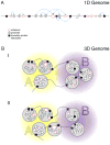
Figure 2. Genomic interactions
Promoter (black circle) and enhancers (red) are represented as circles. The size of enhancers indicates the strength of their activity. Architectural boundary proteins are shown as black squares and interactions relevant to gene expression are shown as dotted blue lines. (A) Linear representation of interactions between genomic elements. (B) Three-dimensional representation of the genome, where interactions are largely confined to TADs (gray circles) and TADs containing elements of similar activity are arranged in compartments (A or B). Situation “I” represents the 3D organization of the linear genome depicted in Figure 2A. Situation “II” represents changing interactions (leading to altered expression) by stochastic cell-to-cell differences (for interactions with promoters 1–6) or increased enhancer activity, leading to altered promoter expression (for interactions with promoter 7) and compartment change. Note that the altered expression does not lead to a change in TAD organization.
Figure 3
Figure 3. The stability and reproducibility of chromosomal interactions
Chromosomal territories and compartments are very stable within on cell cycle, but they are unlikely to be reproduced from one cell cycle to the next. Conversely, interactions between loops (within TADs) will be unstable and variable within each cell cycle, but this “instability” is reproducible from one cell cycle to the next. At the junction between stability and reproducibility, TADs confine looping, while maintaining the possibility of compartmentalization.

References

    1. Abney JR, Cutler B, Fillbach ML, Axelrod D, Scalettar BA. Chromatin dynamics in interphase nuclei and its implications for nuclear structure. The Journal of cell biology. 1997;137:1459–1468. - PMC - PubMed
    1. Bantignies F, Roure V, Comet I, Leblanc B, Schuettengruber B, Bonnet J, Tixier V, Mas A, Cavalli G. Polycomb-dependent regulatory contacts between distant Hox loci in Drosophila. Cell. 2011;144:214–226. - PubMed
    1. Bau D, Marti-Renom MA. Structure determination of genomic domains by satisfaction of spatial restraints. Chromosome research: an international journal on the molecular, supramolecular and evolutionary aspects of chromosome biology. 2011;19:25–35. - PubMed
    1. Bau D, Sanyal A, Lajoie BR, Capriotti E, Byron M, Lawrence JB, Dekker J, Marti-Renom MA. The three-dimensional folding of the alpha-globin gene domain reveals formation of chromatin globules. Nature structural & molecular biology. 2011;18:107–114. - PMC - PubMed
    1. Belton J-M, McCord RP, Gibcus JH, Naumova N, Zhan Y, Dekker J. Hi-C: A comprehensive technique to capture the conformation of genomes. Methods 2012 - PMC - PubMed

Publication types