

Chronic rhinosinusitis with nasal polyps is characterized by B-cell inflammation and EBV-induced protein 2 expression
- PMID: 23473835
- PMCID: PMC3741659
- DOI: 10.1016/j.jaci.2013.01.043
Chronic rhinosinusitis with nasal polyps is characterized by B-cell inflammation and EBV-induced protein 2 expression
Abstract
Background: Despite the high prevalence and morbidity of chronic rhinosinusitis (CRS), little is known about the mechanisms that underlie its pathogenesis. Recent studies have suggested that B cells might play an important role in CRS.
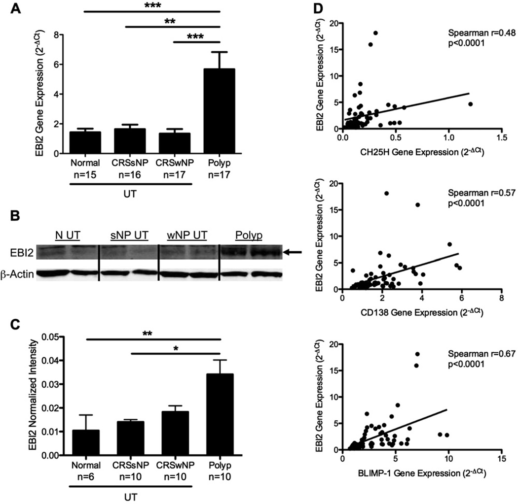
Objective: We sought to thoroughly characterize B lineage cells within sinus tissues of patients with CRS and healthy control subjects and to determine whether levels of EBV-induced protein 2, which is known to play an important role in the development of B-cell responses, were increased in patients with CRS.
Methods: Cells isolated from sinus tissues of patients with CRS and healthy control subjects were characterized by means of flow cytometry and immunohistochemistry. Local production of antibodies was measured in tissue extracts, nasal lavage fluid, and sera by using multiplex bead arrays and ELISA. Quantitative RT-PCR, ELISA, and Western blotting were used to assess gene and protein expression from tissue extracts.
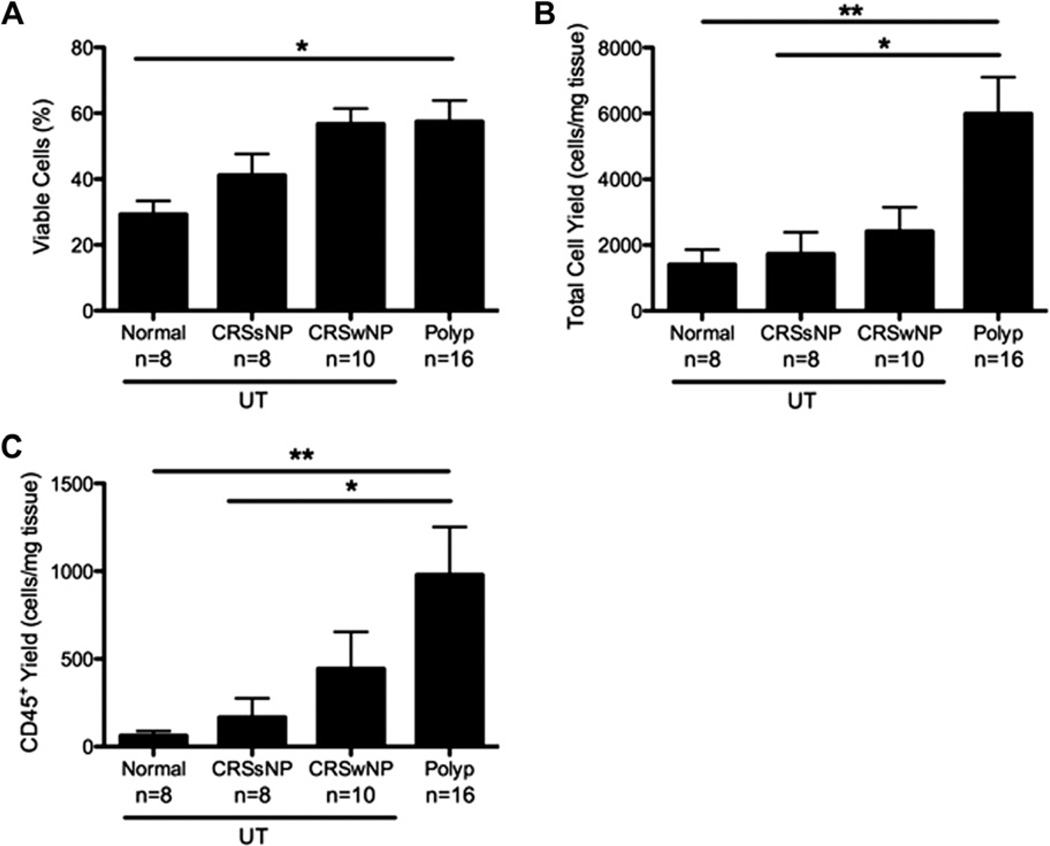
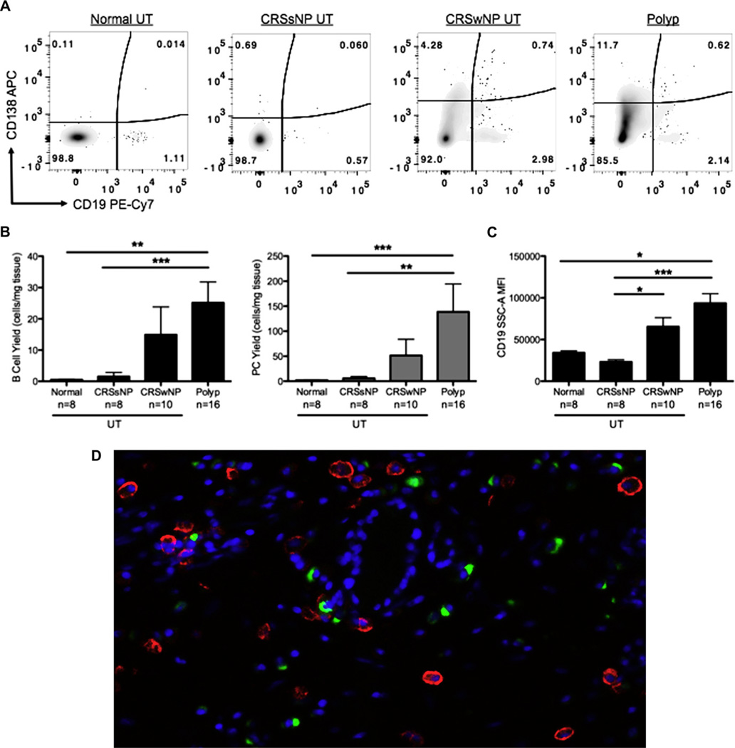
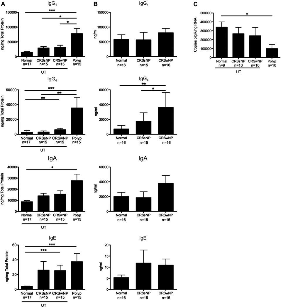
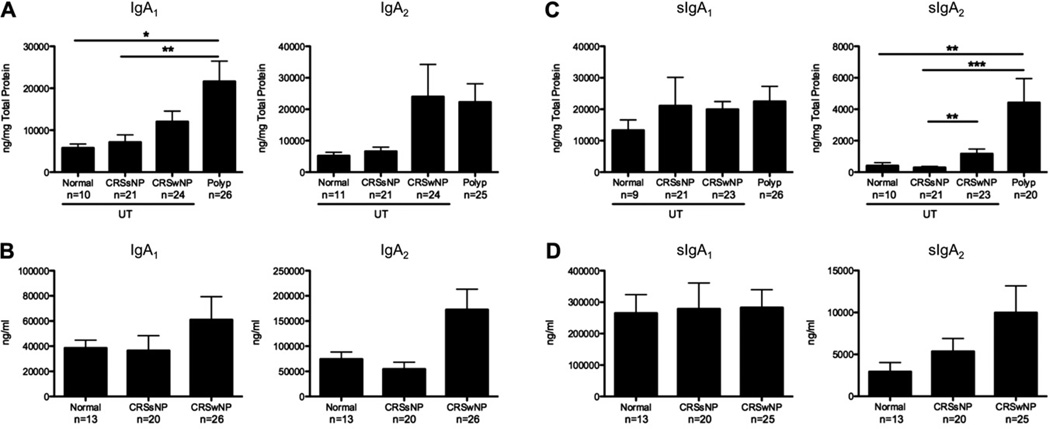
Results: Nasal polyps (NPs) from patients with CRS had increased levels of both B cells and plasma cells compared with uncinate tissue from healthy control subjects (P<.05). NPs also contained significantly increased levels of several antibody isotypes compared with normal uncinate tissue (P<.05), but no differences in circulating antibody levels were found. Interestingly, levels of EBV-induced protein 2 were also increased in NPs (P<.05) and were positively correlated with expression of plasma cell markers (CD138 and B lymphocyte-induced maturation protein) in sinus tissue.
Conclusion: B cells and plasma cells are enriched in NPs, actively produce antibodies locally, and might contribute to chronic inflammation in patients with CRS. Elucidating the mechanisms that underlie this excessive local B-cell response might provide novel insights for the development of improved therapeutic strategies.
Copyright © 2013 American Academy of Allergy, Asthma & Immunology. Published by Mosby, Inc. All rights reserved.
Conflict of interest statement
Disclosure of potential conflict of interest: J. E. Norton, R. K. Chandra, and A. Kato have received research support from the National Institutes of Health (NIH). A. T. Peters has provided expert witness testimony on drug allergy and has received lecture fees from Baxter. L. C. Grammer III has received research and travel support from the NIH; has received the Bazley Foundation grant; has received consultancy fees from Astellas Pharmaceuticals; is employed by Northwestern University and Northwestern Medical Faculty Foundation; has received research support from the NIH, the Food Allergy Network, and S&C Electric; has received lecture fees from the American Academy of Allergy, Asthma & Immunology (AAAAI); and receives royalties from Lippincott, UpToDate, BMU, and Elsevier. R. P. Schleimer has received research support from the NIH; has received consultancy fees from Intersect ENT, GlaxoSmithKline, and Allakos; and has stock/stock options in Allakos and Avrasense. The rest of the authors declare that they have no relevant conflicts of interest.
Figures
References
Publication types
MeSH terms
Substances
Grants and funding
LinkOut - more resources
Full Text Sources
Other Literature Sources
Medical
Miscellaneous
