Review
. 2013 May;21(5):230-7.
doi: 10.1016/j.tim.2013.02.003. Epub 2013 Mar 7.

Bacterial contact-dependent growth inhibition

Affiliations
Review

Bacterial contact-dependent growth inhibition

Zachary C Ruhe et al. Trends Microbiol. 2013 May.

Abstract

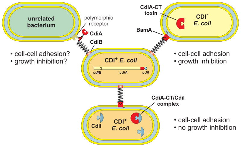
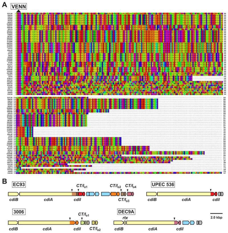
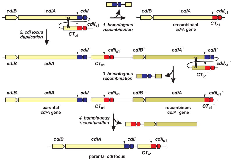
Bacteria cooperate to form multicellular communities and compete against one another for environmental resources. Here, we review recent advances in the understanding of bacterial competition mediated by contact-dependent growth inhibition (CDI) systems. Different CDI+ bacteria deploy a variety of toxins to inhibit neighboring cells and protect themselves from autoinhibition by producing specific immunity proteins. The genes encoding CDI toxin-immunity protein pairs appear to be exchanged between cdi loci and are often associated with other toxin-delivery systems in diverse bacterial species. CDI also appears to facilitate cooperative behavior between kin, suggesting that these systems may have other roles beyond competition.


Figures

Figure 1
Figure 1. Contact-dependent growth inhibition (CDI) in Escherichia coli
CDI+ E. coli express cdiBAI gene clusters and present CdiB/CdiA on the cell surface. CdiA binds receptors on neighboring E. coli cells and delivers a toxin derived from its C-terminus (CdiA-CT) into the target cell. CdiA-CT toxins inhibit the growth of CDI cells, but isogenic CDI+ inhibitors produce CdiI immunity proteins that protect them from toxin activity. The extracellular residues of most outer membrane proteins are highly variable between species, suggesting that a variety of specific receptors may be targeted by CDI.
Figure 2
Figure 2. E. coli CdiA-CT toxin diversity
A) CdiA-CT sequences are presented beginning with the conserved VENN peptide motif. UniProt accession numbers are provided to the left of each sequence and residues are colored according to the Taylor scheme. B) The cdi loci from E. coli EC93, UPEC 536, E. coli 3006 and E. coli O157:H7 DEC9A are depicted with cdiA-CT–cdiI pairs color-coded to indicate family types. The downward pointing arrows indicate VENN-encoding regions. The EC93 orphan-1 (CT/Io1), orphan-2 (CT/Io2) and orphan-4 (CT/Io4) pairs share sequence identity with the main toxin–immunity sequences from UPEC 536, E. coli 3006 and E. coli DEC9A, respectively. Open reading frames in light blue correspond to insertion sequence elements and transposase genes, rtx encodes a predicted toxin acyltransferase family member and genes depicted in gray are unrelated to CDI.
Figure 3
Figure 3. Models of orphan toxin–immunity gene rearrangement
Orphan cdiA-CT genes that contain conserved sequences upstream of the VENN-encoding region (downward arrow) can undergo homologous recombination with the full-length cdiA gene (indicated by cross-over in step 1). Recombination would delete the parental toxin–immunity coding sequences and fuse the orphan module (red) to cdiA. Alternatively, the cdi locus could undergo spontaneous duplication (step 2) followed by homologous recombination (step 3) to generate a recombinant cdiA gene. Further recombination between the orphan cdiA-CT and the recombined cdiA could regenerate the original parental genotype (step 4).
Figure 4
Figure 4. CdiA-CT–CdiI complex structures
A) Structural alignment of the CdiA-CTo11EC869 (maroon) and CdiA-CTIIBp1026b (orange) nuclease domains. The β4-β5 hairpin in CdiA-CTo11EC869 interacts with the CdiIo11EC869 immunity protein. B) CdiIo11EC869 and CdiIIIBp1026b immunity proteins bind to distinct sites on the toxin nuclease domains. Structures correspond to 4G6V and 4G6U in the Protein Data Bank and were rendered using PyMol. C) Alignment of CdiA-CTo11EC869 toxin homologues. The CdiA-CTo11EC869 nuclease domain sequence is aligned with related toxin sequences from the indicated bacterial species. UniProt accession numbers are given to the right of each sequence. Secondary structure elements (blue α-helices and red β-strands) from CdiA-CTo11EC869 are indicated above the alignment. The alignment was rendered with Jalview 2.8 at 30% sequence identity with progressively darker shades of purple indicating greater residue conservation. The conservation index is based on [38] and values are provided below each residue. Predicted toxin active site residues are rendered in red, and the β4-β5 hairpin (boxed) mediates interactions with the CdiIo11EC869 immunity protein.

References

    1. Lopez D, et al. Biofilms. Cold Spring Harb Perspect Biol. 2010;2:a000398. - PMC - PubMed
    1. Ng WL, Bassler BL. Bacterial quorum-sensing network architectures. Annu Rev Genet. 2009;43:197–222. - PMC - PubMed
    1. Destoumieux-Garzon D, et al. Focus on modified microcins: structural features and mechanisms of action. Biochimie. 2002;84:511–519. - PubMed
    1. Cascales E, et al. Colicin biology. Microbiol Mol Biol Rev. 2007;71:158–229. - PMC - PubMed
    1. Aoki SK, et al. Contact-dependent inhibition of growth in Escherichia coli. Science. 2005;309:1245–1248. - PubMed

Publication types

MeSH terms

Substances

LinkOut - more resources