

Differences in brainstem fiber tract response to radiation: a longitudinal diffusion tensor imaging study
- PMID: 23474114
- PMCID: PMC3646932
- DOI: 10.1016/j.ijrobp.2013.01.028
Differences in brainstem fiber tract response to radiation: a longitudinal diffusion tensor imaging study
Abstract
Purpose: To determine whether radiation-induced changes in white matter tracts are uniform across the brainstem.
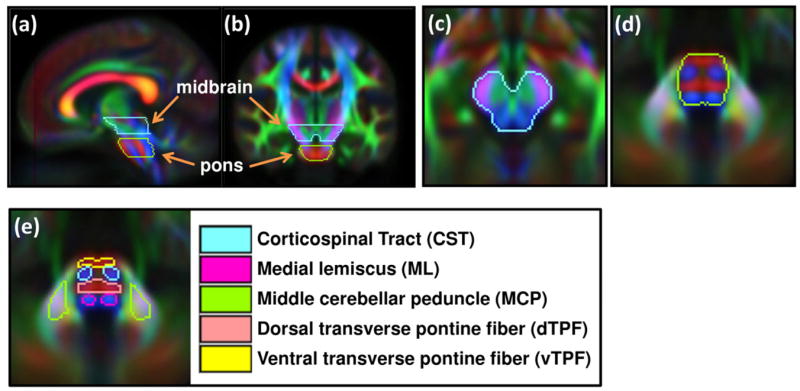
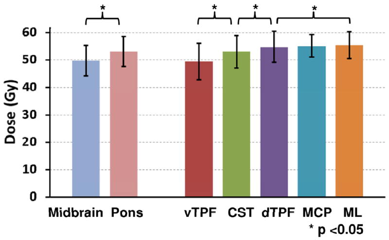
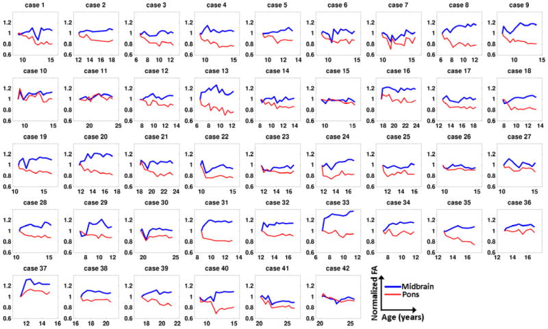
Methods and materials: We analyzed serial diffusion tensor imaging data, acquired before radiation therapy and over 48 to 72 months of follow-up, from 42 pediatric patients (age 6-20 years) with medulloblastoma. FSL software (FMRIB, Oxford, UK) was used to calculate fractional anisotropy (FA) and axial, radial, and mean diffusivities. For a consistent identification of volumes of interest (VOIs), the parametric maps of each patient were transformed to a standard brain space (MNI152), on which we identified VOIs including corticospinal tract (CST), medial lemniscus (ML), transverse pontine fiber (TPF), and middle cerebellar peduncle (MCP) at the level of pons. Temporal changes of DTI parameters in VOIs were compared using a linear mixed effect model.
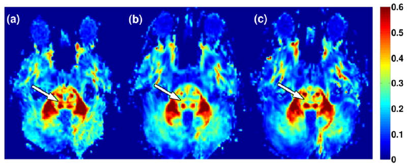
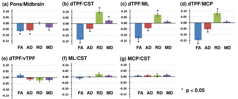
Results: Radiation-induced white matter injury was marked by a decline in FA after treatment. The decline was often accompanied by decreased axial diffusivity, increased radial diffusivity, or both. This implied axonal damage and demyelination. We observed that the magnitude of the changes was not always uniform across substructures of the brainstem. Specifically, the changes in DTI parameters for TPF were more pronounced than in other regions (P<.001 for FA) despite similarities in the distribution of dose. We did not find a significant difference among CST, ML, and MCP in these patients (P>.093 for all parameters).
Conclusions: Changes in the structural integrity of white matter tracts, assessed by DTI, were not uniform across the brainstem after radiation therapy. These results support a role for tract-based assessment in radiation treatment planning and determination of brainstem tolerance.
Copyright © 2013 Elsevier Inc. All rights reserved.
Conflict of interest statement
Conflicts of Interest Notification: No actual or potential conflicts of interest exist.
Figures
Similar articles
-
Brain tumor therapy-induced changes in normal-appearing brainstem measured with longitudinal diffusion tensor imaging.Int J Radiat Oncol Biol Phys. 2012 Apr 1;82(5):2047-54. doi: 10.1016/j.ijrobp.2011.03.057. Epub 2011 Jun 12. Int J Radiat Oncol Biol Phys. 2012. PMID: 21664060 Free PMC article.
-
Vulnerability of white matter to insult during childhood: evidence from patients treated for medulloblastoma.J Neurosurg Pediatr. 2016 Jul;18(1):29-40. doi: 10.3171/2016.1.PEDS15580. Epub 2016 Mar 25. J Neurosurg Pediatr. 2016. PMID: 27015518
-
Diffusion tensor imaging of the brainstem in children with achondroplasia.Dev Med Child Neurol. 2014 Nov;56(11):1085-92. doi: 10.1111/dmcn.12492. Epub 2014 May 14. Dev Med Child Neurol. 2014. PMID: 24825324 Free PMC article.
-
Mapping radiation dose distribution on the fractional anisotropy map: applications in the assessment of treatment-induced white matter injury.Neuroimage. 2006 May 15;31(1):109-15. doi: 10.1016/j.neuroimage.2005.12.007. Epub 2006 Jan 30. Neuroimage. 2006. PMID: 16448821
-
The role of diffusion tensor imaging and fractional anisotropy in the evaluation of patients with idiopathic normal pressure hydrocephalus: a literature review.Neurosurg Focus. 2016 Sep;41(3):E12. doi: 10.3171/2016.6.FOCUS16192. Neurosurg Focus. 2016. PMID: 27581308 Review.
Cited by
-
Spatial Agreement of Brainstem Dose Distributions Depending on Biological Model in Proton Therapy for Pediatric Brain Tumors.Adv Radiat Oncol. 2020 Aug 28;6(1):100551. doi: 10.1016/j.adro.2020.08.008. eCollection 2021 Jan-Feb. Adv Radiat Oncol. 2020. PMID: 33490724 Free PMC article.
-
National Cancer Institute Workshop on Proton Therapy for Children: Considerations Regarding Brainstem Injury.Int J Radiat Oncol Biol Phys. 2018 May 1;101(1):152-168. doi: 10.1016/j.ijrobp.2018.01.013. Int J Radiat Oncol Biol Phys. 2018. PMID: 29619963 Free PMC article. Review.
-
Effects of Surgery and Proton Therapy on Cerebral White Matter of Craniopharyngioma Patients.Int J Radiat Oncol Biol Phys. 2015 Sep 1;93(1):64-71. doi: 10.1016/j.ijrobp.2015.05.017. Epub 2015 May 16. Int J Radiat Oncol Biol Phys. 2015. PMID: 26279025 Free PMC article.
-
Reirradiation of recurrent medulloblastoma: does clinical benefit outweigh risk for toxicity?Cancer. 2014 Dec 1;120(23):3731-7. doi: 10.1002/cncr.28907. Epub 2014 Jul 30. Cancer. 2014. PMID: 25080363 Free PMC article.
-
Associating IDH and TERT Mutations in Glioma with Diffusion Anisotropy in Normal-Appearing White Matter.AJNR Am J Neuroradiol. 2023 May;44(5):553-561. doi: 10.3174/ajnr.A7855. Epub 2023 Apr 27. AJNR Am J Neuroradiol. 2023. PMID: 37105678 Free PMC article.
References
-
- Debus J, Hug EB, Liebsch NJ, et al. Brainstem tolerance to conformal radiotherapy of skull base tumors. Int J Radiat Oncol Biol Phys. 1997;39:967–975. - PubMed
-
- Wang S, Wu EX, Qiu D, et al. Longitudinal diffusion tensor magnetic resonance imaging study of radiation-induced white matter damage in a rat model. Cancer Res. 2009;69:1190–1198. - PubMed
Publication types
MeSH terms
Grants and funding
LinkOut - more resources
Full Text Sources
Other Literature Sources
Miscellaneous
