. 2013 Aug;38(9):1674-84.
doi: 10.1038/npp.2013.65. Epub 2013 Mar 8.

Hyposensitivity to gamma-aminobutyric acid in the ventral tegmental area during alcohol withdrawal: reversal by histone deacetylase inhibitors

Affiliations

Hyposensitivity to gamma-aminobutyric acid in the ventral tegmental area during alcohol withdrawal: reversal by histone deacetylase inhibitors

Devinder S Arora et al. Neuropsychopharmacology. 2013 Aug.

Abstract

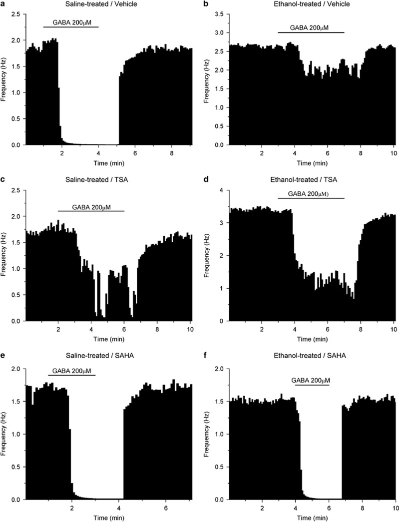
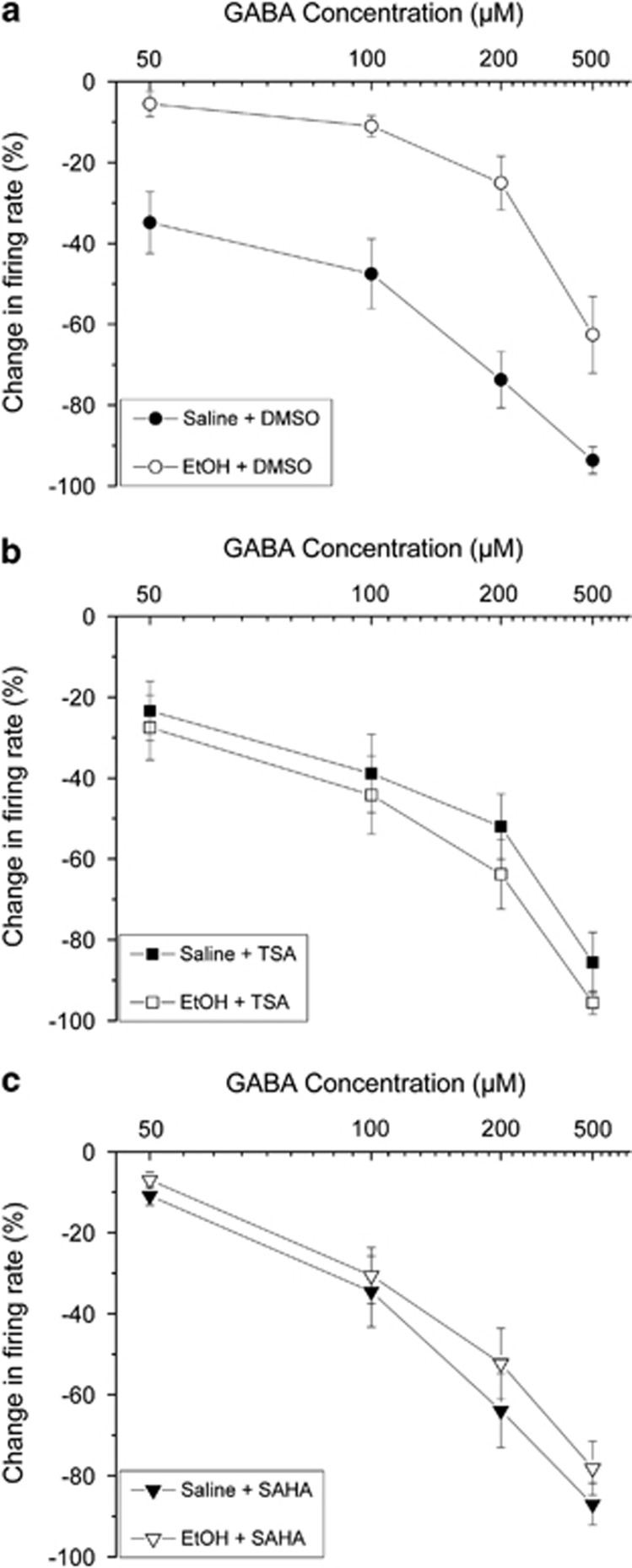
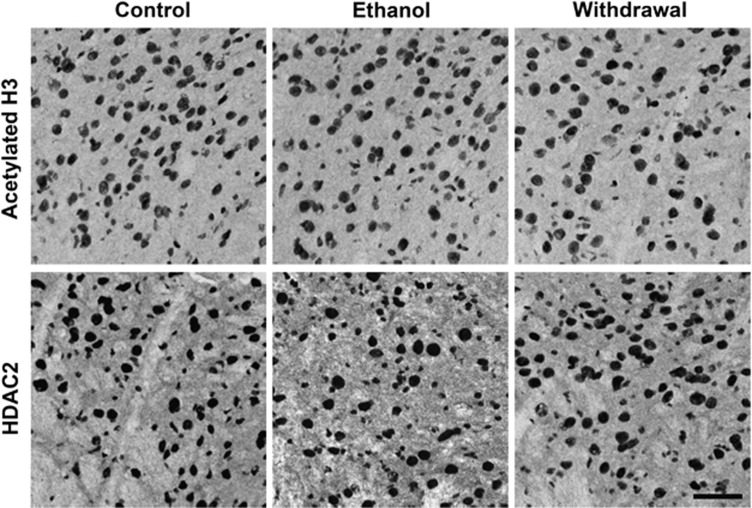
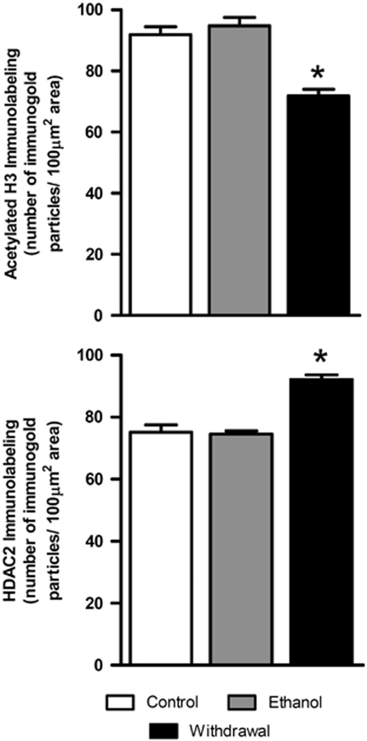
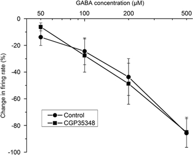
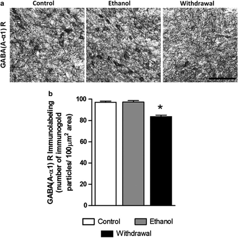
Putative dopaminergic (pDAergic) ventral tegmental area (VTA) neurons have an important role in alcohol addiction. Acute ethanol increases the activity of pDAergic neurons, and withdrawal from repeated ethanol administration produces a decreased sensitivity of pDAergic VTA neurons to GABA. Recent studies show that behavioral changes induced by chronic alcohol are reversed by inhibitors of histone deacetylases (HDACs). Whether HDAC-induced histone modifications regulate changes in GABA sensitivity of VTA pDAergic neurons during withdrawal is unknown. Here, we investigated modulation of withdrawal-induced changes in GABA sensitivity of pDAergic VTA neurons by HDAC inhibitors (HDACi), and also measured the levels of HDAC2, histone (H3-K9) acetylation, and GABA-Aα1 receptor (GABA (A-α1) R) subunit in VTA during ethanol withdrawal. Mice were injected intraperitoneally (ip) with either ethanol (3.5 g/kg) or saline twice daily for 3 weeks. In recordings from pDAergic VTA neurons in brain slices from ethanol-withdrawn mice, sensitivity to GABA (50-500 μM) was reduced. In brain slices from ethanol-withdrawn mice incubated with the HDACi SAHA (vorinostat) or trichostatin A (TSA) for 2 h, the hyposensitivity of pDAergic VTA neurons to GABA was significantly attenuated. There was no effect of TSA or SAHA on GABA sensitivity of pDAergic VTA neurons from saline-treated mice. In addition, ethanol withdrawal was associated with an increase in levels of HDAC2 and a decrease in histone (H3-K9) acetylation and levels of GABA (A-α1) R subunits in the VTA. Therefore, blockade of upregulation of HDAC2 by HDACi normalizes GABA hyposensitivity of pDAergic neurons developed during withdrawal after chronic ethanol treatment, which suggests the possibility that inhibition of HDACs can reverse ethanol-induced neuroadaptational changes in reward circuitry.


Figures

Figure 1
Figure 1
Single neurons: effects of 200 μM GABA. Each figure represents the firing rate of a single neuron of the VTA over time. Vertical bars are proportional to the firing rate over a 5-s interval, horizontal bars represent the duration of application of GABA (200 μM). Neurons were recorded in brain slices from mice treated with saline (a, c, e) or ethanol (b, d, f). Slices were incubated with DMSO (a, b), TSA (c, d), or SAHA (e, f). Addition of 200 μM GABA to the extracellular medium resulted in maximum reductions of firing in these neurons of (a) −99.6%, (b) −22.7%, (c) −76.9%, (d) −70.0%, (e) −99.2%, (f) −98.5%.
Figure 2
Figure 2
Pooled responses: effects of GABA. Pooled concentration–response curves for pDAergic VTA neurons from brain slices from mice treated with repeated injections of either saline (filled symbols) or ethanol (open symbols). Slices were incubated for 2 h with 0.1% DMSO (a), 12.5 μM TSA (b), or 3 μM SAHA (c). (a) Incubation in DMSO (vehicle controls): slices from saline-treated (•) or ethanol-treated mice (○); (b) incubation in TSA: slices from saline-treated (▪) or ethanol-treated mice (□); (c) incubation in SAHA: slices from saline-treated (▾) or ethanol-treated mice (∇).
Figure 3
Figure 3
Representative photomicrographs (scale bar=40 μm) showing acetylated histone H3-K9 (top panel) and HDAC2 (bottom panel) gold immunolabeling in the VTA of repeated saline treatment (Control), repeated ethanol treatment with last ethanol injection 1 h before sacrifice (Ethanol), and repeated ethanol treatment with last ethanol injection >14 h before sacrifice (Withdrawal).
Figure 4
Figure 4
Bar diagrams showing the differences in the acetylated H3 (H3-K9) and HDAC2 protein levels in the VTA of repeated saline treatment (Control, n=6), repeated ethanol treatment with last ethanol injection 1 h before sacrifice (Ethanol, n=6), and repeated ethanol treatment with last ethanol injection >14 h before sacrifice (Withdrawal, n=6). There was a significant decrease in acetylated H3 (*p<0.001) and a reciprocal increase in the levels of HDAC2 (*p<0.001) in the VTA of mice in withdrawal from ethanol (Withdrawal) compared with that of saline-treated controls (Control) or to ethanol-treated mice that received an ethanol injection 1 h before sacrifice (Ethanol).
Figure 5
Figure 5
The effects of GABA-B antagonist CGP35348 on GABA-induced inhibition of pDAergic VTA neurons. Pooled concentration–response curves for responses to GABA of pDAergic VTA neurons from brain slices from mice in the absence (•) or presence (▪) of CGP35348 (1 μM). GABA (50–500 μM) was applied to each neuron, then CGP35348 was added to the medium and the same concentrations of GABA were tested. There was no significant difference in response to GABA between the groups (two-way ANOVA, F1,51=1.4E-5, p>0.99).
Figure 6
Figure 6
(a) Representative low-magnification photomicrographs (scale bar=40 μm) of gold immunolabeling of GABA (A-α1) R subunit in the VTA of repeated saline treatment (Control), repeated ethanol treatment with last ethanol injection 1 h before sacrifice (Ethanol), and repeated ethanol treatment with last ethanol injection >14 h before sacrifice (Withdrawal). (b) Bar diagrams showing the differences in the protein levels of GABA (A-α1) R subunit in the VTA of repeated saline treatment (Control, n=6), repeated ethanol treatment with last ethanol injection 1 h before sacrifice (Ethanol, n=6), and repeated ethanol treatment with last ethanol injection >14 h before sacrifice (Withdrawal, n=6). There was a significant decrease in GABA (A-α1) R subunit (*p<0.001) in the VTA of mice in withdrawal from ethanol (Withdrawal) compared with that of saline-treated controls (Control) or to ethanol-treated mice.

References

    1. Albanese A, Minciacchi D. Organization of the ascending projections from the ventral tegmental area: a multiple fluorescent retrograde tracer study in the rat. J Comp Neurol. 1983;216:406–420. - PubMed
    1. Arora D, Haluk DM, Kourrich S, Pravetoni M, Fernandez-Alacid L, Nicolau JC, et al. Altered neurotransmission in the mesolimbic reward system of Girk mice. J Neurochem. 2010;114:1487–1497. - PMC - PubMed
    1. Bailey CP, Manley SJ, Watson WP, Wonnacott S, Molleman A, Little HJ. Chronic ethanol administration alters activity in ventral tegmental area neurons after cessation of withdrawal hyperexcitability. Brain Res. 1998;803:144–152. - PubMed
    1. Brodie MS. Increased ethanol excitation of dopaminergic neurons of the ventral tegmental area after chronic ethanol treatment. Alcohol Clin Exp Res. 2002;26:1024–1030. - PubMed
    1. Brodie MS, McElvain MA, Bunney EB, Appel SB. Pharmacological reduction of small conductance calcium-activated potassium current (SK) potentiates the excitatory effect of ethanol on ventral tegmental area dopamine neurons. J Pharmacol Exp Ther. 1999a;290:325–333. - PubMed

Publication types

MeSH terms