. 2014 Mar 6;33(10):1297-305.
doi: 10.1038/onc.2013.64. Epub 2013 Mar 11.

Canonical and non-canonical NF-κB signaling promotes breast cancer tumor-initiating cells

Affiliations

Canonical and non-canonical NF-κB signaling promotes breast cancer tumor-initiating cells

M F Kendellen et al. Oncogene. .

Abstract

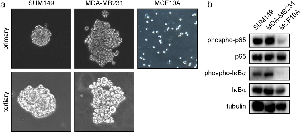
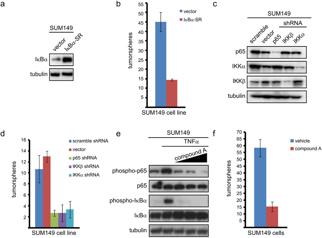
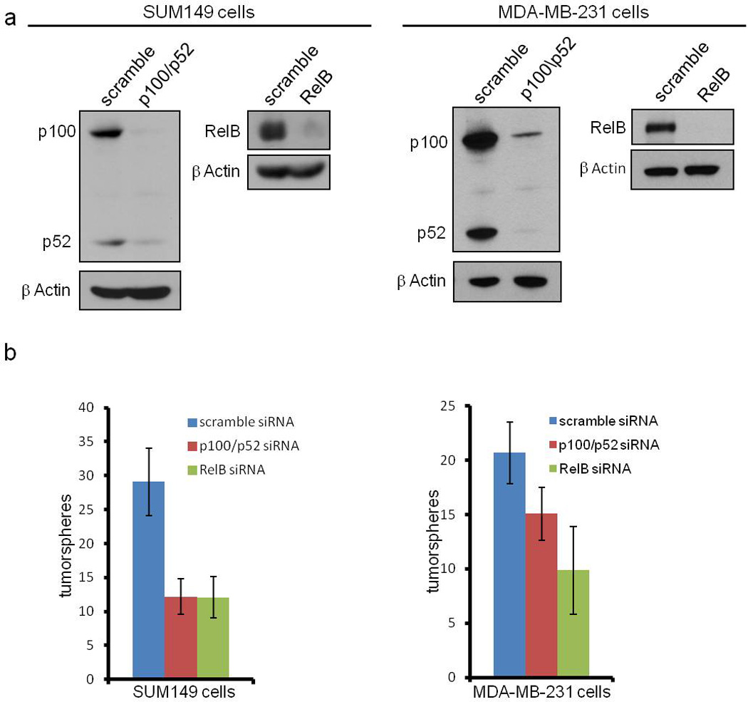
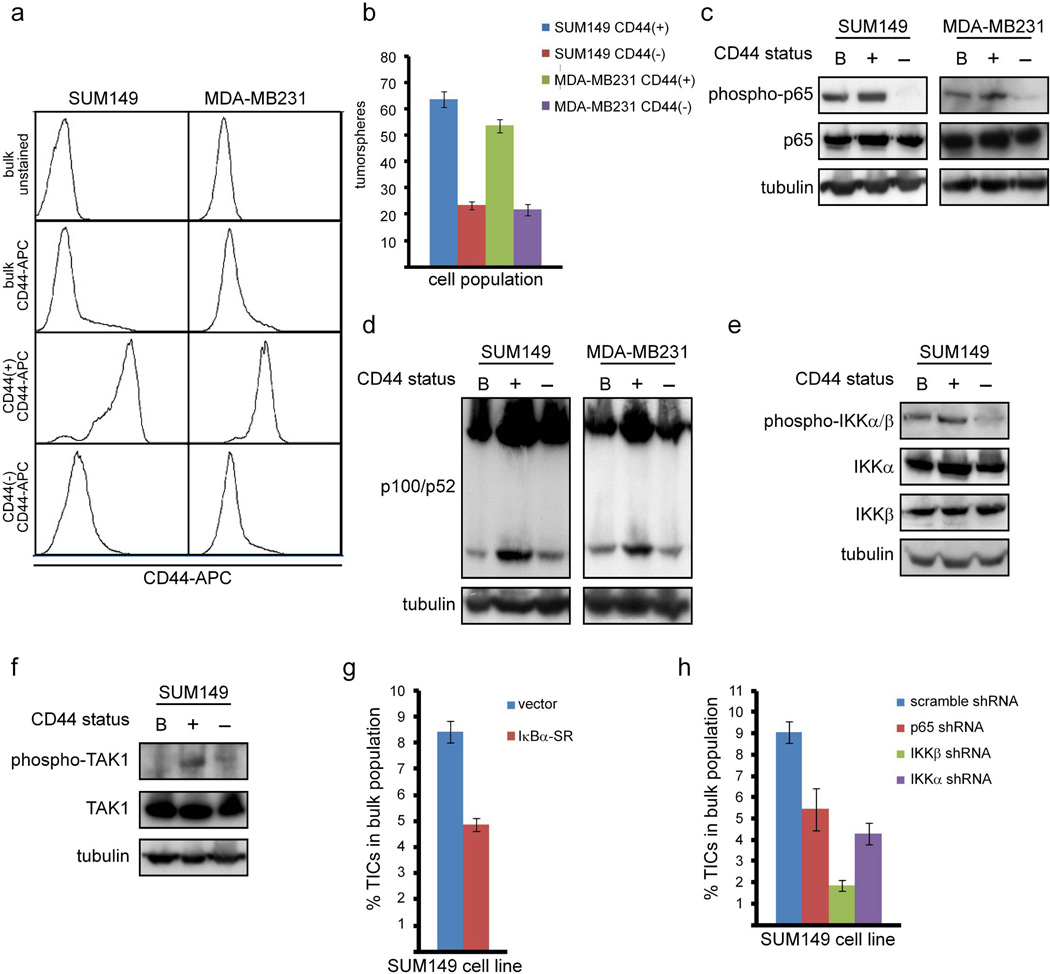
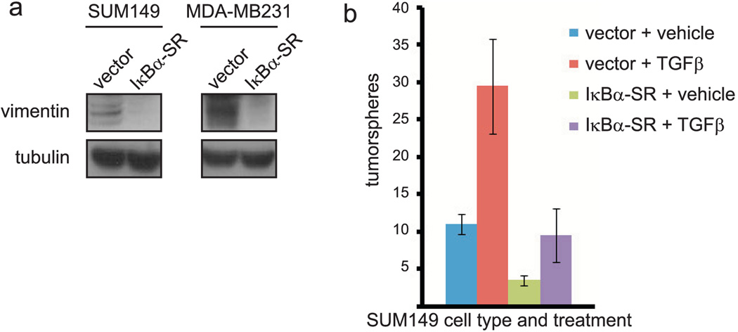
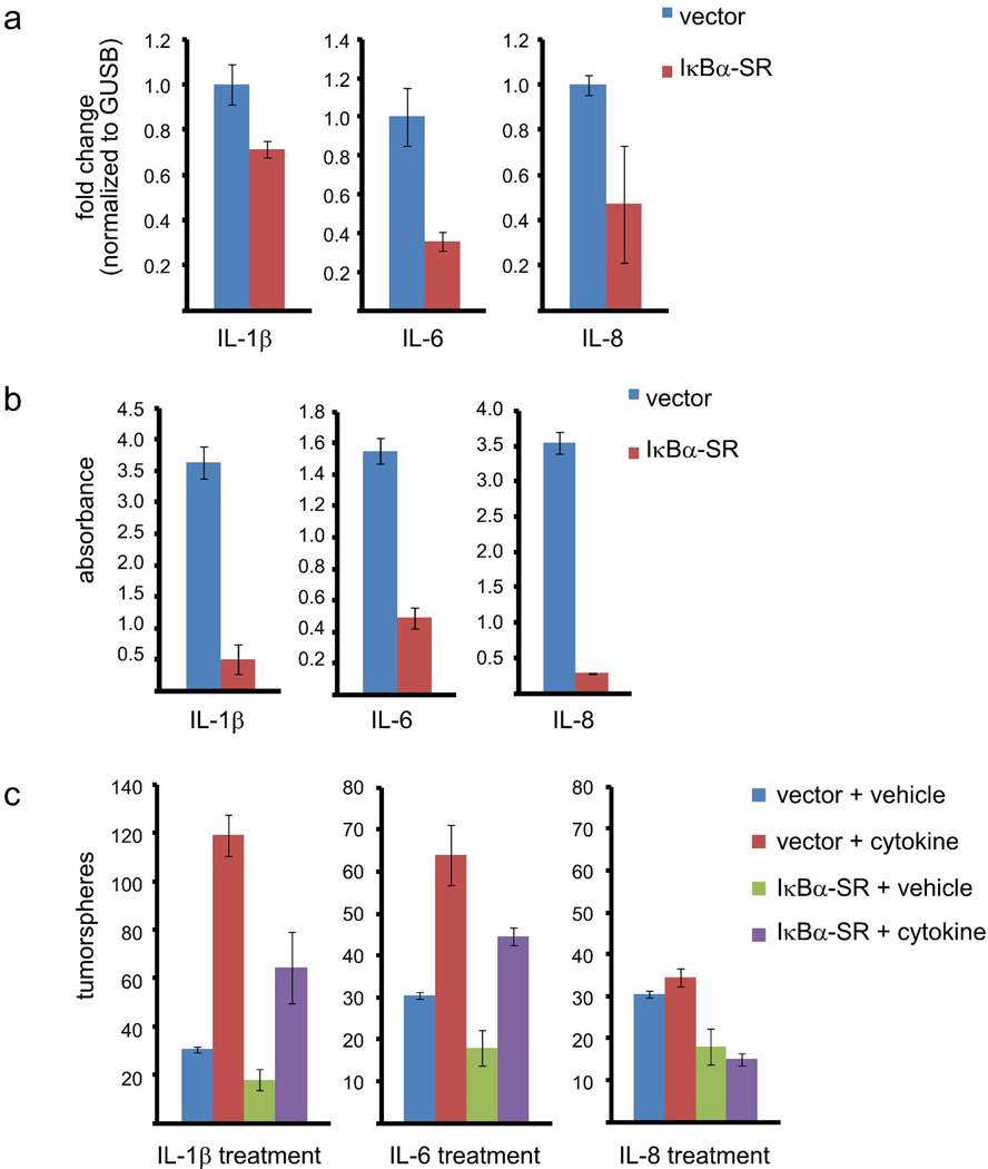
Tumor-initiating cells (TICs) are a sub-population of cells that exhibit a robust ability to self-renew and contribute to the formation of primary tumors, the relapse of previously treated tumors and the development of metastases. TICs have been identified in various tumors including those of the breast, and are particularly enriched in the basal-like and claudin-low subtypes of breast cancer. The signaling pathways that contribute to the function and maintenance of TICs are under intense study. We explored the potential involvement of the nuclear factor-κB (NF-κB) family of transcription factors in TICs in cell lines that are representative of basal-like and claudin-low breast cancer. NF-κB was found to be activated in breast cancer cells that form tumorspheres efficiently. Moreover, both canonical and non-canonical NF-κB signaling is required for these cells to self-renew in vitro and to form xenograft tumors efficiently in vivo using limiting dilutions of cells. Consistent with this fact, canonical and non-canonical NF-κB signaling is activated in TICs isolated from breast cancer cell lines. Experimental results indicate that NF-κB promotes the function of TICs by stimulating epithelial-to-mesenchymal transition and by upregulating the expression of the inflammatory cytokines interleukin-1β and interleukin-6. The results suggest the use of NF-κB inhibitors for clinical therapy of certain breast cancers.


Conflict of interest statement

Competing Interests/Conflicts: The authors declare that they have no competing or conflicting interests.

Figures

Figure 1
Figure 1. NF-κB signaling is preferentially activated in tumorsphere-forming breast cancer cells
(A) Primary and tertiary tumorspheres formed by the indicated bulk populations of basal-like and claudin-low breast cancer cells in serum-free culture on low-adhesion plates. (B) Phosphorylation of p65 and IκBα as markers of NF-κB activation in the indicated bulk populations of breast cancer cells (SUM149 and MDA-MB231) or immortalized breast (MCF10A) cells.
Figure 2
Figure 2. Canonical NF-κB signaling is required for basal-like breast cancer cells to efficiently self-renew
(A) Immunoblot of the indicated proteins in SUM149 cells stably expressing an empty vector or IκBα-SR. (B) Quantification of tumorspheres formed by 100 SUM149 cells expressing empty vector or IκBα-SR. (C) Immunoblot of the indicated proteins by 100 SUM149 cells stably infected with the indicated shRNA constructs. (D) Quantification of tumorspheres formed by 100 SUM149 cells stably expressing the indicated shRNA constructs. (E) Phosphorylation of p65 and IκBα as markers of activation of canonical NF-κB signaling in SUM149 cells pre-treated with increasing doses of compound A and then treated with TNFα. (F) Quantification of tumorspheres formed by 100 SUM149 cells treated daily with 5 µM compound A.
Figure 3
Figure 3. Non-canonical NF-κB signaling is required for basal-like breast cancer cells to self-renew
(A) Immunoblot of the indicated proteins in SUM149 and MDA-MB231 cells expressing scrambled siRNA or siRNA targeting p100/p52 or RelB using the indicated antibodies. (B) Quantification of tumorspheres formed by 100 SUM149 or MDA-MB231 cells expressing the indicated siRNA constructs. p values for the right panel of (B) are: p = 0.0413 for scramble siRNA compared to p100/p52 siRNA and p = 0.0011 for scramble siRNA compared to RelB siRNA.
Figure 4
Figure 4. Canonical and non-canonical NF-κB signaling is preferentially activated in TICs and required for the maintenance of TICs in breast cancer cells
(A) FACS analysis of the indicated populations of SUM149 cells stained with CD44-APC. (B) Quantification of tumorspheres formed by 100 cells from the indicated cell populations of SUM149 or MDA-MB231 cells. (C) Phosphorylation of p65 as a marker of activation of canonical NF-κB signaling in the indicated populations of SUM149 and MDA-MB231 cells. (D) Cleavage of p100 to p52 as a marker of activation of non-canonical NF-κB signaling in the indicated populations of SUM149 and MDA-MB231 cells. (E) Phosphorylation of IKKα and IKKβ in the indicated populations of SUM149 cells. (F) Phosphorylation of TAK1 in the indicated populations of SUM149 cells. (G, H) Percentage of TICs isolated from SUM149 cells stably expressing IκBα-SR (G) or the indicated shRNA constructs (H).
Figure 5
Figure 5. NF-κB activation promotes expression of markers of EMT in TICs and TGFβ-induced self-renewal of basal-like breast cancer cells
(A) Immunoblot of vimentin in SUM149 or MDA-MB231 cells stably expressing an empty vector or IκBα-SR. (B) Quantification of tumorspheres formed by 100 SUM149 cells stably expressing empty vector or IκBα-SR, followed by treatment with vehicle control or TGFβ.
Figure 6
Figure 6. IL-1β and IL-6 stimulate the self-renewal of basal-like breast cancer cells downstream of NF-κB
(A) Real-time PCR showing expression of IL-1β, IL-6, or IL-8 in the bulk population of SUM149 cells stably expressing an empty vector or IκBα-SR. (B) ELISA analysis showing the abundance of IL-1β, IL-6, or IL-8 in the media of SUM149 cells stably expressing empty vector or IκBα-SR. (C) Quantification of tumorspheres formed by 100 SUM149 cells expressing empty vector or IκBα-SR, followed by treatment with IL-1β, IL-6, IL-8 or vehicle control.

References

    1. Reya T, Morrison SJ, Clarke MF, Weissman IL. Stem cells, cancer, and cancer stem cells. Nature. 2001;414:105–111. - PubMed
    1. Charafe-Jauffret E, Ginestier C, Birnbaum D. Breast cancer stem cells: tools and models to rely on. BMC Cancer. 2009;9:202. - PMC - PubMed
    1. Ponti D, Costa A, Zaffaroni N, Pratesi G, Petrangolini G, Coradini D, et al. Isolation and in vitro propagation of tumorigenic breast cancer cells with stem/progenitor cell properties. Cancer Res. 2005;65:5506–5511. - PubMed
    1. Croker AK, Allan AL. Cancer stem cells: implications for the progression and treatment of metastatic disease. J Cell Mol Med. 2008;2:374–390. - PMC - PubMed
    1. Lawson JC, Blatch GL, Edkins AL. Cancer stem cells in breast cancer and metastasis. Breast Cancer Res Treat. 2009;2:241–254. - PubMed

Publication types

MeSH terms