. 2014 Mar 6;33(10):1306-15.
doi: 10.1038/onc.2013.59. Epub 2013 Mar 11.

Protein kinase Cδ is required for ErbB2-driven mammary gland tumorigenesis and negatively correlates with prognosis in human breast cancer

Affiliations

Protein kinase Cδ is required for ErbB2-driven mammary gland tumorigenesis and negatively correlates with prognosis in human breast cancer

B L Allen-Petersen et al. Oncogene. .

Abstract

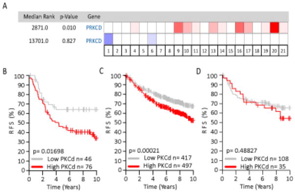
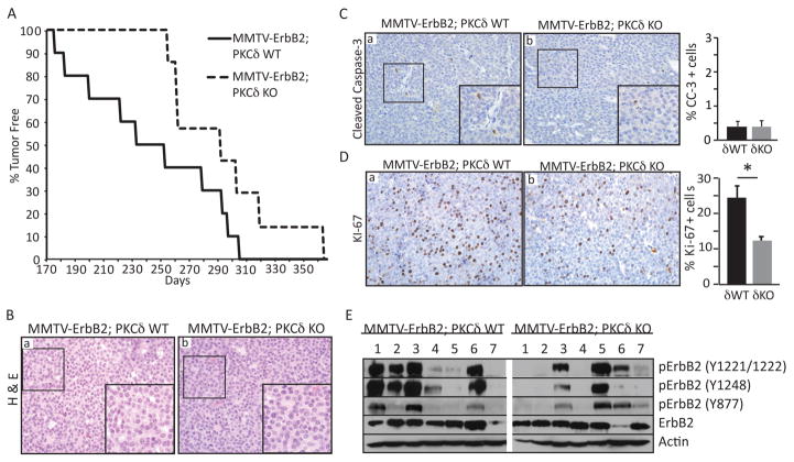
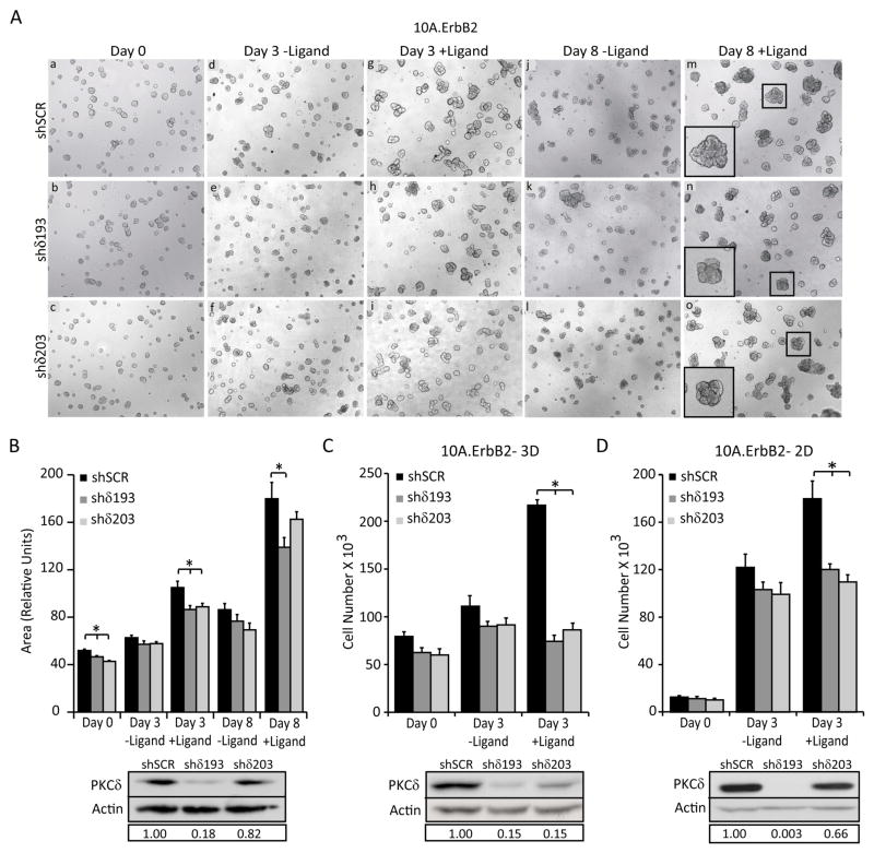
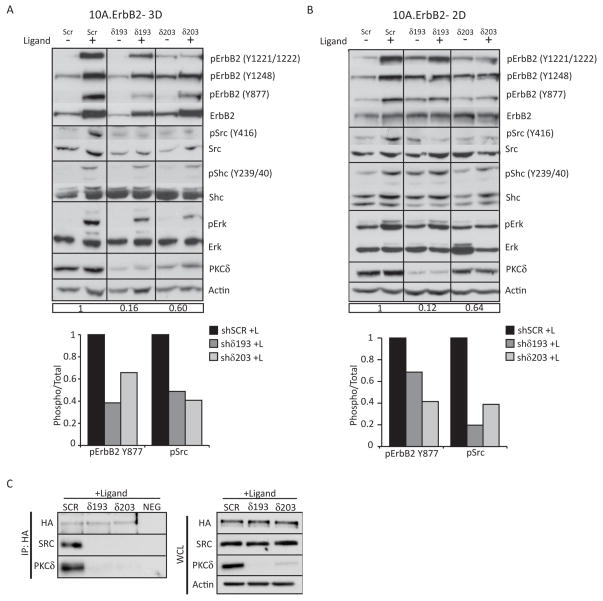
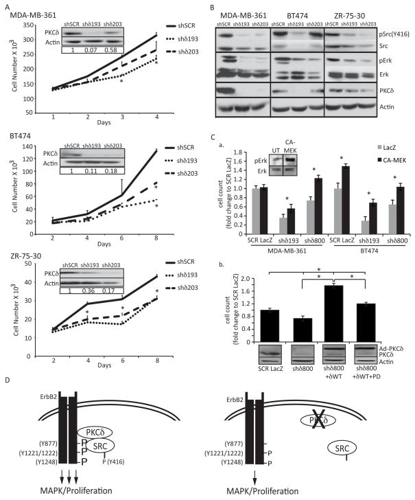
Protein kinase C δ (PKCδ) regulates apoptosis in the mammary gland, however, the functional contribution of PKCδ to the development or progression of breast cancer has yet to be determined. Meta-analysis of ErbB2-positive breast cancers shows increased PKCδ expression, and a negative correlation between PKCδ expression and prognosis. Here, we present in-vivo evidence that PKCδ is essential for the development of mammary gland tumors in a ErbB2-overexpressing transgenic mouse model, and in-vitro evidence that PKCδ is required for proliferative signaling downstream of the ErbB2 receptor. Mouse mammary tumor virus (MMTV)-ErbB2 mice lacking PKCδ (δKO) have increased tumor latency compared with MMTV-ErbB2 wild-type (δWT) mice, and the tumors show a dramatic decrease in Ki-67 staining. To explore the relationship between PKCδ and ErbB2-driven proliferation more directly, we used MCF-10A cells engineered to express a synthetic ligand-inducible form of the ErbB2 receptor. Depletion of PKCδ with short hairpin RNA inhibited ligand-induced growth in both two-dimensional (2D) (plastic) and three-dimensional (3D) (Matrigel) culture, and correlated with decreased phosphorylation of the ErbB2 receptor and reduced activation of Src and MAPK/ERK pathways. Similarly, in human breast cancer cell lines in which ErbB2 is overexpressed, depletion of PKCδ suppresses proliferation, Src and ERK activation. PKCδ appears to drive proliferation through the formation of an active ErbB2/PKCδ/Src signaling complex, as depletion of PKCδ disrupts association of Src with the ErbB2 receptor. Taken together, our studies present the first evidence that PKCδ is a critical regulator of ErbB2-mediated tumorigenesis, and suggest further investigation of PKCδ as a target in ErbB2-positive breast cancer.


Conflict of interest statement

Conflict of interest: The authors declare no conflict of interest.

Figures

Figure 1
Figure 1. Increased expression of PKCδ negatively correlates with prognosis in ErbB2 positive human breast cancer
A. Overexpression (red) and underexpression (blue) of PKCδ in 21 human breast cancer data sets. Intensity of the cell color corresponds to the top percentage of ranked genes, with 50% (white) representing no change between tumor sets. The median rank for PKCδ overexpression in ErbB2 positive tumors across each data set is 2,871 (P=0.01), while the underexpression median rank is 13,701 (P=0.827). B, C, and D. Kaplan-Meier analysis, using relapse-free survival as endpoint, for (B) ErbB2 positive tumors (four data sets, n= 122, P=0.017), (C) All molecular subtypes (four data sets, n= 914, P=0.0002), and (D) Triple-negative (ER-/PR-/ErbB2-)(four data sets, n=143, P=0.488), stratified into two quantiles base on high (red line) or low (gray line) PKCδ expression.
Figure 2
Figure 2. Loss of PKCδ increases tumor latency in MMTV-ErbB2 transgenic mice
A. MMTV-ErbB2;δWT (n=10) and δKO (n=7) mice were palpated weekly to detect mammary tumor growth. Graph shows the number of days to tumor detection verses the percentage of tumor-free mice for each genotype; P=0.03. B. MMTV-ErbB2;δWT and MMTV-ErbB2;δKO tumor sections were stained with hematoxylin and eosin (a, b). Histology- 20X, insets depict 60X enlargement of boxed region. C and D. Cleaved caspase-3 (C) and Ki-67 (D) immunohistochemistry on MMTV-ErbB2;δWT and MMTV-ErbB2;δKO tumor sections. Histology- 20X, insets depict 60X enlargement of boxed region. Graphs depict quantification of the percent of cleaved caspase-3 or Ki-67 positive cells over 6–8 fields per tumor, per genotype; n=5–8 MMTV-ErbB2;δWT, 4–7 MMTV-ErbB2;δKO; *P=0.01. E. Tumor lysates from MMTV-ErbB2;δWT (7) and δKO (7) mice were immunoblotted with the following antibodies to determine the levels of ErbB2 receptor phosphorylation: pErbB2 (Y877, Y1221/1221, Y1248); ErbB2; and actin.
Figure 3
Figure 3. PKCδ is required for ErbB2-driven proliferation
For all panels: PKCδ was depleted using lentiviral shRNA constructs (shδ193 and shδ203) and compared to control shRNA (shSCR) as described in Materials and Methods. A. 10A.ErbB2 cells depleted of PKCδ using shRNA (shδ193 and shδ203) were grown on Matrigel for 6 days (a, b, c). Cells were then left untreated (d, e, f; j, k, l) or treated with 1μM ligand for 3–8 days (g, h, i; m, n, o). Representative images of three separate experiments taken at 5X magnification are shown. Inset shows digital enlargement to show structure morphology. B. Quantification of structure area from Figure 2A, using Metamorph software. Representative of three experiments shown; *P=<0.02. D. and E. 10A.ErbB2 cells depleted of PKCδ were plated at equal densities in 3D (D) or 2D (E) growth assays. For 3D growth, cells were plated on Matrigel and grown for 6 days. Ligand was added for 3 days after which cells were tryspinized and counted; *P=<0.0001. For 2D growth, cells were treated with or without ligand for 4 days, trypsinized, and counted; *P=<0.04. B, C, and D. Insets show depletion of PKCδ by immunoblot with actin as a loading control. Boxed region below blots shows relative PKCδ knockdown compared to shSCR controls. Representative graphs of three separate experiments are shown.
Figure 4
Figure 4. Loss of PKCδ attenuates ErbB2 signal transduction
For all panels: PKCδ was depleted using lentiviral shRNA constructs (shδ193 and shδ203) and compared to control shRNA (shSCR) as described in Materials and Methods. A. and B. 10A.ErbB2 cells depleted of PKCδ were plated in 3D culture (A) or 2D culture (B) and then treated for 3 days or 24 hours with ligand respectively. Protein expression and phosphorylation in whole cell lysates was evaluated by immunobloting. The following antibodies were used: pErbB2 (Y877, Y1221/1221, Y1248); ErbB2; pSrc (Y416); Src; pShc (Y239/240); Shc, pErk1/2 (T202/Y204); Erk1/2; PKCδ; and actin. Immunoblots probed for phosphorylated proteins were stripped and probed with antibodies to total protein, as indicated. Blots were probed for actin as a loading control. Relative PKCδ knockdown compared to shSCR is shown in boxed region below blots. Immunoblots were quantified by densitometry; graphs depict the levels of phosphorylated pErbB2 Y877 and pSrc Y416 over total protein levels upon ligand treatment. Representative experiments shown; experiments were repeated a minimum of three times. C. 10A.ErbB2 cells depleted of PKCδ were treated for 24 hours with ligand. Cells were lysed and ErbB2 was immunoprecipitated with anti-HA primary antibody. Immunoprecipitated protein was resolved on an SDS-page gel and probed for HA, PKCδ and Src. Lysates were incubated with a non-specific mouse IgG as a negative control (NEG). Whole cell lysates (WCL) were probed for HA, PKCδ, and Src. Actin was used as a loading control. Representative experiments shown; experiments were repeated a minimum of three times.
Figure 5
Figure 5. PKCδ regulates proliferation in ErbB2 overexpressing human breast cancer cells
A. MDA-MB-361, BT-474, and ZR-75-30 cells were depleted of PKCδ as described in Materials and Methods. Cells were plated at equal densities and counted at four time points. Representative experiments shown; experiments were repeated a minimum of three times, p=<0.05. Insets show depletion of PKCδ by immunoblot with actin as a loading control. B. Whole cell lysates from MDA-MB-361, BT-474, and ZR-75-30 cells were resolved on an SDS-page gel and probed with the following antibodies: pSrc (Y416); Src; pERK1/2 (T202/Y204); ERK1/2; PKCδ. Immunoblots probed for phosphorylated proteins were stripped and probed with antibodies to total protein, as indicated. Blots were probed for actin as a loading control. Representative experiments are shown; experiments were repeated a minimum of three times. C., panel a, MDA-MB-361 and BT474 cells with PKCδ knockdown (shδ193 or shδ800) were transduced with an adenovirus encoding either LacZ or a constitutively activated MEK (CA-MEK) MOI=100). Cells were counted on day 5 (BT-474) or day 6 (MDA-MB-361). Inset shows pERK activation upon 48 hours of CA-MEK expression in BT-474 cells. Panel b, MDA-MB-361 cells (SCR and shδ800) were transduced with adenovirus encoding either LacZ or GFP-PKCδWT (MOI=100). On the subsequent day media was replaced and 10uM PD98059 was added to the indicated wells; cells were counted on day 6. Lysates were probed for PKCδ and actin. D. Model: Loss of PKCδ decreases the phosphorylation and binding of Src to the ErbB2 receptor, resulting in decreased ERK signaling and abrogated ErbB2 driven proliferation. See text for detailed discussion.

References

    1. Slamon DJ, Clark GM, Wong SG, Levin WJ, Ullrich A, McGuire WL. Human breast cancer: correlation of relapse and survival with amplification of the HER-2/neu oncogene. Science. 1987;235:177–82. - PubMed
    1. Slamon DJ, Godolphin W, Jones LA, Holt JA, Wong SG, Keith DE, et al. Studies of the HER-2/neu proto-oncogene in human breast and ovarian cancer. Science. 1989;244:707–12. - PubMed
    1. Graus-Porta D, Beerli RR, Daly JM, Hynes NE. ErbB-2, the preferred heterodimerization partner of all ErbB receptors, is a mediator of lateral signaling. EMBO J. 1997;16:1647–55. - PMC - PubMed
    1. Jones JT, Akita RW, Sliwkowski MX. Binding specificities and affinities of egf domains for ErbB receptors. FEBS Lett. 1999;447:227–31. - PubMed
    1. Reyland ME. Protein kinase C isoforms: Multi-functional regulators of cell life and death. Front Biosci. 2009;14:2386–99. - PMC - PubMed