. 2013 Jul 1;22(13):2642-51.
doi: 10.1093/hmg/ddt113. Epub 2013 Mar 7.

Increased genome instability in human DNA segments with self-chains: homology-induced structural variations via replicative mechanisms

Affiliations

Increased genome instability in human DNA segments with self-chains: homology-induced structural variations via replicative mechanisms

Weichen Zhou et al. Hum Mol Genet. .

Abstract

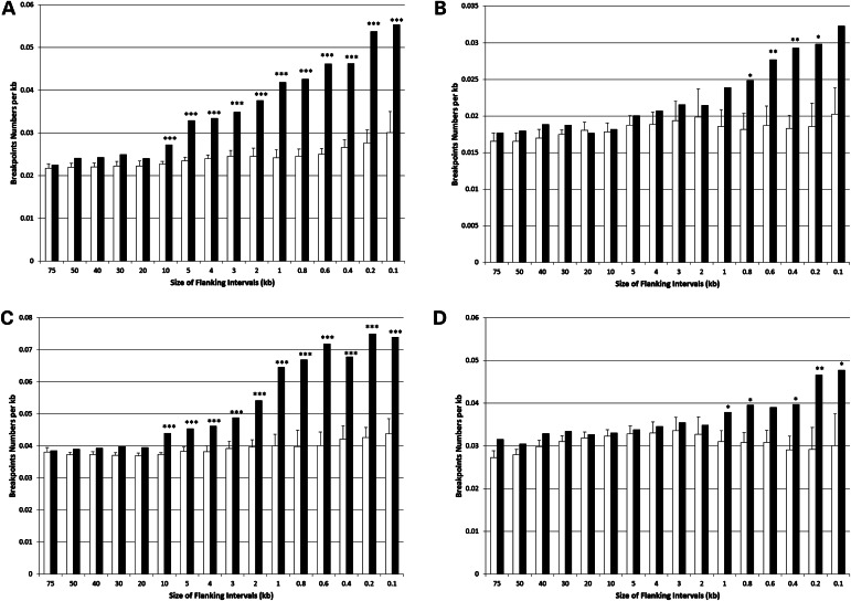
Environmental factors including ionizing radiation and chemical agents have been known to be able to induce DNA rearrangements and cause genomic structural variations (SVs); however, the roles of intrinsic characteristics of the human genome, such as regional genome architecture, in SV formation and the potential mechanisms underlying genomic instability remain to be further elucidated. Recently, locus-specific observations showed that 'self-chain' (SC), a group of short low-copy repeats (LCRs) in the human genome, can induce autism-associated SV mutations of the MECP2 and NRXN1 genes. In this study, we conducted a genome-wide analysis to investigate SCs and their potential roles in genomic SV formation. Utilizing a vast amount of human SV data, we observed a significant biased distribution of human germline SV breakpoints to SC regions. Notably, the breakpoint distribution pattern is different between SV types across deletion, duplication, inversion and insertion. Our observations were coincident with a mechanism of SC-induced DNA replicative errors, whereas SC may sporadically be used as substrates of nonallelic homologous recombination (NAHR). This contention was further supported by our consistent findings in somatic SV mutations of cancer genomes, suggesting a general mechanism of SC-induced genome instability in human germ and somatic cells.


Figures

Figure 1.
Figure 1.
Genomic rearrangements and homology-driven mechanisms of SV formation in the human genome. (A) NAHR between repeats (dark and light blue arrowed bars) in direct orientation can cause reciprocal duplications (dup) and deletions (del). The cross depicts a DNA recombination event. (B) The recombination between inverted repeats can lead to inversions (inv). The likely involvement of inverted (C) and direct repeats (D) in DNA replicative mechanisms and SV mutations. The thick lines and arrows depict single DNA strands and short thin lines represent the annealing between Watson–Crick base pairs (blue-yellow or red-green). During DNA replication, adjacent short repeats could lead to secondary structures and consequently cause replication fork stalling. The newly synthesized DNA strands are shown by dashed lines. Based on the replicative mechanisms (20), DNA template switching can occur to resume replication and generate SVs as well. SV types and template switching patterns: deletions, switching forward; duplication, switching to the opposite strand and backward; insertion (ins), switching to a template in another replicon (shown by a grey line) and backward; inversion, switching between leading and lagging strands via homology and/or microhomology. The dotted lines represent the DNA template switching events.
Figure 2.
Figure 2.
Counting SV breakpoints in the flanking regions of SCRs. The example, in which the starting size of 1 kb and the increment of 1 kb were adopted for SCR-flanking regions, was illustrated. (A) Five non-overlapping SCRs (SCR-1 to SCR-5) are shown. Triangles depict SV breakpoints; black depicts breakpoints outside SCRs; grey depicts breakpoints in SCRs. (B) Before counting SV breakpoints, we check whether any two of the SCR-flanking regions overlap. Here, we show the example that the 1 kb flanking regions of SCR-2 and SCR-3 overlap each other. (C) To avoid counting the breakpoints between SCR-2 and SCR-3 twice, we merge these two SCRs into a new SCR (SCR-2–3). (D) The numbers of the SV breakpoints in 1 kb SCR-flanking regions are counted (shown by red triangles). (E) Then, the size of SCR-flanking regions for investigation is increased to 2 kb. Check whether any two of 2 kb flanking regions overlap. (F) Merge any two SCRs whether their flanking regions overlap each other. (G) The numbers of the SV breakpoints in 2 kb SCR-flanking regions are counted. (H) Increase the size of SCR-flanking regions for investigation again and repeat the steps E-G till the size of SCR-flanking regions reaches N kb.
Figure 3.
Figure 3.
Both germline SVs in human populations and somatic SVs in cancer genomes have a significant biased breakpoint distribution to SCRs. (A) Breakpoint distribution of the SVs resolved by microarray and/or NGS read-depth methods in human populations. (B) Breakpoint distribution of the SVs resolved by NGS split-read and/or assembly methods in human populations. (C) Breakpoint distribution of the SVs resolved by microarray methods in cancer genomes. X-axis, size of SCR-flanking regions. From left to right, the narrowing-down of SCR-flanking regions. Y-axis, SV breakpoint density (number per kb). Black columns, SCRs; open columns, simulated control regions. The significant differences in breakpoint density between the flanking regions of SCRs and those of control regions are indicated by asterisks (*P < 0.05; **P < 0.01; ***P < 0.001).
Figure 4.
Figure 4.
The correlations of breakpoint distributions between SV types and SC orientations. Based on the germline SVs identified by NGS split-read and/or assembly methods, deletion breakpoints have a biased distribution to both +SCRs (A) and −SCRs (B). Duplication breakpoints also have a biased distribution to both +SCRs (C) and −SCRs (D). Distribution of inversion breakpoints in flanking regions of +SCRs (E) and −SCRs (F). The significant differences in breakpoint density between the regions flanking SCRs (black columns) and those flanking control regions (open columns) are indicated by asterisks (*P < 0.05; **P < 0.01; ***P < 0.001).
Figure 5.
Figure 5.
Biased breakpoint distributions of the somatic SVs identified by microarray methods in cancer genomes. The breakpoints of deletions (i.e. copy number losses) in the regions flanking +SCRs (A) and −SCRs (B). The breakpoints of duplications and insertions (i.e. copy number gains) in the regions flanking +SCRs (C) and −SCRs (D). The significant differences in breakpoint density between the regions flanking SCRs (black columns) and those flanking control regions (open columns) are indicated by asterisks (*P < 0.05; **P < 0.01; ***P < 0.001).

References

    1. Stankiewicz P., Lupski J.R. Structural variation in the human genome and its role in disease. Annu. Rev. Med. 2010;61:437–455. - PubMed
    1. Alkan C., Coe B.P., Eichler E.E. Genome structural variation discovery and genotyping. Nat. Rev. Genet. 2011;12:363–376. doi:10.1080/13803390701336411. - DOI - PMC - PubMed
    1. Liu P., Carvalho C.M., Hastings P., Lupski J.R. Mechanisms for recurrent and complex human genomic rearrangements. Curr. Opin. Genet. Dev. 2012;22:211–220. - PMC - PubMed
    1. The 1000 Genomes Project Consortium. A map of human genome variation from population-scale sequencing. Nature. 2010;467:1061–1073. doi:10.1080/13803390701346113. - DOI - PMC - PubMed
    1. Lupski J.R. Genomic disorders: structural features of the genome can lead to DNA rearrangements and human disease traits. Trends Genet. 1998;14:417–422. doi:10.1002/(SICI)1099-1166(199706)12:6<619::AID-GPS554>3.0.CO;2-H. - DOI - PubMed

Publication types