

Review
doi: 10.1007/s00018-013-1282-8.
Epub 2013 Mar 12.
Vascular endothelial growth factors (VEGFs) and stroke
Affiliations
- PMID: 23475070
- PMCID: PMC3634892
- DOI: 10.1007/s00018-013-1282-8
Item in Clipboard
Review
Vascular endothelial growth factors (VEGFs) and stroke
Cell Mol Life Sci.
2013 May.
Abstract
Vascular endothelial growth factors (VEGFs) have been shown to participate in atherosclerosis, arteriogenesis, cerebral edema, neuroprotection, neurogenesis, angiogenesis, postischemic brain and vessel repair, and the effects of transplanted stem cells in experimental stroke. Most of these actions involve VEGF-A and the VEGFR-2 receptor, but VEGF-B, placental growth factor, and VEGFR-1 have been implicated in some cases as well. VEGF signaling pathways represent important potential targets for the acute and chronic treatment of stroke.
Figures
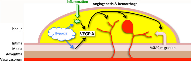
VEGF-A and atherosclerosis. Hypoxia and inflammation in the atherosclerotic plaque trigger VEGF-A expression in vascular smooth muscle cells (VSMC) and macrophages. VEGF-A, in turn, acts on vasa vasorum to promote angiogenesis, which may be associated with hemorrhage, and promotes migration of VSMC from the tunica media of the vessel wall into the plaque
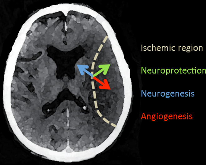
VEGF effects in acute ischemic stroke. A horizontal section of the human brain shows an acute infarct within the territory of the middle cerebral artery (dotted line). VEGF-A is induced in the ischemic border zone and acts on local neurons and endothelial cells to promote neuroprotection (green arrow) and angiogenesis (red arrow). VEGF-A also stimulates neurogenesis (blue arrow) in the subventricular zone, from which new neurons migrate to the site of ischemia. Other VEGF family members, including VEGF-B and PlGF, share some of these effects
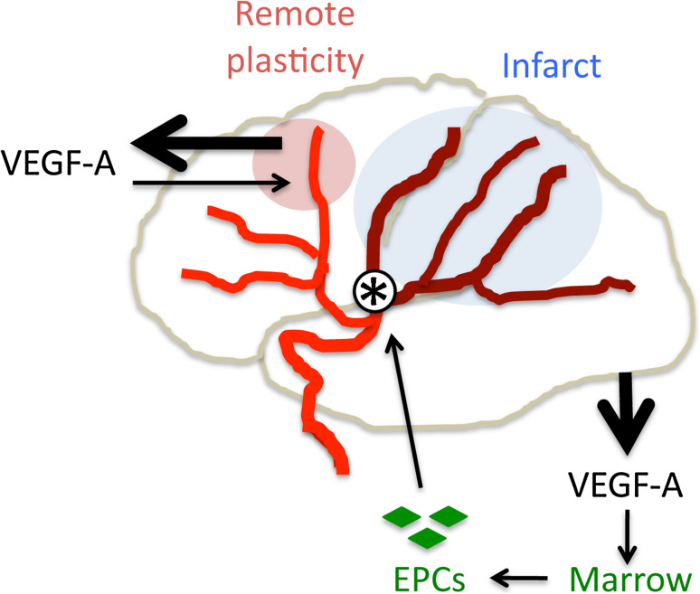
VEGF-A-mediated postischemic repair. VEGF-A appears to be a mediator of postischemic brain and vessel repair. Following cerebral infarction (blue) due to arterial occlusion (asterisk), VEGF-A expression is increased (thick arrow, top left) in nonischemic brain regions that exhibit remote plasticity (pink); VEGF-A acting here (thin arrow, top left) may help to restore brain function. Increased serum VEGF-A levels after stroke (thick arrow, bottom right) also mobilize endothelial progenitor cells (EPCs, green) from the bone marrow, enhancing repair of ischemia-damaged vessels (thin arrows, bottom right)
References
-
- Hirashima M. Regulation of endothelial cell differentiation and arterial specification by VEGF and notch signaling. Anat Sci Int. 2009;84:95–101. - PubMed
-
- Shibuya M. Brain angiogenesis in developmental and pathological processes: therapeutic aspects of vascular endothelial growth factor. FEBS J. 2009;276:4636–4643. - PubMed
-
- Mackenzie F, Ruhrberg C. Diverse roles for VEGF-A in the nervous system. Development. 2012;139:1371–1380. - PubMed
-
- Sluimer JC, Daemen MJ. Novel concepts in atherogenesis: angiogenesis and hypoxia in atherosclerosis. J Pathol. 2009;218:7–29. - PubMed
Publication types
MeSH terms
Substances
Grants and funding
LinkOut - more resources
Full Text Sources
Other Literature Sources
Medical
