Review
. 2013 Jun;9(3):360-72.
doi: 10.1007/s12015-013-9433-8.

Mesenchymal stem cells as a treatment for peripheral arterial disease: current status and potential impact of type II diabetes on their therapeutic efficacy

Affiliations
Review

Mesenchymal stem cells as a treatment for peripheral arterial disease: current status and potential impact of type II diabetes on their therapeutic efficacy

Jinglian Yan et al. Stem Cell Rev Rep. 2013 Jun.

Abstract

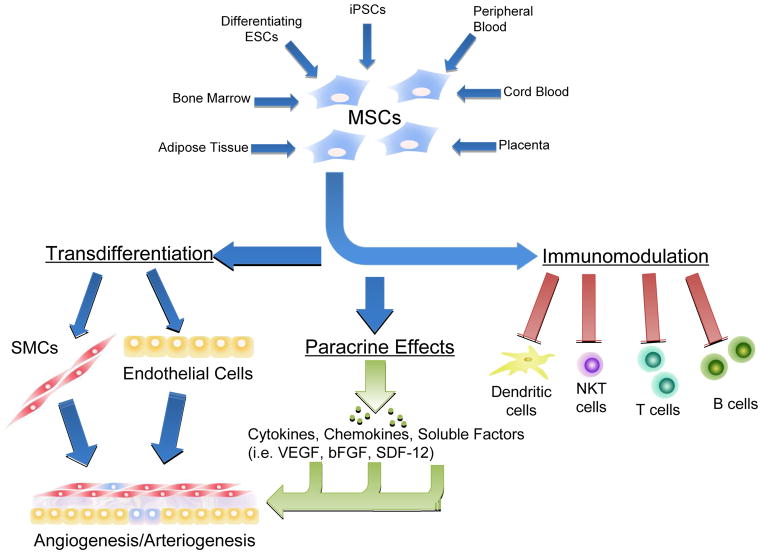
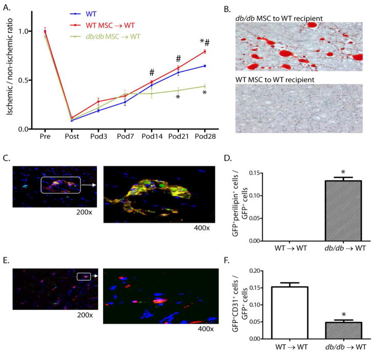
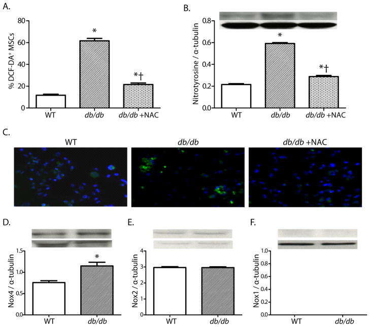
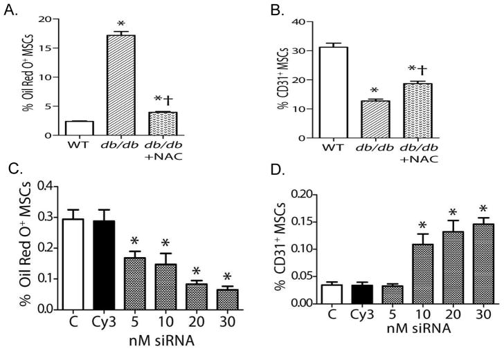
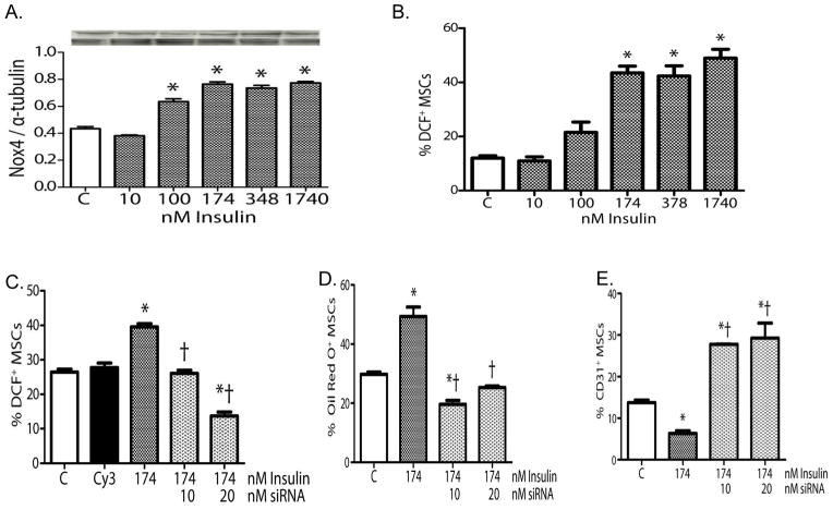
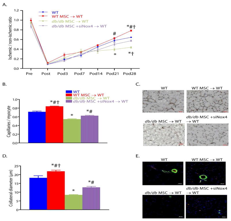
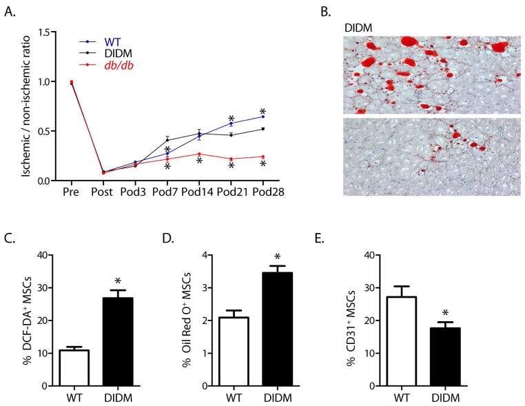
Mesenchymal stem cells (MSCs), due to their paracrine, transdifferentiation, and immunosuppressive effects, hold great promise as a therapy for peripheral arterial disease. Diabetes is an important risk factor for peripheral arterial disease; however, little is known of how type II diabetes affects the therapeutic function of MSCs. This review summarizes the current status of preclinical and clinical studies that have been performed to determine the efficacy of MSCs in the treatment of peripheral arterial disease. We also present findings from our laboratory regarding the impact of type II diabetes on the therapeutic efficacy of MSCs neovascularization after the induction of hindlimb ischemia. In our studies, we documented that experimental type II diabetes in db/db mice impaired MSCs' therapeutic function by favoring their differentiation towards adipocytes, while limiting their differentiation towards endothelial cells. Moreover, type II diabetes impaired the capacity of MSCs to promote neovascularization in the ischemic hindlimb. We further showed that these impairments of MSC function and multipotency were secondary to hyperinsulinemia-induced, Nox4-dependent oxidant stress in db/db MSCs. Should human MSCs display similar oxidant stress-induced impairment of function, these findings might permit greater leverage of the potential of MSC transplantation, particularly in the setting of diabetes or other cardiovascular risk factors, as well as provide a therapeutic approach by reversing the oxidant stress of MSCs prior to transplantation.


Conflict of interest statement

Conflict of interest

The authors declare no potential conflicts of interest.

Figures

Figure 1
Figure 1
Biological sources and activity of Mesenchymal Stem Cells. MSCs can be isolated from multiple sources, and exert therapeutic effects on multiple systems to contribute to the therapy of peripheral arterial disease.
Figure 2
Figure 2
Foot blood flow recovery, muscle histology, and co-localization studies in WT mice transplanted with db/db or WT MSC after induction of hindlimb ischemia. (A) Foot blood flow recovery by LDPI (mean±SEM; n=6; *P<0.05 vs. WT; #P<0.05 vs. db/db MSC → WT transplant group). (B) Histology of gastrocnemius muscle from the ischemic hindlimb (Oil Red O for identification of adipocytes, hematoxylin counter stain, 200x). (C) Representative confocal images (GFP for identification of MSC [green] and perilipin for identification of adipocytes [red]. (D) Ratio of GFP+periplipin+ cells to GFP+ cells in the ischemic hindlimb muscle (mean±SEM; n=5; *P<0.05). (E) Representative confocal images (GFP for the identification of MSC [green] and CD31 for the identification of endothelial cells [red]). (F) Ratio of GFP+CD31+ cells to GFP+ cells in the ischemic hindlimb muscle (mean±SEM; n=5; *P<0.05). Reprinted with permission from JAHA with the specific citation[43].
Figure 3
Figure 3
Oxidant stress, and NADPH oxidase expression in db/db and WT MSCs. (A, B) Oxidant levels, as determined by DCF staining and nitrotyrosine accumulation in MSCs (mean±SEM; n=6; *P<0.05 vs. WT; †P<0.05 vs. db/db). (C) Representative photomicrographs of DCF staining in MSCs (200x). (D–F) Quantitative expressions of Nox4, Nox2, and Nox1 proteins in MSC cell lysates (D–E: mean±SEM; n=6; *P<0.05; F: n=6). Reprinted with permission from JAHA with the specific citation[43].
Figure 4
Figure 4
In vitro responses of MSCs to selective differentiation media. (A, B) Quantification of MSC differentiation towards adipocytes(A)or endothelial cells(B) (For panels A,B: mean±SEM; n=6; *P<0.05 vs. WT; †P<0.05 vs. db/db.) (C) Nox4 siRNA reduced differentiation of db/db MSCs into an adipocyte phenotype. (D) Nox4 siRNA increased differentiation of db/db MSCs into an endothelial phenotype. (For panels C and D: mean±SEM, n=6; *P<0.05 vs. control [C]; Cy3 is the siRNA control). Reprinted with permission from JAHA with the specific citation[43].
Figure 5
Figure 5
Effects of insulin on WT MSCs. (A) Insulin increased Nox4 expression in WT MSCs. (B) Insulin increased oxidant levels in WT MSCs. (C) Pretreatment of WT MSCs with Nox4 siRNA blocked the pro-oxidant effects of insulin in WT MSCs. (D) Pretreatment of WT MSCs with Nox4 siRNA blocked the effects of insulin on adipocyte differentiation. (E) Pretreatment of WT MSCs with Nox4 siRNA blocked the effects of insulin on endothelial differentiation. (For all panels: mean±SEM, n=6; *P<0.05 vs. control [C]; †P<0.05 vs. insulin alone; Cy3 was used as the siRNA control.). Reprinted with permission from JAHA with the specific citation[43].
Figure 6
Figure 6
Reversal of Nox4 induced oxidant stress in type 2 diabetic MSCs restores their capacity to augment post-ischemic neovascularization in db/db mice. (A) Foot blood flow recovery measurement by LDPI. (B, C) Quantification (B) of capillary density by CD31 immunostaining and representative images (C). (D, E) Quantification (D) of collateral diameter by CD31 and alpha-SMA double staining and representative images (E). (For all panels: mean±SEM; n=6; *P<0.05 vs. WT; †P<0.05 vs. db/db MSC +siNox4 → WT transplant group; #P<0.05 vs. db/db MSC→ WT transplant group; scale bar, 50 μm.). Reprinted with permission from JAHA with the specific citation[43].
Figure 7
Figure 7
Diet-induced diabetes (DIDM) impairs recovery from hindlimb ischemia, induces adipocyte differentiation in ischemic muscle, and restricts MSC multipotency. (A) Foot blood flow recovery in DIDM mice (mean±SEM, n=6; *P<0.05 vs. DIDM; note that the WT data shown here were generated specifically for comparison to DIDM mice and are different from those displayed in Figure 3). (B) Intramuscular adipocyte infiltration within the ischemic hindlimb muscle from DIDM mice. (C) Oxidant levels, as evidenced by DCF staining (n=7). (D) MSCs from DIDM demonstrated increased differentiation to an adipocyte phenotype (n=8). (E) MSCs from DIDM mice demonstrated reduced differentiation to an endothelial phenotype (n=7–8). (For panels C–E: mean±SEM; *P<0.05.). Reprinted with permission from JAHA with the specific citation[43].

References

    1. Takahashi TKC, Masuda H, Chen D, Silver M, Kearney M, Magner M, Isner JM, Asahara T. Ischemia- and cytokine-induced mobilization of bone marrow-derived endothelial progenitor cells for neovascularization. Nat Med. 1999;5(4):434–438. - PubMed
    1. Kinnaird TSE, Burnett MS, Epstein SE. Bone-marrow-derived cells for enhancing collateral development: mechanisms, animal data, and initial clinical experiences. Circ Res. 2004;95(4):354–363. - PubMed
    1. Asahara TMT, Sullivan A, Silver M, van der Zee R, Li T, Witzenbichler B, Schatteman G, Isner JM. Isolation of putative progenitor endothelial cells for angiogenesis. Science. 1997;275(5302):964–967. - PubMed
    1. Crosby KW, JR, Schatteman G, Martin PJ, Raines EW, Seifert RA, Bowen-Pope DF. Endothelial cells of hematopoietic origin make a significant contribution to adult blood vessel formation. Circ Res. 2000;87(9):728–30. - PubMed
    1. Sneider EB, NP, Messina LM. Regenerative medicine in the treatment of peripheral arterial disease. J Cell Biochem. 2009;108(4):753–61. - PubMed

Publication types