. 2013 May 15;304(10):L646-56.
doi: 10.1152/ajplung.00164.2012. Epub 2013 Mar 8.

Hydrogen gas reduces hyperoxic lung injury via the Nrf2 pathway in vivo

Affiliations

Hydrogen gas reduces hyperoxic lung injury via the Nrf2 pathway in vivo

Tomohiro Kawamura et al. Am J Physiol Lung Cell Mol Physiol. .

Abstract

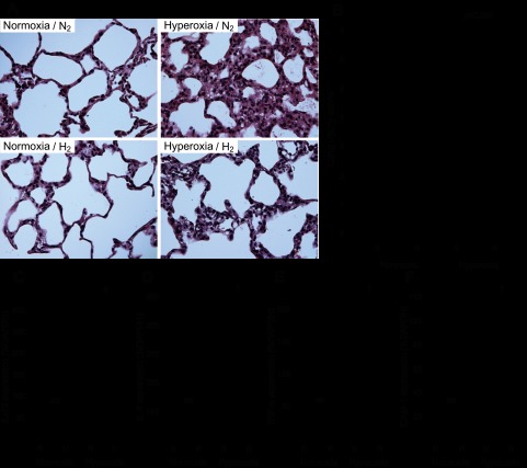
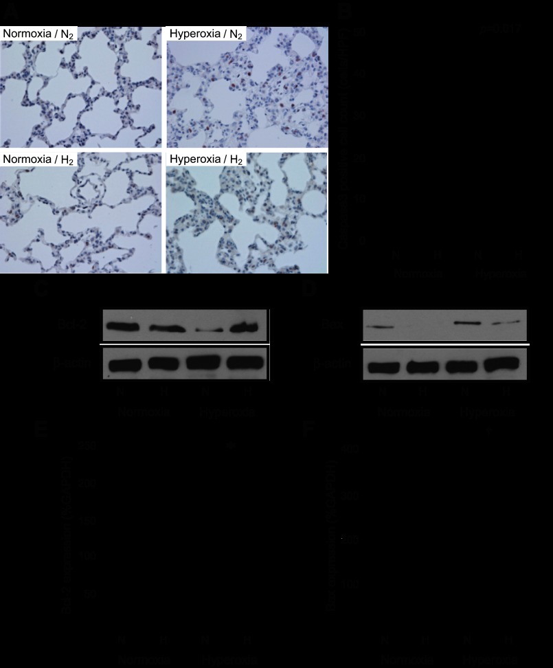
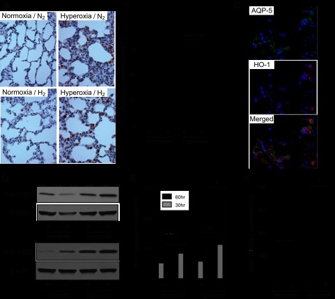
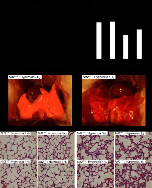
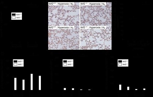
Hyperoxic lung injury is a major concern in critically ill patients who receive high concentrations of oxygen to treat lung diseases. Successful abrogation of hyperoxic lung injury would have a huge impact on respiratory and critical care medicine. Hydrogen can be administered as a therapeutic medical gas. We recently demonstrated that inhaled hydrogen reduced transplant-induced lung injury and induced heme oxygenase (HO)-1. To determine whether hydrogen could reduce hyperoxic lung injury and investigate the underlying mechanisms, we randomly assigned rats to four experimental groups and administered the following gas mixtures for 60 h: 98% oxygen (hyperoxia), 2% nitrogen; 98% oxygen (hyperoxia), 2% hydrogen; 98% balanced air (normoxia), 2% nitrogen; and 98% balanced air (normoxia), 2% hydrogen. We examined lung function by blood gas analysis, extent of lung injury, and expression of HO-1. We also investigated the role of NF-E2-related factor (Nrf) 2, which regulates HO-1 expression, by examining the expression of Nrf2-dependent genes and the ability of hydrogen to reduce hyperoxic lung injury in Nrf2-deficient mice. Hydrogen treatment during exposure to hyperoxia significantly improved blood oxygenation, reduced inflammatory events, and induced HO-1 expression. Hydrogen did not mitigate hyperoxic lung injury or induce HO-1 in Nrf2-deficient mice. These findings indicate that hydrogen gas can ameliorate hyperoxic lung injury through induction of Nrf2-dependent genes, such as HO-1. The findings suggest a potentially novel and applicable solution to hyperoxic lung injury and provide new insight into the molecular mechanisms and actions of hydrogen.

Keywords: NF-E2-related factor 2; heme oxygenase; hydrogen; inflammation.


Figures

Fig. 1.
Fig. 1.
Physiological effects of hydrogen on hyperoxic lung injury and assessment of lung permeability and edema in rats. A: blood oxygenation levels in blood drawn from the abdominal aorta of rats after 60 h of exposure to normoxic or hyperoxic conditions; n = 8 experiments, †P < 0.01 and *P < 0.05. B: body weight loss. Rats were weighed immediately before and immediately after 60 h of exposure to normoxic or hyperoxic conditions; n = 8, †P < 0.01 and *P < 0.05. C: animal survival during prolonged exposure to hyperoxic conditions. Survival was assessed every 8 h; n = 5 for each group, log-rank test, P = 0.0027.
Fig. 2.
Fig. 2.
Assessment of lung permeability and edema in rats. A: pleural effusion volume after 60 h of normoxia or hyperoxia; n = 8 for each group, *P < 0.05. B: wet-to-dry ratio of the lungs after 60 h of normoxia or hyperoxia; n = 5 for each group, †P < 0.01 and *P < 0.05. C: cell counts in the bronchoalveolar lavage fluid (BALF); n = 5 for each group, †P < 0.01 and *P < 0.05. D: protein concentration in BALF; n = 5 for each group, †P < 0.01 and *P < 0.05.
Fig. 3.
Fig. 3.
Assessment of pulmonary inflammation. A: representative images of hematoxylin-eosin (H&E) staining of the lung. Left: lungs exposed to normoxic conditions with 2% nitrogen or 2% hydrogen. Right: Lungs exposed to hyperoxic conditions with 2% nitrogen or 2% hydrogen. Magnification ×400. B: lung injury scores of H&E-stained lungs. Acute lung injury was scored according to 1) thickness of the alveolar wall, 2) infiltration or aggregation of neutrophils in airspace, the alveolar wall, or the vessel wall, and 3) alveolar congestion, and each item was graded on a four-point scale. Each component ranged from 0 to 3, with higher scores indicating more severe damage. A total lung injury score was calculated as the sum of the three components (from 0 to 9). At least 10 fields (median 17, range 13–20 fields) were chosen randomly from each section and were examined at ×400 magnification. Normoxia n = 4, hyperoxia n = 6. CF: real-time RT-PCR for inflammatory mediators in lung tissue after 60 h normoxia or hyperoxia exposure. Relative levels of the mRNAs for interleukin (IL)-1β (C), IL-6 (D), tumor necrosis factor (TNF)-α (E), and intercellular adhesion molecule (ICAM)-1 (F) were normalized to glyceraldehyde-3-phosphate dehydrogenase (GAPDH) mRNA; n = 4 for each group, †P < 0.01 and *P < 0.05. NS, not significant.
Fig. 4.
Fig. 4.
Assessment of pulmonary apoptosis. A: pulmonary epithelial cell apoptosis was determined by immunohistochemistry for cleaved caspase 3 after 60 h of exposure to normoxia or hyperoxia. Left: lungs exposed to normoxic conditions with 2% nitrogen or 2% hydrogen. Right: lungs exposed to hyperoxic conditions with 2% nitrogen or 2% hydrogen. Magnification ×400. B: cleaved caspase 3-positive cells were counted with the samples' identities masked and expressed as the number of positive cells per high-power field (HPF). More than 10 fields were chosen randomly from each section for quantitation. Normoxia n = 4, hyperoxia n = 6. C: Western blots for B cell lymphoma-2 (Bcl-2) and β-actin on protein extracts from lungs after a 60-h exposure to normoxia or hyperoxia with 2% nitrogen or hydrogen. The images are representative of 3 independent experiments; n = 4 for each group. D: Western blots for Bcl-2-associated X-protein (Bax) and β-actin on protein extracts from lungs after a 60-h exposure to normoxia or hyperoxia with 2% nitrogen or hydrogen. The images are representative of 3 independent experiments; n = 4 for each group. E: real-time RT-PCR for Bcl-2 mRNA in lung tissue after 60 h of normoxia or hyperoxia; n = 4 for each group, *P < 0.05. Expression was normalized to GAPDH mRNA. F: real-time RT-PCR for Bax mRNA in lung tissue after 60 h of normoxia or hyperoxia; n = 4 for each group, †P < 0.01. Expression was normalized to GAPDH mRNA.
Fig. 5.
Fig. 5.
Hydrogen increased heme oxygenase (HO)-1 in the lung. A: representative images of immunohistochemistry for HO-1 in the lungs after 60 h of normoxia or hyperoxia. Magnification = ×400. B: HO-1-positive cells were counted with the samples' identities masked and expressed as the number of positive cells per HPF. The cell count was analyzed with Tukey-Kramer methods. Normoxia n = 4, hyperoxia n = 6; >10 fields were chosen randomly from each section. C: representative images of immunofluorescent staining for aquaporin-5 (AQP-5), a marker for lung epithelial cells, and HO-1 in the lungs after 60 h of hyperoxia in the presence of 2% hydrogen (hyperoxia/H2). Top, AQP-5-positive cells. Middle, HO-1-positive cells. Bottom, merged image. D: Western blots for HO-1 and β-actin on protein extracts from the lungs taken after 30 and 60 h of normoxia or hyperoxia. The images are representative of 3 independent experiments; n = 4 for each group. E: real-time RT-PCR for HO-1 mRNA in lung tissues after 30 or 60 h normoxia or hyperoxia exposure. Gene expression was normalized to GAPDH mRNA; n = 4 for each group, †P < 0.01 and *P < 0.05. F: effect of hydrogen on HO-1 activity in the rat lung. HO-1 activity was expressed as pmol of bilirubin formed·mg of protein−1·60 min−1. *P < 0.05.
Fig. 6.
Fig. 6.
Assessment of the NF-E2-related factor (Nrf) 2-dependent mRNA expression in the lung after 60 h of normoxia/hyperoxia exposure. Expression of Nrf2-dependent genes in lung tissues after 60 h of normoxia or hyperoxia. The levels of mRNAs for NAD(P)H dehydrogenase quinone (Nqo) 1 (A), GSTA2 (B), UDP-glucuronosyl transferase (UGT) 1A6 (C), and peroxiredoxin (Prdx) 1 (D) were quantitated by real-time RT-PCR. Gene expression was normalized to GAPDH mRNA; n = 4 for each group, †P < 0.01 and *P < 0.05.
Fig. 7.
Fig. 7.
Assessment of the effects of hydrogen on hyperoxic lung injury in Nrf2−/− mice. A and B: blood oxygenation levels were measured in the blood drawn from the abdominal aorta of the Nrf2+/+ (A) and Nrf2−/− (B) mice after 60 h of exposure to normoxia or hyperoxia; n = 4 for each group, *P < 0.05. C and D: representative images of the gross anatomy of lungs from the Nrf2+/+ (C) and Nrf2−/− (D) mice 60 h after hyperoxia with 2% hydrogen. E and F: representative images of H&E staining of lungs from Nrf2+/+ and Nrf2−/− mice after 60 h of normoxia (E) or hyperoxia (F) with and without hydrogen.
Fig. 8.
Fig. 8.
Assessment of the oxidative stress markers and the Nrf2-dependent mRNA expression in Nrf2−/− mice. A: assessment of malondialdehyde (MDA) in the lung tissue of Nrf2+/+ and Nrf2−/− mice after 60 h normoxia or hyperoxia exposure; n = 4 for each group, *P < 0.05. B: representative images of immunohistochemistry for 8-hydroxy-2′-deoxyguanosine (8-OHdG) in the lungs after 60 h of hyperoxia in Nrf2+/+ and Nrf2−/− mice. Magnification ×400. C: 8-OHdG-positive cells were counted with the samples' identities masked and expressed as the number of positive cells per HPF. More than 10 fields were chosen randomly from each section for quantitation; n = 4 for each group, †P < 0.01. DF: assessment of the Nrf2-dependent mRNA expression in Nrf2+/+ and Nrf2−/− mice after 60 h normoxia/hyperoxia exposure. Nrf2-dependent genes, HO-1 (D), Nqo1 (E), and GSTA2 (F), were quantitated by real-time RT-PCR after 60 h of exposure to hyperoxia; n = 4 for each group, †P < 0.01. Gene expression was normalized to hypoxanthine phosphoribosyltransferase (HPRT) mRNA.

References

    1. Alam J, Cook JL. How many transcription factors does it take to turn on the heme oxygenase-1 gene? Am J Respir Cell Mol Biol 36: 166–174, 2007 - PubMed
    1. Alam J, Cook JL. Transcriptional regulation of the heme oxygenase-1 gene via the stress response element pathway. Curr Pharm Des 9: 2499–2511, 2003 - PubMed
    1. Altemeier WA, Sinclair SE. Hyperoxia in the intensive care unit: why more is not always better. Curr Opin Crit Care 13: 73–78, 2007 - PubMed
    1. Awasthi S, Gyurasics A, Knight SA, Welty SE, Smith CV. Protein oxidation biomarkers in hyperoxic lung injury in rats: effects of U-74389. Toxicol Lett 95: 47–61, 1998 - PubMed
    1. Baird L, Dinkova-Kostova AT. The cytoprotective role of the Keap1-Nrf2 pathway. Arch Toxicol 85: 241–272, 2011 - PubMed

Publication types

MeSH terms

LinkOut - more resources