. 2013 May;195(10):2244-54.
doi: 10.1128/JB.02011-12. Epub 2013 Mar 8.

A mutant with aberrant extracellular LcrV-YscF interactions fails to form pores and translocate Yop effector proteins but retains the ability to trigger Yop secretion in response to host cell contact

Affiliations

A mutant with aberrant extracellular LcrV-YscF interactions fails to form pores and translocate Yop effector proteins but retains the ability to trigger Yop secretion in response to host cell contact

Dana E Harmon et al. J Bacteriol. 2013 May.

Abstract

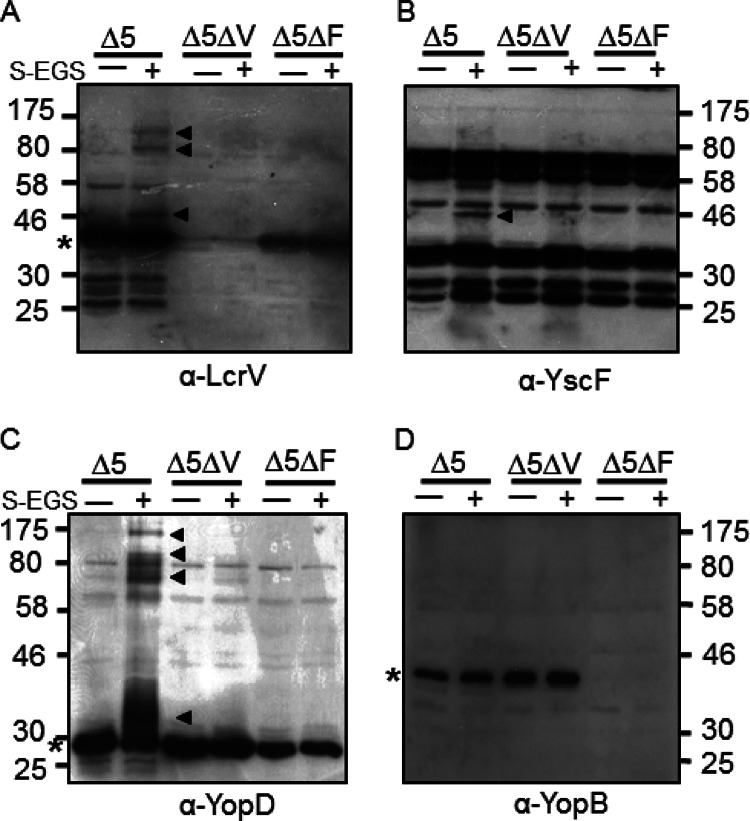
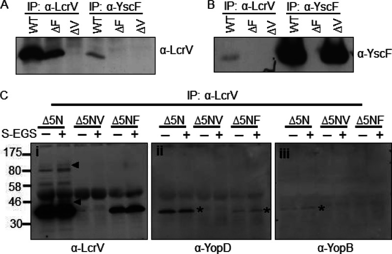
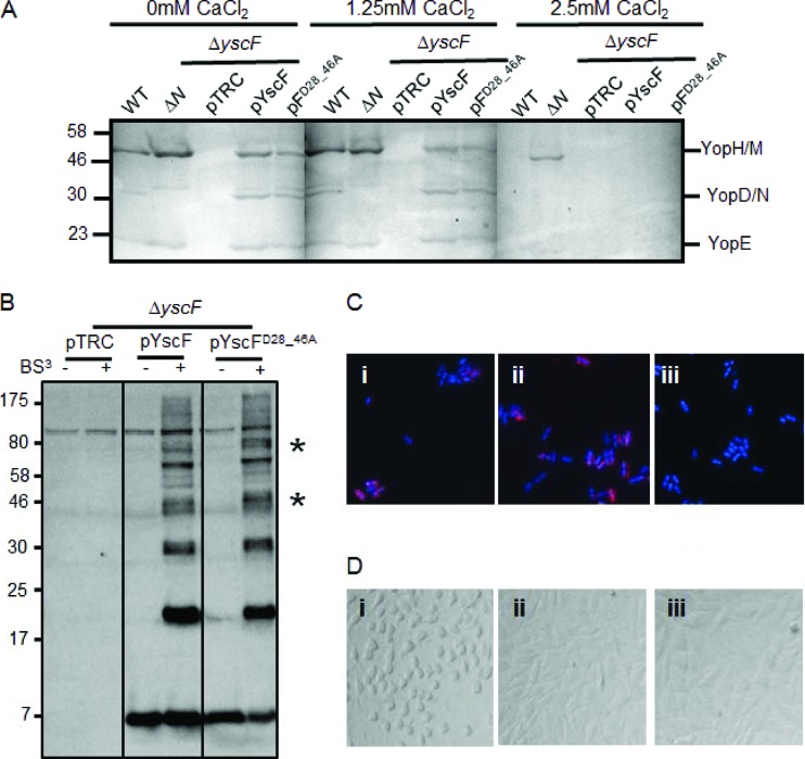
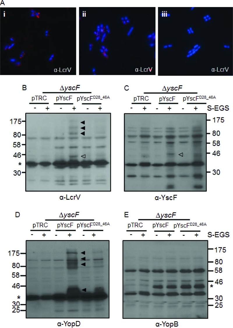
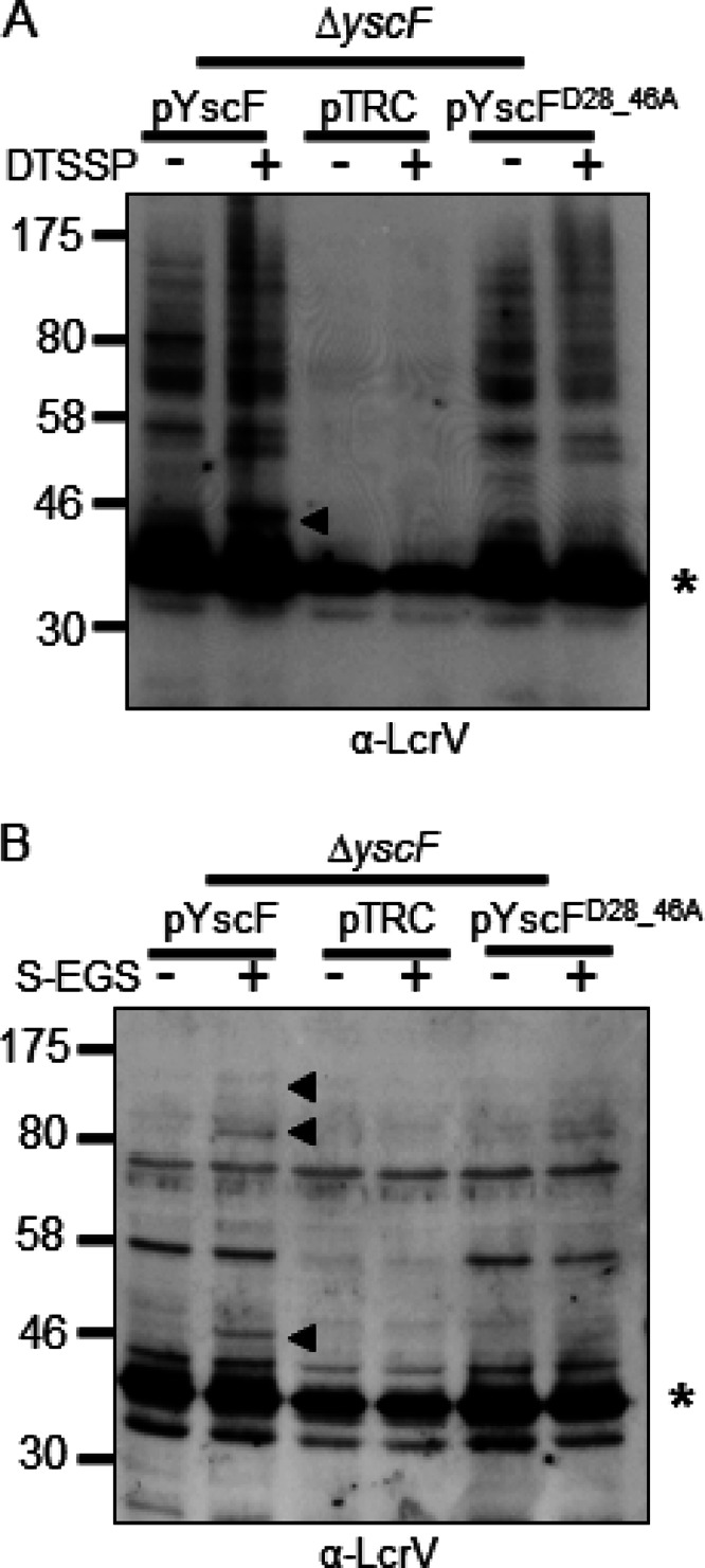
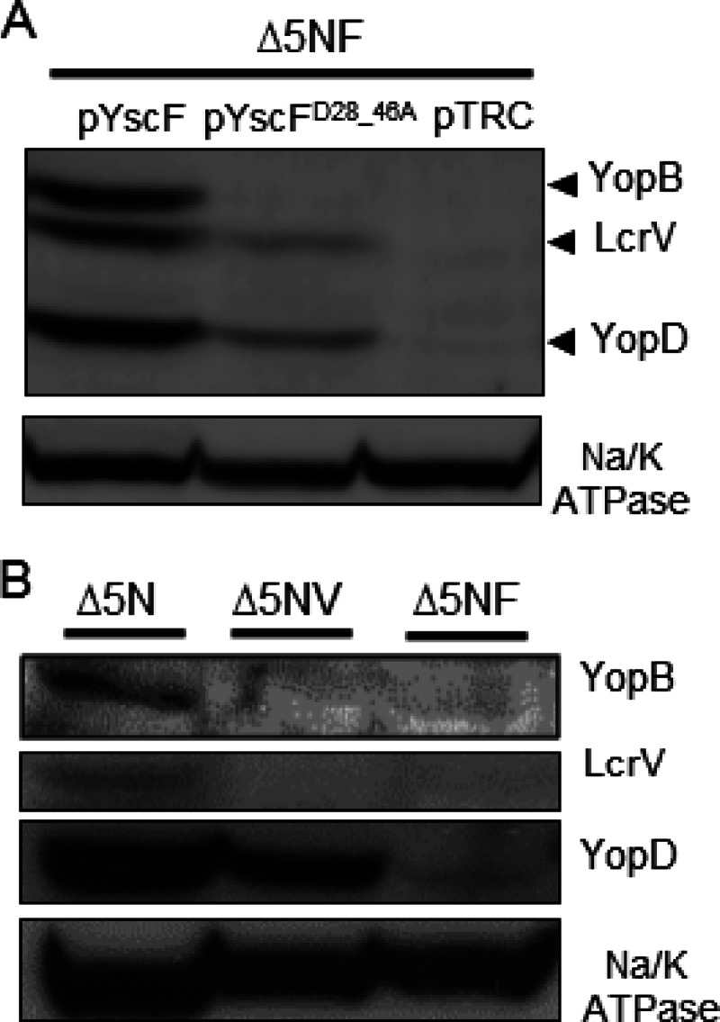
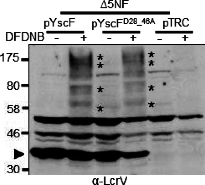
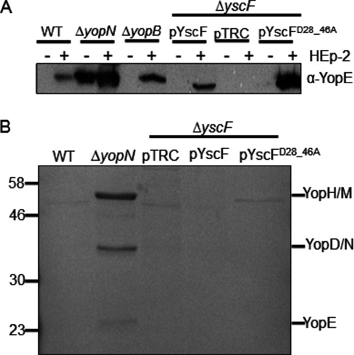
The plasmid-encoded type three secretion system (TTSS) of Yersinia spp. is responsible for the delivery of effector proteins into cells of the innate immune system, where these effectors disrupt the target cells' activity. Successful translocation of effectors into mammalian cells requires Yersinia to both insert a translocon into the host cell membrane and sense contact with host cells. To probe the events necessary for translocation, we investigated protein-protein interactions among TTSS components of the needle-translocon complex using a chemical cross-linking-based approach. We detected extracellular protein complexes containing YscF, LcrV, and YopD that were dependent upon needle formation. The formation of these complexes was evaluated in a secretion-competent but translocation-defective mutant, the YscFD28AD46A strain (expressing YscF with the mutations D28A and D46A). We found that one of the YscF and most of the LcrV and YopD cross-linked complexes were nearly absent in this mutant. Furthermore, the YscFD28AD46A strain did not support YopB insertion into mammalian membranes, supporting the idea that the LcrV tip complex is required for YopB insertion and translocon formation. However, the YscFD28AD46A strain did secrete Yops in the presence of host cells, indicating that a translocation-competent tip complex is not required to sense contact with host cells to trigger Yop secretion. In conclusion, in the absence of cross-linkable LcrV-YscF interactions, translocon insertion is abolished, but Yersinia still retains the ability to sense cell contact.


Figures

Fig 1
Fig 1
Cross-linking analysis of secreted but cell-associated LcrV, YscF, YopD, and YopB. The ΔyopHEMOJ (Δ5), ΔyopHEMOJ ΔlcrV (Δ5ΔV), or ΔyopHEMOJ ΔyscF (Δ5ΔF) Y. pseudotuberculosis strain was induced for Yop secretion for 1.5 h at 37°C, washed once in PBS, and then exposed to sulfo-EGS for 30 min at 37°C. Whole cells were solubilized in sample buffer and then subjected to Western blot analysis with antibodies against LcrV (A), YscF (B), YopD (C), or YopB (D). Filled arrowheads indicate cross-linked bands; asterisks indicate the monomeric protein. YscF is not visible on the gel. Molecular mass (kDa) is indicated on the sides of the gels.
Fig 2
Fig 2
YscF and LcrV can be immunoprecipitated (IP) after cross-linking. (A and B) A WT, ΔyscF (ΔF), or ΔlcrV (ΔV) Y. pseudotuberculosis strain was exposed to DTSSP and then lysed. Lysates were split and immunoprecipitated with LcrV or YscF antibodies. Cross-links within the immunoprecipitates were cleaved with BME. Proteins were separated by SDS-PAGE and analyzed by Western blotting with anti-LcrV (A) or anti-YscF (B). (C) LcrV was immunoprecipitated from lysates of Y. pseudotuberculosis exposed to sulfo-EGS with anti-LcrV antibody. Proteins were eluted from protein A beads by boiling in sample buffer. Proteins were analyzed by Western blotting with anti-LcrV (i), anti-YopD (ii), and anti-YopB (iii) antibodies. Each experiment was repeated twice, and representative blots are shown. Asterisks indicate incompletely stripped LcrV. Δ5N, ΔyopHEMOJN; Δ5NV, ΔyopHEMOJN ΔlcrV; Δ5NF, ΔyopHEMOJN ΔyscF; α, anti.
Fig 3
Fig 3
Characterization of the YscFD28AD46A needle in Y. pseudotuberculosis. (A) The WT IP2666, ΔyopN, or ΔyscF strain carrying pTRC99A (pTRC), pTRC99A-yscF (pYscF), or pTRC99A-yscFD28AD46A (pFD28_46A) was grown in 2× YT medium plus 5 mM EGTA and 20 mM MgCl2 supplemented with the indicated amounts of CaCl2 for 2 h at 26°C. Expression of YscF from pTRC99A was induced with the addition of IPTG when bacteria were shifted to 37°C. Supernatants from cultures grown for 2 h at 37°C were precipitated with TCA. Secreted proteins were separated by SDS-PAGE and visualized by Coomassie blue. (B) Bacteria were grown under secretion-inducing conditions for 90 min and then exposed to 1 mM BS3 or water for 30 min. Whole cells were solubilized in sample buffer and subjected to Western blot analysis with antibody against YscF. Asterisks indicate HMW bands that vary in intensity between the WT YscF- and YscFD28AD46A-expressing strains. pYscFD28_46A, pTRC99A-yscFD28AD46A. (C) Y. pseudotuberculosis cells were grown under secretion-inducing conditions for 1.5 h and then fixed and mounted onto coverslips. Cells were labeled with anti-YscF antibody and visualized with Alexa Fluor-594-conjugated anti-rabbit secondary (red). Bacteria were counterstained with DAPI (blue). Frame i, ΔyscF(pTRC99A-yscF); frame ii, ΔyscF (pTRC99A-yscFD28AD46A); frame iii, ΔyscF(pTRC99A). (D) HEp-2 cells were seeded into 96-well plates and then infected with Y. pseudotuberculosis at an MOI of 50:1. Images were taken after 1 h of incubation at 37°C. Frame i, ΔyscF(pTRC99A-yscF); frame ii, ΔyscF(pTRC99A-yscFD28AD46A); frame iii, ΔyscF(pTRC99A). Each experiment was repeated a minimum of two times. Representative experiments are shown for each.
Fig 4
Fig 4
LcrV cross-linked complexes are altered on Y. pseudotuberculosis expressing YscFD28AD46A polymers. (A) Y. pseudotuberculosis cells were grown under secretion-inducing conditions for 1.5 h, fixed, mounted onto coverslips, and labeled with anti-LcrV antibody and then with Alexa Fluor-594-conjugated anti-rabbit secondary antibody (red). Frame i, ΔyscF(pTRC99A-yscF); frame ii, ΔyscF(pTRC99A-yscFD28AD46A); frame iii, ΔyscF(pTRC99A). (B to E) ΔyscF strains expressing pTRC99A, pTRC99A-yscF, or pTRC99A-yscFD28AD46A were grown in secretion medium at 37°C. Expression of YscF was induced with the addition of 30 μM IPTG followed by incubation at 37°C. Cross-linking with sulfo-EGS (S-EGS) was performed as described in the legend for Fig. 1. Proteins were detected with anti-LcrV (B), anti-YscF (C), anti-YopD (D), or anti-YopB (E) antibodies. Open arrowheads indicate the YscF-LcrV complex; filled arrowheads indicate HMW cross-linked bands. Asterisks indicate the monomeric protein; YscF is not visible on the gel. The experiment was repeated twice, and a representative blot is shown.
Fig 5
Fig 5
YscF-LcrV and HMW LcrV complexes from a YscFD28AD46A strain are poorly detected with DTSSP or when grown in minimal medium. (A) A ΔyscF strain expressing pTRC99A (pTRC), pTRC99A-yscF (pYscF), or pTRC99A-yscFD28AD46A (pYscFD28_46A) was grown in secretion medium at 37°C. Expression from pTRC99A was induced with the addition of 30 μM IPTG followed by incubation at 37°C for 1.5 h. Cells were incubated with 1 mM DTSSP or water for 30 min at 37°C. Bacteria were solubilized, and proteins were analyzed by Western blotting with anti-LcrV antibody. (B) Strains expressing pTRC99A, pTRC99A-yscF, or pTRC99A-yscFD28AD46A were grown in M9 minimal medium supplemented with 0.4% glucose and 1% defined amino acid mix lacking arginine, glutamine, lysine, and asparagine. Bacteria were incubated for 2 h at 26°C; 30 μM IPTG was added, and bacteria were grown at 37°C for 3 h. Cultures were exposed to 1 mM sulfo-EGS for 30 min at 37°C. Proteins were analyzed by Western blot analysis. Arrows indicate YscF-LcrV and HMW complexes formed by the addition of cross-linking agents. Asterisks indicate monomeric LcrV. Each experiment was repeated twice and a representative blots are shown.
Fig 6
Fig 6
Association of LcrV, YopB, and YopD with HEp-2 plasma membranes after infection with the WT YscF or YscFD28AD46A strain. HEp-2 cells were infected with Y. pseudotuberculosis strains at an MOI of 50:1. After 1 h, the plasma membranes were collected, and the proteins were separated by SDS-PAGE. Proteins were visualized by Western blot analysis with anti-YopB, anti-LcrV, anti-YopD, and anti-Na/K ATPase antibodies. (A) Strains lacking yopHEMOJN yscF (Δ5NF) and expressing pYscF, pYscFD28AD46A, or pTRC99A were induced in 30 μM IPTG at 37°C. (B) Western blot analysis of the ΔyopHEMOJN (Δ5N), ΔyopHEMOJN ΔlcrV (Δ5NV), and ΔyopHEMOJN ΔyscF (Δ5NF) strains. The experiments were performed twice, and a representative blot is shown.
Fig 7
Fig 7
Membrane-associated LcrV forms oligomers in both WT YscF- and YscFD28AD46A-expressing strains. Y. pseudotuberculosis ΔyopHEMOJN ΔyscF (Δ5NF) expressing pTRC99A, pTRC99A-yscF, or pTRC99A-yscFD28AD46A was grown at 37°C for 1.5 h with 30 μM IPTG to induce expression of YscF. The bacteria were then used to infect HEp-2 cells at an MOI of 50:1. After a 1-h incubation at 37°C, HEp-2 cells were collected, and the bacteria were removed by centrifugation. Lysates were incubated with 500 μM DFDNB before plasma membrane vesicles were isolated. Proteins were separated by SDS-PAGE and analyzed by Western blotting with anti-LcrV antibody. Asterisks indicate HMW LcrV complexes formed by the addition of DFDNB. The experiment was repeated twice, and a representative blot is shown.
Fig 8
Fig 8
YscFD28AD46A secretes Yops into the cell supernatant in the presence of HEp-2 cells. A WT, ΔyopN, ΔyopB, or ΔyscF strain expressing pTRC99A, pTRC99A-yscF, or pTRC99A-yscFD28AD46A was grown in 2× YT medium plus 5 mM CaCl2 for 2 h at 26°C and then grown at 37°C for 2 h. Strains carrying pTRC99A or plasmid derivatives were exposed to IPTG to induce expression of YscF. (A) HEp-2 cells were infected at an MOI of 50:1 (+). Equivalent numbers of bacteria were added to wells lacking HEp-2 cells (−). After a 1-h incubation at 37°C, supernatants were collected, and proteins were analyzed by TCA precipitation and separated by SDS-PAGE. Proteins were visualized by Western blot analysis with anti-YopE antibody. (B) After growth at 37°C, bacteria were gently spun and grown for an additional hour in RPMI medium. Supernatants were collected, and proteins were precipitated with TCA, separated by SDS-PAGE, and visualized by Coomassie blue dye. Each experiment was repeated at least twice, and a representative blot is shown.

References

    1. Mueller CA, Broz P, Cornelis GR. 2008. The type III secretion system tip complex and translocon. Mol. Microbiol. 68:1085–1095 - PubMed
    1. Putzker M, Sauer H, Sobe D. 2001. Plague and other human infections caused by Yersinia species. Clin. Lab. 47:453–466 - PubMed
    1. Huang XZ, Nikolich MP, Lindler LE. 2006. Current trends in plague research: from genomics to virulence. Clin. Med. Res. 4:189–199 - PMC - PubMed
    1. Cornelis GR, Boland A, Boyd AP, Geuijen C, Iriarte M, Neyt C, Sory MP, Stainier I. 1998. The virulence plasmid of Yersinia, an antihost genome. Microbiol. Mol. Biol. Rev. 62:1315–1352 - PMC - PubMed
    1. Durand EA, Maldonado-Arocho FJ, Castillo C, Walsh RL, Mecsas J. 2010. The presence of professional phagocytes dictates the number of host cells targeted for Yop translocation during infection. Cell Microbiol. 12:1064–1082 - PMC - PubMed

Publication types

MeSH terms

LinkOut - more resources