Review
. 2013 Mar 26;110(13):4886-92.
doi: 10.1073/pnas.1302018110. Epub 2013 Mar 8.

Stiffened lipid platforms at molecular force foci

Affiliations
Review

Stiffened lipid platforms at molecular force foci

Andriy Anishkin et al. Proc Natl Acad Sci U S A. .

Abstract

How mechanical forces are sensed remains largely mysterious. The forces that gate prokaryotic and several eukaryotic channels were found to come from the lipid membrane. Our survey of animal cells found that membrane force foci all have cholesterol-gathering proteins and are reinforced with cholesterol. This result is evident in overt force sensors at the tips of stereocilia for vertebrate hearing and the touch receptor of Caenorhabditis elegans and mammalian neurons. For less specialized cells, cadherins sustain the force between neighboring cells and integrins between cells and matrix. These tension bearers also pass through and bind to a cholesterol-enriched platform before anchoring to cytoskeleton through other proteins. Cholesterol, in alliance with sphingomyelin and specialized proteins, enforces a more ordered structure in the bilayer. Such a stiffened platform can suppress mechanical noise, redirect, rescale, and confine force. We speculate that such platforms may be dynamic. The applied force may allow disordered-phase lipids to enter the platform-staging channel opening in the thinner mobile neighborhood. The platform may also contain specialized protein/lipid subdomains enclosing mechanosensitive channels to open with localized tension. Such a dynamic stage can mechanically operate structurally disparate channels or enzymes without having to tie them directly to cadherin, integrin, or other protein tethers.


Conflict of interest statement

The authors declare no conflict of interest.

Figures
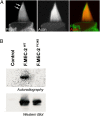
Fig. 1.
Fig. 1.
Enrichment of cholesterol at foci of force perception. (A) Filipin, a fluorescent antibiotic that binds cholesterol, preferentially stains the tips of bullfrog stereocilia. (Reproduced with permission from ref. .) (B) MEC-2, which complexes with the ion channel in C. elegans touch receptor, binds cholesterol. [3H]photocholesterol-treated MEC-2s from expressing HEK-cell lysate were immunoprecipitated and electrophoresed. For similar protein amount (Lower) wild-type but not MEC-2 from a touch-blind mutant captures cholesterol (Upper). (Reproduced from ref. .)
Fig. 2.
Fig. 2.
A generic diagram of a force-bearing focus in animals. (A) Representation of the components of such a focal ensemble with protein a, which has an extracellular domain that anchors outward. It makes a single pass per protein through the membrane and binds protein b, which then binds other proteins, including those (c) that recruit cholesterol and those (e) that bind the cytoskeleton (f) as well as enzymes (g) or channels (h). In type 1, the cell–cell junctions, a is cadherin, b is catenin, c is an SPFH-domain protein such as stomatin, and e can be vinculin that binds F-actin (f). In type 2, the cell-matrix junctions, a is the αβ integrin dimer, b includes talin, c is an SPFH protein such as flotillin, and e includes α-actinin and filamin that binds F-actin (f). The SPFH proteins gather cholesterol to produc-the ordered lipid platform (yellow) distinct from the disordered lipid membrane (blue). The entire ensemble bears a constitutive ∼picoNewton stretch (red arrow) at rest. Not drawn to scale. (B) Representation of such a focus at its constitutive state (Upper) and under added tension (Lower). The tension on the tether (upward arrow) is redirected into membrane stretch (two-headed arrows) by and confined within the ordered lipid platform (yellow).
Fig. 3.
Fig. 3.
Structures of Stomatin-family proteins. (A) Averaged images of some ring complexes of purified yeast prohibitins observed by single particle electron microscopy. (Reproduced from ref. .) (B) Crystal-structure diagram of the mouse stomatin dimer, the smallest functional unit. [Reprinted by permission from Macmillan Publishers Ltd: The EMBO Journal (ref. 66), copyright (2012).]
Fig. 4.
Fig. 4.
Speculative dynamic staging for channel activation by cholesterol-enriched platforms. (A) Such a nano-platform comprises lipids enriched with cholesterol and sphingolipids outwardly enclosed by multimers of SPFH proteins (e.g., stomatins) and inwardly surround the channel (e.g., ASIC). (Left) Such enrichment enforces order in the stiff platform, suppressing mechanical noises and making the membrane thicker to ensure channel closure. (Right) Upon a stretch force transmitted through the bilayer or the direct connections of the frame to the external proteins, the SPFH proteins are displaced, allowing common phospholipids to enter the platform. The thinner and more liquid bilayer and possibly the stretch, reaching the channel through the disordered lipids, open the channel. On tension release the system might return to the closed arrangement because of propensity of the raft lipids to phase separation and selective contacts with the channel. (B) An alternative scheme has the ordered lipids (the entire square) outside the SPFH-protein ring, which corrals a puddle of ordinary lipids surrounding the channel. (Left) Noise is suppressed and channel closed by the indirectly ordered lipids in the absence of added tension. (Right) Under external forces the stiffened frame expands, produces local stretch in the confined puddle, and opens the channel without lipid diffusion. The SPFH protein may be also attached to the channel through linkers as implied by the zigzag line. Diagrams here are representatives of many possible variations (e.g., direct and completely surrounding SPFH-channel binding without a lipid puddle inbetween). In cases where membrane stretch originates from a protein tether (Fig. 2), the stiffened platform also serves to redirect and confine the stretch force. See Mechanical Roles of Cholesterol-rich Platform. CH, channel; chol, cholessterol; PL, phospholipids; SM, sphingomyelin.

References

    1. Martinac B, Buechner M, Delcour AH, Adler J, Kung C. Pressure-sensitive ion channel in Escherichia coli. Proc Natl Acad Sci USA. 1987;84(8):2297–2301. - PMC - PubMed
    1. Martinac B, Adler J, Kung C. Mechanosensitive ion channels of E. coli activated by amphipaths. Nature. 1990;348(6298):261–263. - PubMed
    1. Sukharev SI, Blount P, Martinac B, Kung C. Mechanosensitive channels of Escherichia coli: The MscL gene, protein, and activities. Annu Rev Physiol. 1997;59:633–657. - PubMed
    1. Sukharev SI, Blount P, Martinac B, Blattner FR, Kung C. A large-conductance mechanosensitive channel in E. coli encoded by mscL alone. Nature. 1994;368(6468):265–268. - PubMed
    1. Anishkin A, Kung C. Microbial mechanosensation. Curr Opin Neurobiol. 2005;15(4):397–405. - PubMed

Publication types

LinkOut - more resources