

The segmental morphometric properties of the horse cervical spinal cord: a study of cadaver
- PMID: 23476145
- PMCID: PMC3582170
- DOI: 10.1155/2013/734923
The segmental morphometric properties of the horse cervical spinal cord: a study of cadaver
Abstract
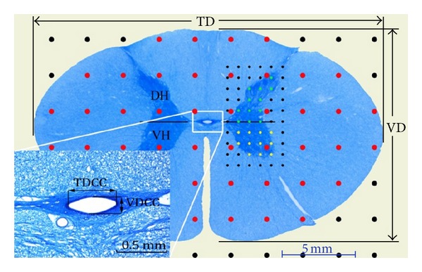
Although the cervical spinal cord (CSC) of the horse has particular importance in diseases of CNS, there is very little information about its segmental morphometry. The objective of the present study was to determine the morphometric features of the CSC segments in the horse and possible relationships among the morphometric features. The segmented CSC from five mature animals was used. Length, weight, diameter, and volume measurements of the segments were performed macroscopically. Lengths and diameters of segments were measured histologically, and area and volume measurements were performed using stereological methods. The length, weight, and volume of the CSC were 61.6±3.2 cm, 107.2±10.4 g, and 95.5±8.3 cm3, respectively. The length of the segments was increased from C1 to C3, while it decreased from C3 to C8. The gross section (GS), white matter (WM), grey matter (GM), dorsal horn (DH), and ventral horn (VH) had the largest cross-section areas at C8. The highest volume was found for the total segment and WM at C4, GM, DH, and VH at C7, and the central canal (CC) at C3. The data obtained not only contribute to the knowledge of the normal anatomy of the CSC but may also provide reference data for veterinary pathologists and clinicians.
Figures


Similar articles
-
Morphometry of cervical segments grey matter in the male rat spinal cord.J Neurosci Methods. 2004 Oct 30;139(2):217-29. doi: 10.1016/j.jneumeth.2004.04.031. J Neurosci Methods. 2004. PMID: 15488235
-
Characterization and limitations of diffusion tensor imaging metrics in the cervical spinal cord in neurologically intact subjects.J Magn Reson Imaging. 2013 Oct;38(4):861-7. doi: 10.1002/jmri.24039. Epub 2013 Feb 6. J Magn Reson Imaging. 2013. PMID: 23389869
-
Feasibility of grey matter and white matter segmentation of the upper cervical cord in vivo: a pilot study with application to magnetisation transfer measurements.Neuroimage. 2012 Nov 15;63(3):1054-9. doi: 10.1016/j.neuroimage.2012.07.048. Epub 2012 Jul 28. Neuroimage. 2012. PMID: 22850571
-
Endoscopic anatomy of the cervical vertebral canal in the horse: a cadaver study.Equine Vet J. 2011 May;43(3):317-23. doi: 10.1111/j.2042-3306.2010.00170.x. Epub 2010 Aug 26. Equine Vet J. 2011. PMID: 21492209
-
Morphometry of the normal cadaveric cervical spinal cord.Spine (Phila Pa 1976). 1994 Sep 15;19(18):2077-81. doi: 10.1097/00007632-199409150-00013. Spine (Phila Pa 1976). 1994. PMID: 7825049
Cited by
-
Anatomical and Embryological Development of the Chick Cerebrum in Different Embryonic Periods.Vet Med Sci. 2025 Jan;11(1):e70124. doi: 10.1002/vms3.70124. Vet Med Sci. 2025. PMID: 39792061 Free PMC article.
-
Sex differences in cervical spinal cord and spinal canal development in Thoroughbred horses.J Vet Med Sci. 2022 Sep 21;84(10):1363-1367. doi: 10.1292/jvms.22-0234. Epub 2022 Aug 10. J Vet Med Sci. 2022. PMID: 35944983 Free PMC article.
References
-
- Ko HY, Park JH, Shin YB, Baek SY. Gross quantitative measurements of spinal cord segments in human. Spinal Cord. 2004;42(1):35–40. - PubMed
-
- Thomas CE, Combs CM. Spinal cord segments. B. Gross structure in the adult monkey. The American Journal of Anatomy. 1965;116:205–216. - PubMed
-
- Braun A. Der segmentale feinbau des rückenmarks des pferdes. Acta Anatomica. 1950;12:1–76. - PubMed
-
- Turgut M, Tunc AT, Aslan H, Yazici AC, Kaplan S. Effect of pinealectomy on the morphology of the chick cervical spinal cord: a stereological and histopathological study. Brain Research. 2007;1129(1):166–173. - PubMed
-
- da Costa RC, Parent JM, Partlow G, Dobson H, Holmberg DL, LaMarre J. Morphologic and morphometric magnetic resonance imaging features of Doberman Pinschers with and without clinical signs of cervical spondylomyelopathy. American Journal of Veterinary Research. 2006;67(9):1601–1612. - PubMed
MeSH terms
LinkOut - more resources
Full Text Sources
Other Literature Sources
Medical
Miscellaneous
