

Tualang honey promotes apoptotic cell death induced by tamoxifen in breast cancer cell lines
- PMID: 23476711
- PMCID: PMC3586458
- DOI: 10.1155/2013/989841
Tualang honey promotes apoptotic cell death induced by tamoxifen in breast cancer cell lines
Abstract
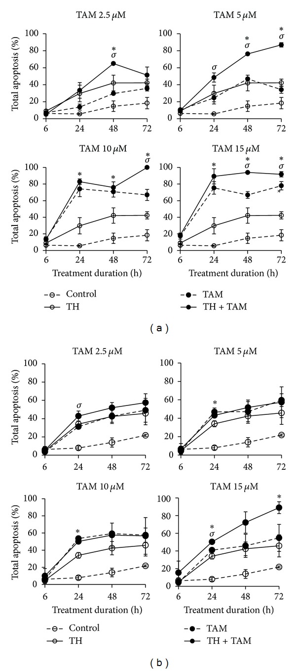
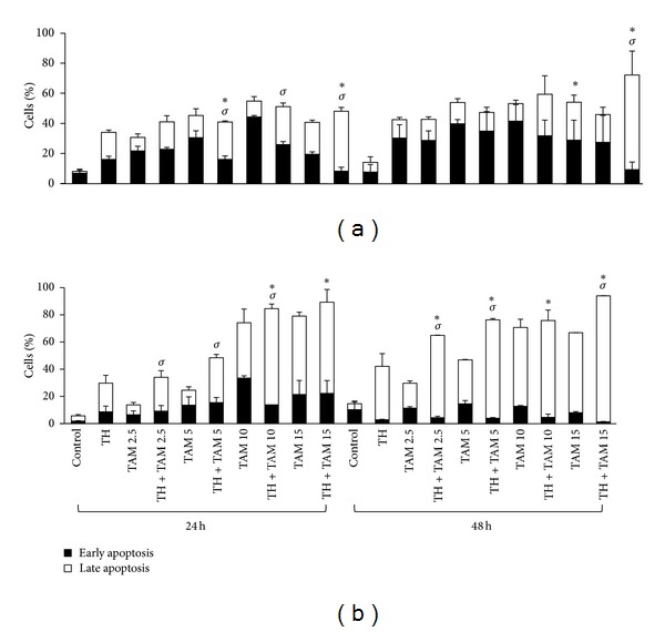
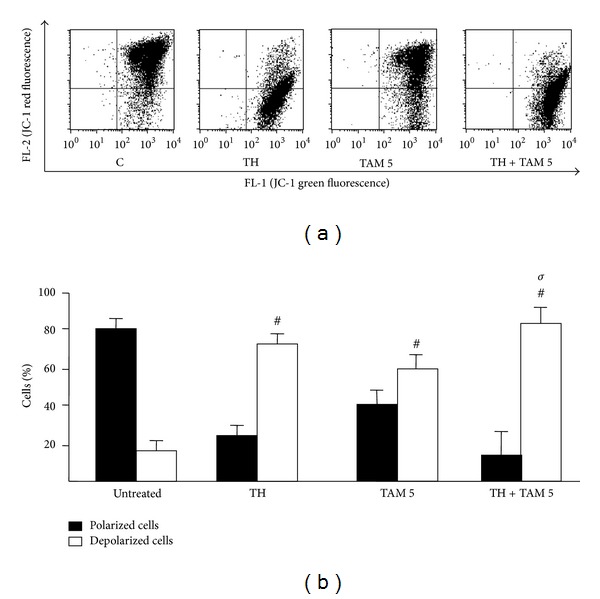
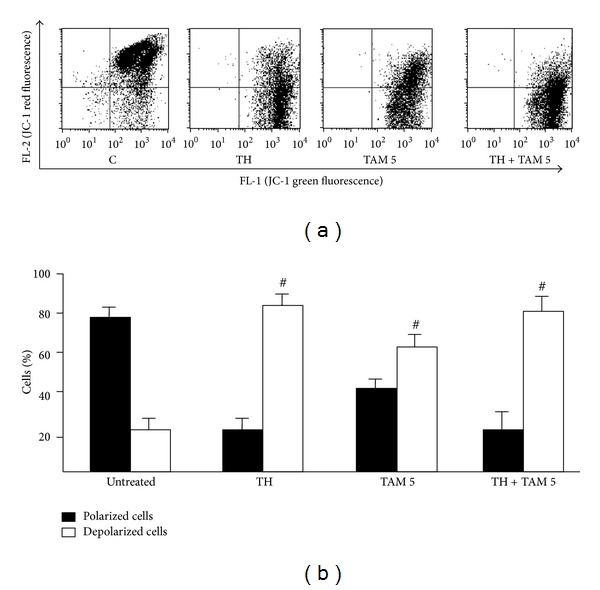
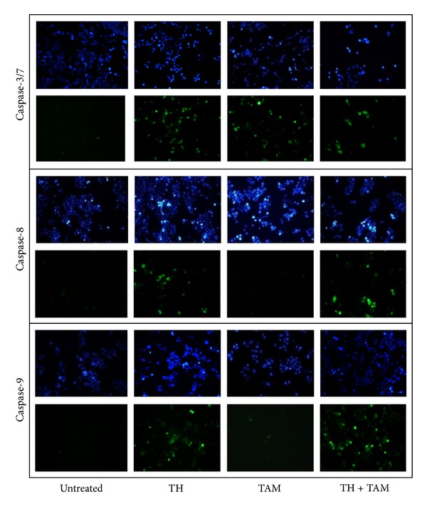
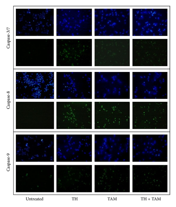
Tualang honey (TH) is rich in flavonoids and phenolic acids and has significant anticancer activity against breast cancer cells comparable to the effect of tamoxifen (TAM), in vitro. The current study evaluated the effects of TH when used in combination with TAM on MCF-7 and MDA-MB-231 cells. We observed that TH promoted the anticancer activity of TAM in both the estrogen receptor-(ER-)responsive and ER-nonresponsive human breast cancer cell lines. Flow cytometric analyses indicated accelerated apoptosis especially in MDA-MB-231 cells and with the involvement of caspase-3/7, -8 and -9 activation as shown by fluorescence microscopy. Depolarization of the mitochondrial membrane was also increased in both cell lines when TH was used in combination with TAM compared to TAM treatment alone. TH may therefore be a potential adjuvant to be used with TAM for reducing the dose of TAM, hence, reducing TAM-induced adverse effects.
Figures
Similar articles
-
Comparison of cytotoxicity and genotoxicity of 4-hydroxytamoxifen in combination with Tualang honey in MCF-7 and MCF-10A cells.BMC Complement Altern Med. 2014 Mar 19;14:106. doi: 10.1186/1472-6882-14-106. BMC Complement Altern Med. 2014. PMID: 24646375 Free PMC article.
-
Tualang honey induces apoptosis and disrupts the mitochondrial membrane potential of human breast and cervical cancer cell lines.Food Chem Toxicol. 2011 Apr;49(4):871-8. doi: 10.1016/j.fct.2010.12.010. Epub 2010 Dec 16. Food Chem Toxicol. 2011. PMID: 21167897
-
Tamoxifen-induced rapid death of MCF-7 breast cancer cells is mediated via extracellularly signal-regulated kinase signaling and can be abrogated by estrogen.Endocrinology. 2007 Jun;148(6):2764-77. doi: 10.1210/en.2006-1269. Epub 2007 Mar 15. Endocrinology. 2007. PMID: 17363451
-
Role of mitochondria in tamoxifen-induced rapid death of MCF-7 breast cancer cells.Apoptosis. 2005 Dec;10(6):1395-410. doi: 10.1007/s10495-005-2137-z. Apoptosis. 2005. PMID: 16215679
-
[Cytotoxicity of tamoxifen and its principal metabolites in human breast cancer cell lines].Bull Cancer. 1996 Oct;83(10):808-15. Bull Cancer. 1996. PMID: 8952630 Review. French.
Cited by
-
Physicochemical Characteristics and Bioactive Compounds of Different Types of Honey and Their Biological and Therapeutic Properties: A Comprehensive Review.Antibiotics (Basel). 2023 Feb 6;12(2):337. doi: 10.3390/antibiotics12020337. Antibiotics (Basel). 2023. PMID: 36830249 Free PMC article. Review.
-
Zeolite X from coal fly ash inhibits proliferation of human breast cancer cell lines (MCF-7) via induction of S phase arrest and apoptosis.Mol Biol Rep. 2018 Dec;45(6):2063-2074. doi: 10.1007/s11033-018-4363-9. Epub 2018 Sep 11. Mol Biol Rep. 2018. PMID: 30206739
-
Comparison of cytotoxicity and genotoxicity of 4-hydroxytamoxifen in combination with Tualang honey in MCF-7 and MCF-10A cells.BMC Complement Altern Med. 2014 Mar 19;14:106. doi: 10.1186/1472-6882-14-106. BMC Complement Altern Med. 2014. PMID: 24646375 Free PMC article.
-
Monofloral Honeys as a Potential Source of Natural Antioxidants, Minerals and Medicine.Antioxidants (Basel). 2021 Jun 25;10(7):1023. doi: 10.3390/antiox10071023. Antioxidants (Basel). 2021. PMID: 34202118 Free PMC article. Review.
-
Cancer Patients' Use of Apitherapy as Supportive Care: An Exploratory Study.Eurasian J Med. 2024 Feb;56(1):15-20. doi: 10.5152/eurasianjmed.2024.22185. Eurasian J Med. 2024. PMID: 39128068 Free PMC article.
References
-
- Gottesman MM. Mechanisms of cancer drug resistance. Annual Review of Medicine. 2002;53:615–627. - PubMed
-
- Li CI, Daling JR, Malone KE. Incidence of invasive breast cancer by hormone receptor status from 1992 to 1998. Journal of Clinical Oncology. 2003;21(1):28–34. - PubMed
-
- Ravdin PM, Cronin KA, Howlader N, et al. The decrease in breast-cancer incidence in 2003 in the United States. The New England Journal of Medicine. 2007;356(16):1670–1674. - PubMed
-
- Osborne CK. Tamoxifen in the treatment of breast cancer. The New English Journal of Medicine. 1998;339:1609–1617. - PubMed
-
- Cersosimo RJ. Tamoxifen for prevention of breast cancer. The Annals of Pharmacotherapy. 2003;37(2):268–273. - PubMed
LinkOut - more resources
Full Text Sources
Other Literature Sources
Research Materials
Miscellaneous
