

doi: 10.1007/s12177-012-9077-y.
Epub 2012 Mar 8.
Alterations in "in vivo" histology of retina in bilateral chronic central serous chorioretinopathy after intravitreal bevacizumab
Affiliations
- PMID: 23476721
- PMCID: PMC3389553
- DOI: 10.1007/s12177-012-9077-y
Item in Clipboard
Alterations in "in vivo" histology of retina in bilateral chronic central serous chorioretinopathy after intravitreal bevacizumab
J Ocul Biol Dis Infor.
2011 Dec.
No abstract available
Figures

a Color fundus photograph of the right eye shows retinal pigment epithelium atrophy, few superficial and deep hemorrhages and hard exudates at the posterior pole. b Color fundus photograph of the left eye shows retinal pigment epithelium atrophy tract, few superficial and deep hemorrhages, and hard exudates at the posterior pole
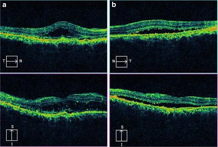
a Spectral domain optical coherence tomography of the right eye shows neurosensory detachment of the macula with retinal pigment epithelial detachment. Granularity in the outer photoreceptor layer and proliferative retinal pigment epithelial cells are also observed. The retinal pigment epithelium is hyperplastic. The photoreceptor IS–OS junction is not visualized. b Spectral domain optical coherence tomography of the left eye shows neurosensory detachment of the macula with retinal pigment epithelial detachment. Granularity in the outer photoreceptor layer and proliferative retinal pigment epithelial cells are also observed. The retinal pigment epithelium is hyperplastic. The photoreceptor IS–OS junction is not visualized
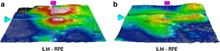
a Three-dimensional internal limiting membrane-retinal pigment epithelium map of the right eye shows increased macular thickness. Central sub field thickness is 444 μm. b Three-dimensional internal limiting membrane-retinal pigment epithelium map of the left eye shows increased macular thickness. Central sub field thickness was 324 μm
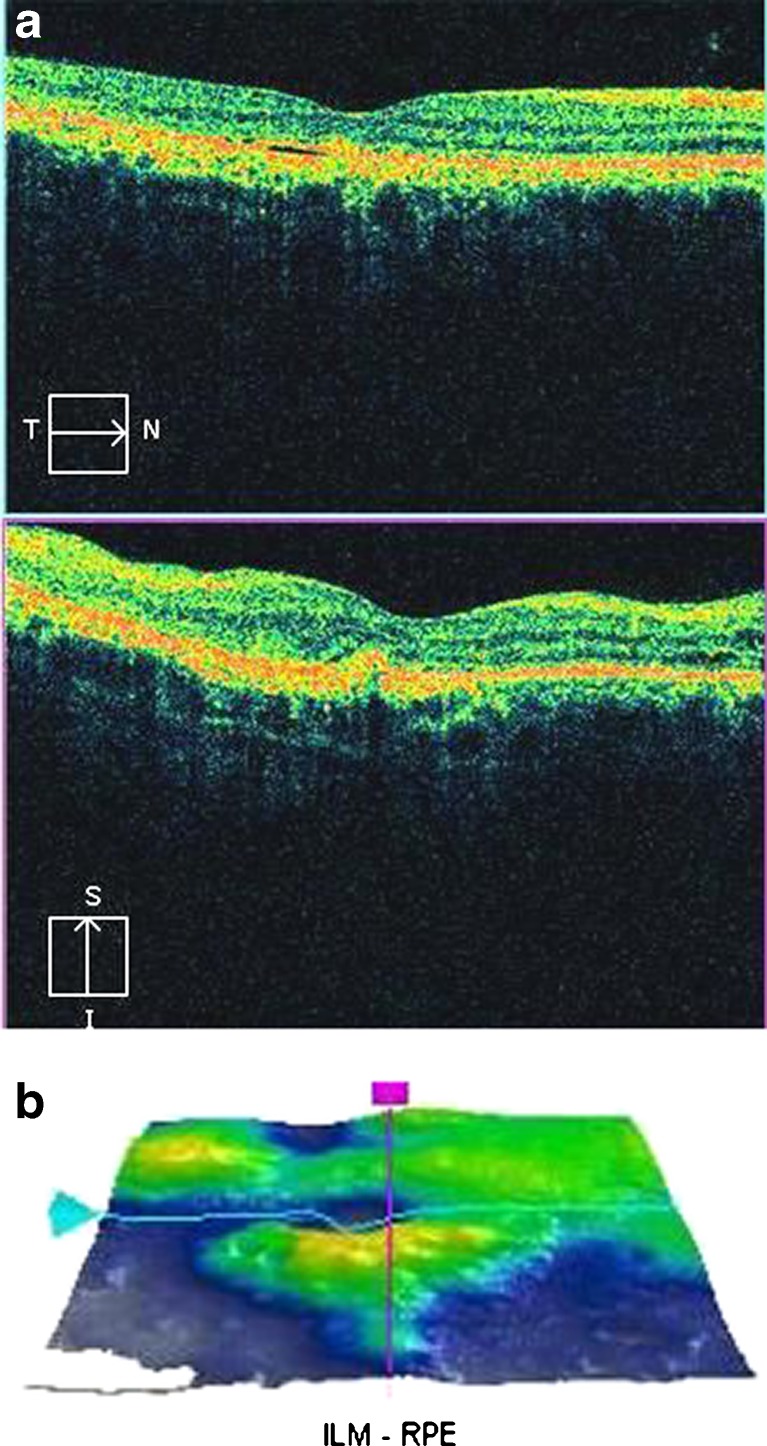
a Post intravitreal bevacizumab injection, spectral domain optical coherence tomography of the right eye shows resolution of subretinal fluid with persistent pigment epithelial detachment. Foveal contour is restored. Residual granularity at outer photoreceptor layer and proliferative retinal pigment epithelial cells are visualized. Small cystic spaces are also observed. The photoreceptor IS–OS junction is not visible in the subfoveal area. The retinal pigment epithelium layer remains hyperplastic. b Three-dimensional internal limiting membrane-retinal pigment epithelium map of the right eye shows decreased macular thickness. Central subfield thickness is 216 μm
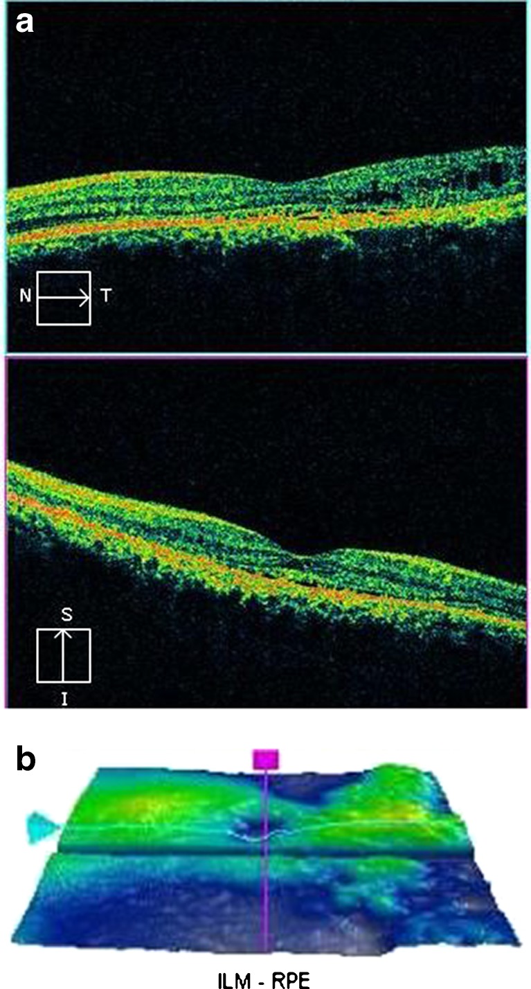
a Post intravitreal bevacizumab injection, spectral domain optical coherence tomography of the left eye shows resolution of subretinal fluid. Foveal contour is restored. Residual granularity at outer photoreceptor layer and proliferative retinal pigment epithelial cells are visualized. The photoreceptor IS–OS junction is visualized in the subfoveal area. b Three-dimensional internal limiting membrane-retinal pigment epithelium map of the left eye shows decreased macular thickness. Central subfield thickness is 238 μm
Similar articles
-
[Recurrent and chronic central serous chorioretinopathy. Retina thickness evaluation one month after intravitreal bevacizumab injection].Arch Soc Esp Oftalmol. 2011 Dec;86(12):407-11. doi: 10.1016/j.oftal.2011.05.021. Epub 2011 Oct 11. Arch Soc Esp Oftalmol. 2011. PMID: 22117740 Clinical Trial. Spanish.
-
Intravitreal Bevacizumab and Ranibizumab in the Treatment of Acute Central Serous Chorioretihopathy: A Single Center Retrospective Study.Semin Ophthalmol. 2018;33(2):265-270. doi: 10.1080/08820538.2016.1228985. Epub 2016 Nov 14. Semin Ophthalmol. 2018. PMID: 27841949
-
Intravitreal triamcinolone acetonide for cystoid macular edema secondary to central serous chorioretinopathy.Retin Cases Brief Rep. 2009 Summer;3(3):319-22. doi: 10.1097/ICB.0b013e31819b19ee. Retin Cases Brief Rep. 2009. PMID: 25389597
-
Central Serous Chorioretinopathy: Pathogenesis and Management.Clin Ophthalmol. 2019 Dec 2;13:2341-2352. doi: 10.2147/OPTH.S220845. eCollection 2019. Clin Ophthalmol. 2019. PMID: 31819359 Free PMC article. Review.
-
[Central serous chorioretinopathy in a pregnant woman].J Fr Ophtalmol. 1996;19(3):216-21. J Fr Ophtalmol. 1996. PMID: 8731772 Review. French.
Cited by
-
Photoreceptor inner segment ellipsoid band integrity on spectral domain optical coherence tomography.Clin Ophthalmol. 2014 Dec 9;8:2507-22. doi: 10.2147/OPTH.S72132. eCollection 2014. Clin Ophthalmol. 2014. PMID: 25525329 Free PMC article. Review.
References
-
- Gass JDM. Pathogenesis of disciform detachment of the neuro-epithelium. II. Idiopathic central serous choroidopathy. Am J Ophthalmol. 1960;63:587–615.
-
- Yannuzzi LA, Shakin JL, Fisher YL, Altomonte MA. Peripheral retinal detachments and retinal pigment epithelial atrophic tracts secondary to central serous pigment epitheliopathy. Ophthalmology. 1984;91:1554–1572. - PubMed
-
- Gomolin JE. Choroidal neovascularization and central serous chorioretinopathy. Can J Ophthalmol. 1989;24:20–23. - PubMed
LinkOut - more resources
Full Text Sources