

Are the Mesothelial-to-Mesenchymal Transition, Sclerotic Peritonitis Syndromes, and Encapsulating Peritoneal Sclerosis Part of the Same Process?
- PMID: 23476771
- PMCID: PMC3582112
- DOI: 10.1155/2013/263285
Are the Mesothelial-to-Mesenchymal Transition, Sclerotic Peritonitis Syndromes, and Encapsulating Peritoneal Sclerosis Part of the Same Process?
Abstract
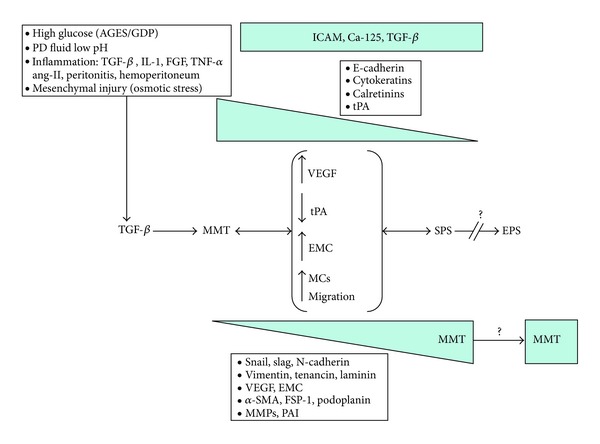
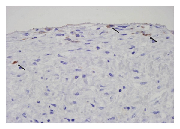
Mesothelial-to-mesenchymal transition (MMT) is an autoregulated physiological process of tissue repair that in uncontrolled conditions, such as peritoneal dialysis (PD), can lead to peritoneal fibrosis. The maximum expression of sclerotic peritoneal syndromes (SPS) is the encapsulating peritoneal sclerosis (EPS) for which no specific treatment exists. The SPS includes a wide range of peritoneal fibrosis that appears progressively and is considered as a reversible process, while EPS does not. EPS is a serious complication of PD characterized by a progressive intra-abdominal inflammatory process that results in bridles and severe fibrous tissue formation which cover and constrict the viscera. Recent studies show that transdifferentiated mesothelial cells isolated from the PD effluent correlate very well with the clinical events such as the number of hemoperitoneum and peritonitis, as well as with PD function (lower ultrafiltration and high Cr-MTC). In addition, in peritoneal biopsies from PD patients, the MMT correlates very well with anatomical changes (fibrosis and angiogenesis). However, the pathway to reach EPS from SPS has not been fully and completely established. Herein, we present important evidence pointing to the MMT that is present in the initial peritoneal fibrosis stages and it is perpetual over time, with at least theoretical possibility that MMT initiated the fibrosing process to reach EPS.
Figures
References
-
- Selgas R, Bajo MA, Del Peso G, Jimenez C. Preserving the peritoneal dialysis membrane in long-term peritoneal dialysis patients. Seminars in Dialysis. 1995;8:326–332.
-
- Williams JD, Craig KJ, Topley N, et al. Morphologic changes in the peritoneal membrane of patients with renal disease. Journal of the American Society of Nephrology. 2002;13(2):470–479. - PubMed
-
- Krediet RT, Lindholm B, Rippe B. Pathophysiology of peritoneal membrane failure. Peritoneal Dialysis International. 2000;20(supplement 4):S22–S42. - PubMed
-
- Pecoits-Filho R, Araújo MRT, Lindholm B, et al. Plasma and dialysate IL-6 and VEGF concentrations are associated with high peritoneal solute transport rate. Nephrology Dialysis Transplantation. 2002;17(8):1480–1486. - PubMed
-
- Zweers MM, Struijk DG, Smit W, Krediet RT. Vascular endothelial growth factor in peritoneal dialysis: a longitudinal follow-up. Journal of Laboratory and Clinical Medicine. 2001;137(2):125–132. - PubMed
LinkOut - more resources
Full Text Sources
Other Literature Sources
