

Ductal carcinoma in situ: what the pathologist needs to know and why
- PMID: 23476791
- PMCID: PMC3580892
- DOI: 10.1155/2013/914053
Ductal carcinoma in situ: what the pathologist needs to know and why
Abstract
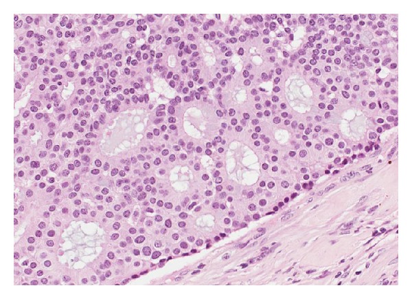
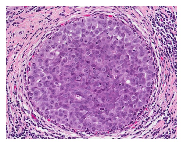
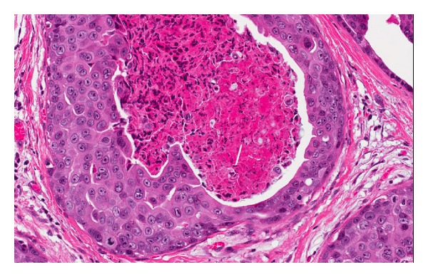
Ductal carcinoma in situ is a proliferation of malignant epithelial cells confined to the ductolobular system of the breast. It is considered a pre-cursor lesion for invasive breast cancer and when identified patients are treated with some combination of surgery, +/- radiation therapy, and +/adjuvant tamoxifen. However, no good biomarkers exist that can predict with accuracy those cases of DCIS destined to progress to invasive disease or once treated those patients that are likely to suffer a recurrence; thus, in the era of screening mammography it seems likely that many patients with DCIS are overtreated. This paper details the parameters that should be included in a pathology report for a case of DClS with some explanations as to their importance for good clinical decision making.
Figures
References
-
- Dupont WD, Page DL. Risk factors for breast cancer in women with proliferative breast disease. The New England Journal of Medicine. 1985;312(3):146–151. - PubMed
-
- Ernster VL, Ballard-Barbash R, Barlow WE, et al. Detection of ductal carcinoma in situ in women undergoing screening mammography. Journal of the National Cancer Institute. 2002;94(20):1546–1554. - PubMed
-
- May DS, Lee NC, Richardson LC, Giustozzi AG, Bobo JK. Mammography and breast cancer detection by race and Hispanic ethnicity: results from a national program (United States) Cancer Causes and Control. 2000;11(8):697–705. - PubMed
-
- Kerlikowske K, Miglioretti DL, Buist DSM, Walker R, Carney PA. Declines in invasive breast cancer and use of postmenopausal hormone therapy in a screening mammography population. Journal of the National Cancer Institute. 2007;99(17):1335–1339. - PubMed
-
- Li CI, Daling JR, Malone KE. Age-specific incidence rates of in situ breast carcinomas by histologic type, 1980 to 2001. Cancer Epidemiology Biomarkers and Prevention. 2005;14(4):1008–1011. - PubMed
LinkOut - more resources
Full Text Sources
Other Literature Sources
