

doi: 10.1155/2013/381583.
Epub 2013 Feb 17.
Simultaneous Larva Migrans and Larva Currens Caused by Strongyloides stercoralis: A Case Report
Affiliations
- PMID: 23476820
- PMCID: PMC3588403
- DOI: 10.1155/2013/381583
Item in Clipboard
Simultaneous Larva Migrans and Larva Currens Caused by Strongyloides stercoralis: A Case Report
Case Rep Dermatol Med.
2013.
Abstract
Strongyloidiasis is an infectious disease caused by the Strongyloides stercoralis larvae, which penetrate the skin, go through the lymphatic circulation, and migrate to the lungs before reaching the intestines. They mature and may cause cutaneous strongyloidiasis, known as larva currens because of the quick migratory rate of the larva. The authors describe a case in which the larvae did not follow their natural lymph route, and after penetrating into the intertriginous area, they migrated to the dermis, developing larva migrans in the early phase, and later associated with the typical lesions of larva currens. The diagnosis was confirmed by the presence of larva in the skin biopsy.
Figures

Area larvae penetration. Hemorrhagic blisters in the area: the larvae penetrated the skin and the purpuric serpiginous lesions in the back of the left foot.

Larva migrans. Diffuse purpuric lesions showing the various routes the larvae took in the left thigh.
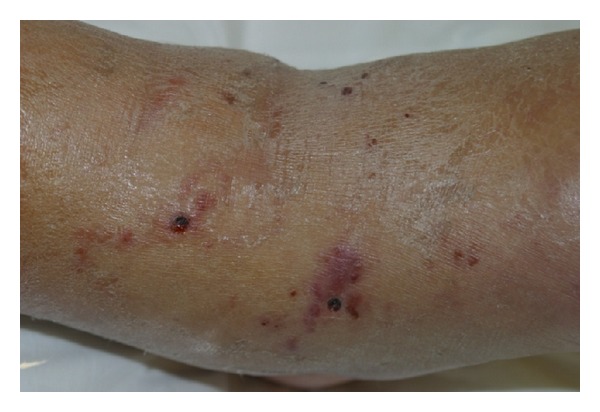
Biopsy site in the popliteal region.

Larva currens. Erythematous, edematous urticarial lesions in the back and the abdomen.

Pathology. PAS 10x: presence of larva PAS positive in stratum corneum.

Strongyloides stercoralis. Presence of larva in the corneal layer.
References
-
- Neves PD, Melo LA, Linardi MP, Vitor AWR. Parasitologia Humana. 11th edition. Atheneu; 2005.
-
- Neto AV, Baldy SLJ, Ramos CM, Branchini NLM. Parasitoses Intestinais. 3rd edition. Sarvier; 1989.
-
- Ly MN, Bethel SL, Usmani AS, Lambert DR. Cutaneous Strongyloides stercoralis infection: an unusual presentation. Journal of the American Academy of Dermatology. 2003;49(supplement 2):S157–S160. - PubMed
-
- Gaus B, Toberer F, Kapaun A, Hartmann M. Chronic strongyloides stercoralis infection. Larva currens as skin manifestation. Hautarzt. 2011;62(5):380–383. - PubMed
-
- Corti M, Villafañe MF, Trione N, Risso D, Abuín JC, Palmieri O. Infection due to Strongyloides stercoralis: epidemiological, clinical, diagnosis findings and outcome in 30 patients. Revista Chilena de Infectologia. 2011;28(3):217–222. - PubMed
LinkOut - more resources
Full Text Sources
Other Literature Sources
