. 2013 Aug 1;22(15):2211-20.
doi: 10.1089/scd.2012.0611. Epub 2013 Apr 9.

Nkx2-5 mediates differential cardiac differentiation through interaction with Hoxa10

Affiliations

Nkx2-5 mediates differential cardiac differentiation through interaction with Hoxa10

Ann N Behrens et al. Stem Cells Dev. .

Abstract

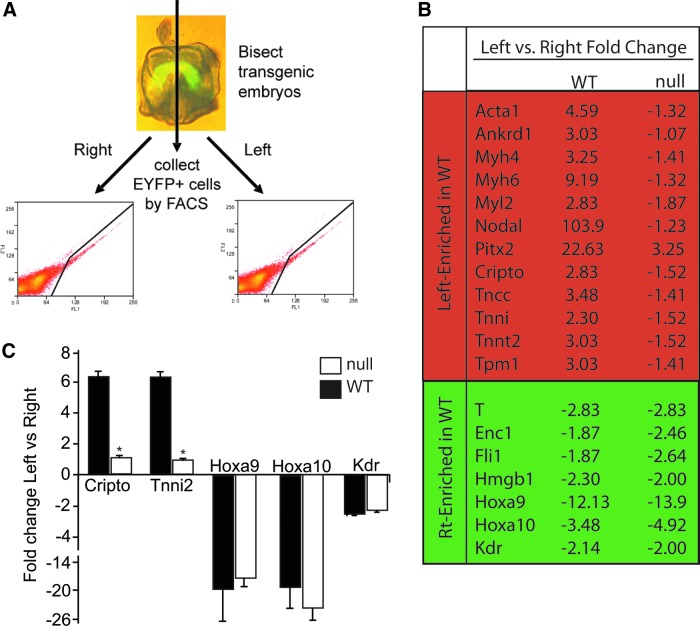
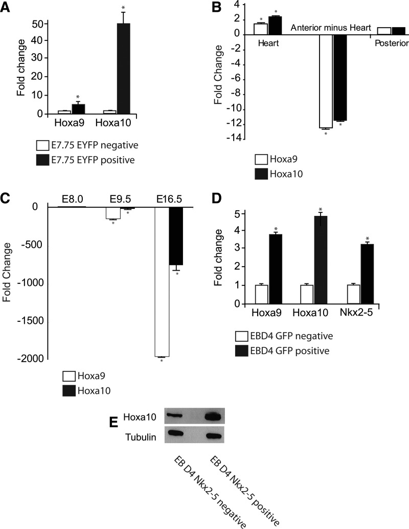
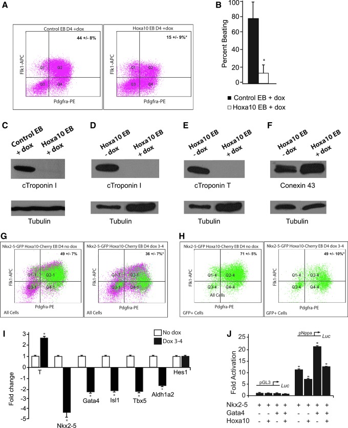
The regulation of cardiac differentiation is complex and incompletely understood. Recent studies have documented that Nkx2-5-positive cells are not limited to the cardiac lineage, but can give rise to endothelial and smooth muscle lineages. Other work has elucidated that, in addition to promoting cardiac development, Nkx2-5 plays a larger role in mesodermal patterning although the transcriptional networks that govern this developmental patterning are undefined. By profiling early Nkx2-5-positive progenitor cells, we discovered that the progenitor pools of the bisected cardiac crescent are differentiating asynchronously. This asymmetry requires Nkx2-5 as it is lost in the Nkx2-5 mutant. Surprisingly, the posterior Hox genes Hoxa9 and Hoxa10 were expressed on the right side of the cardiac crescent, independently of Nkx2-5. We describe a novel, transient, and asymmetric cardiac-specific expression pattern of the posterior Hox genes, Hoxa9 and Hoxa10, and utilize the embryonic stem cell/embryoid body (ES/EB) model system to illustrate that Hoxa10 impairs cardiac differentiation. We suggest a model whereby Hoxa10 cooperates with Nkx2-5 to regulate the timing of cardiac mesoderm differentiation.


Figures

FIG. 1.
FIG. 1.
Hoxa9 and Hoxa10 are expressed in early cardiac progenitor cells of the cardiac crescent. (A) Individual WT and Nkx2-5 null cardiac crescent stage embryos were bisected and EYFP-positive cells were collected from the right and left side of the crescent. Left WT mean=2044, SD=499, n=5; left null mean=1967, SD=200, n=4; right WT mean=2263, SD=294, n=5; right null mean=2167, SD=252, n=4. (B) Transcriptome analysis of the WT and Nkx2-5 null left versus right side of the crescent. Note a significant induction of cardiac structural transcripts in the left versus right WT crescent. This left-sided enrichment is absent in the Nkx2-5 null crescent. The right-sided enrichment in expression is largely unaltered in the absence of Nkx2-5. Significant differential transcript expression is denoted by a green highlight (upregulated) or a red highlight (downregulated). (C) quantitative reverse transcription (qRT)-polymerase chain reaction (PCR) of selected trannscripts in WT (black bar) and null (open bar) right and left crescent.
FIG. 2.
FIG. 2.
Characterization of cardiac Hoxa10 expression. (A) Analyses of enrichment of Hoxa9 and Hoxa10 in the EYFP-positive Nkx2-5 expressing cells versus EYFP-negative cells collected from the cardiac crescent using qRT-PCR; *P<0.05, n=3. (B) qRT-PCR reveals Hoxa9 and Hoxa10 are enriched in the E8.25 (5–7 somite) heart compared to the remaining segments of the embryo; *P<0.05, n=3. (C) Cardiac Hoxa9 and Hoxa10 expression is rapidly downregulated during cardiac development as assessed by qRT-PCR; *P<0.05, n=3. (D) Analyses of fold change in the expression of the Hoxa9, Hoxa10, and Nkx2-5 from GFP-positive Nkx2-5 expressing cells versus GFP negative obtained from embryoid bodies (EBs) harvested at day 4 using qRT-PCR; *P<0.05, n=3. (E) Western analysis of EB day 4 GFP-positive Nkx2-5 expressing cells confirm Hoxa10 protein expression.
FIG. 3.
FIG. 3.
Hoxa10 inhibits cardiogenesis. (A) Percentage of EB day 4, Flk-1-PDGFRα double-positive cardiac progenitor cells are significantly decreased following Hoxa10 overexpression for 48 h (day 2–4) of EB formation; 15%±9% versus 43.6%±7.9%, *P<0.05, n=8. (B) Reduction of beating in EBs overexpressing Hoxa10 following induction with doxycycline (Dox) on days 3–6 versus control EBs plus doycycline; 12.5±10.3 versus 78.4±21.7, *P<0.05, n=8. (C) Western analysis reveals absence of cTroponin I in day 12 EBs overexpressing Hoxa10 following the induction with doxycycline (Dox) on days 3–6 versus day 12 control EBs treated with doxycycline (Dox) on days 3–6). Western analysis reveals absence of cTroponin I (D) and cTroponin T (E) in day 12 Hoxa10 doxycycline-inducible EBs following the induction with doxycycline (Dox) on days 3–6 versus no doxycycline. (F) Protein expression of Connexin 43 is preserved in day 12 EBs following Hoxa10 induction. (G) The Nkx2-5-GFP reporter ES cell line was infected with Hoxa10 or empty lentivirus. Overexpression of Hoxa10 for 24 h (day 3–4 of EB formation) in cardiac crescent equivalent progenitors results in a reduction of total Flk-1/PDGFRα double-positive cardiac progenitor cells; 35.5%±7% versus 48.6%±7%; *P<0.05, n=5. (H) Percentage of Nkx2-5 expressing GFP-positive cells double-positive for Flk-1 and PDGFRα is also significantly decreased with Hoxa10 overexpression; 49%±10% versus 71%±5%; *P<0.05, n=5. (I) qRT-PCR analyses revealed an increase in Brachyury expression and reduction in the cardiac transcription factors Nkx2-5, Gata-4, Tbx-5, and Isl-1 as well as a small decrease in Aldh1a2 in GFP-positive cells collected from day 4 Nkx2-5-GFP EBs following the induction of Hoxa10 with doxycycline (Dox) for 24 h (day 3–4); *P<0.05, n=3. Hes1 expression, however, was not significantly altered. (J) The Nppa promoter was fused to the luciferase (luc) reporter gene and was transfected into Cos7 cells with and without addition of the Nkx2-5, Gata4, or Hoxa10 expression plasmids. Empty pGL3 and Nppa promoter-driven luciferase expression is shown. Nppa promoter revealed a 10.65±0.44-fold activation with Nkx2-5, which was reduced to 7.41±0.50 with addition of Hoxa10 and a 20.55±0.49-fold activation with Nkx2-5 and Gata4, which was reduced to 12.62±0.17 with the addition of Hoxa10; *P<0.05, n=3.

References

    1. Hoffman JI. Incidence of congenital heart disease: II. Prenatal incidence. Pediatr Cardiol. 1995;16:155–165. - PubMed
    1. Olson EN. Srivastava D. Molecular pathways controlling heart development. Science. 1996;272:671–676. - PubMed
    1. Harvey RP. Patterning the vertebrate heart. Nat Rev Genet. 2002;3:544–556. - PubMed
    1. Olson EN. Schneider MD. Sizing up the heart: development redux in disease. Genes Dev. 2003;17:1937–1956. - PubMed
    1. Brand T. Heart development: molecular insights into cardiac specification and early morphogenesis. Dev Biol. 2003;258:1–19. - PubMed

Publication types