. 2013 Mar 28;3(3):651-60.
doi: 10.1016/j.celrep.2013.02.005. Epub 2013 Mar 7.

Homologous recombination DNA repair genes play a critical role in reprogramming to a pluripotent state

Affiliations

Homologous recombination DNA repair genes play a critical role in reprogramming to a pluripotent state

Federico González et al. Cell Rep. .

Abstract

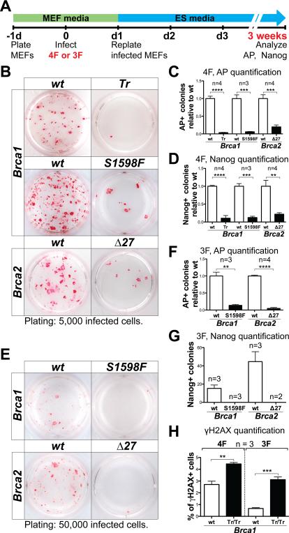
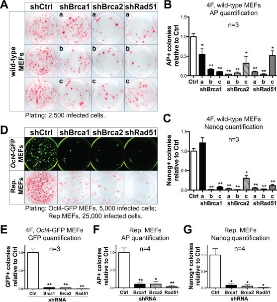
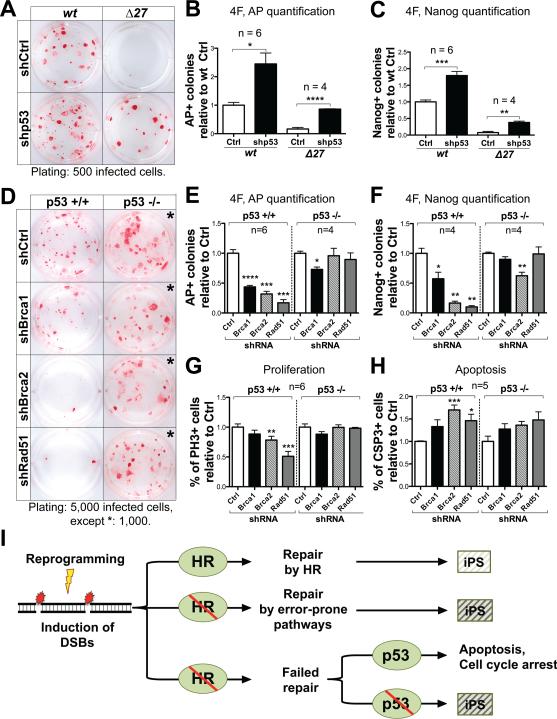
Induced pluripotent stem cells (iPSCs) hold great promise for personalized regenerative medicine. However, recent studies show that iPSC lines carry genetic abnormalities, suggesting that reprogramming may be mutagenic. Here, we show that the ectopic expression of reprogramming factors increases the level of phosphorylated histone H2AX, one of the earliest cellular responses to DNA double-strand breaks (DSBs). Additional mechanistic studies uncover a direct role of the homologous recombination (HR) pathway, a pathway essential for error-free repair of DNA DSBs, in reprogramming. This role is independent of the use of integrative or nonintegrative methods in introducing reprogramming factors, despite the latter being considered a safer approach that circumvents genetic modifications. Finally, deletion of the tumor suppressor p53 rescues the reprogramming phenotype in HR-deficient cells primarily through the restoration of reprogramming-dependent defects in cell proliferation and apoptosis. These mechanistic insights have important implications for the design of safer approaches to creating iPSCs.


Figures

Figure 1
Figure 1. Reprogramming induces DSBs and apoptosis
(A) Representative Fluorescence-activated cell sorting (FACS) plots of 4F- and 3F-infected wild-type MEFs stained for γH2AX, SSEA1 and Annexin V after cells were cultured with or without doxycycline (DOX) for 5 days. Numbers indicate percentages of positive cells. PI: Propidium Iodide; DAPI: 4',6-diamidino-2-phenylindole; L: alive; EA: early apoptotic; LA: late apoptotic; N: necrotic. (B) Quantification of the percentage of γH2AX+ cells in wild-type MEFs infected with reprograming genes in combination or individually. OS: Oct4-Sox2; O: Oct4; S: Sox2; K: Klf4; M: c-Myc. (C, D) Quantification of the percentage of SSEA1+ (C) and Annexin V+ (D) cells in wild-type MEFs transduced with 4F and 3F. Apoptotic cells are the sum of EA and LA cells. (E) Time-lapse, flow cytometric quantification of γH2AX+ cells present in reprogrammable MEFs with or without DOX treatment; cells were separated based on the expression of SSEA1. In all column graphs of this study, error bars indicate SEM, and p values by two-tailed student t-test <0.05, 0.01, 0.001 and 0.0001 are indicated by one, two, three or four asterisks, respectively.
Figure 2
Figure 2. Reprogramming is impaired in Brca1 and Brca2 mutant MEFs
(A) Schematics of all virus-mediated reprogramming experiments in this study. MEFs were infected with 4F or 3F 1-day after plating, and replated in 12-well dishes the next day on irradiated MEFs at densities specified (indicated bellow the AP-staining pictures for all figures in this study). AP and Nanog staining was performed after 3 weeks (unless otherwise noted). (B-D) Representative AP staining (B), and quantification of AP+ (C) and Nanog+ (D) colonies generated with 4F-reprogramming from Brca1Tr/Tr, Brca1S1598F/S1598F and Brca2Δ27/Δ27 MEFs, compared with wild-type (wt) MEFs from littermate controls. (E-G) Representative AP staining (E), and quantification of AP+ (F) and Nanog+ (G) colonies generated with 3F-reprogramming. (H) Quantification of the percentage of γH2AX+ cells in 4F- and 3F-infected, Brca1Tr/Tr mutant and control wild-type MEFs after 5 days of DOX treatment. See also Figure S1.
Figure 3
Figure 3. HR genes are directly required during reprogramming
(A-C) Representative AP staining (A), and quantification of AP+ (B) and Nanog+ (C) colonies generated with 4F-reprogramming and a panel of shRNAs targeting Brca1 (shBrca1-a, b, c), Brca2 (shBrca2-a, b, c) and Rad51 (shRad51-a, b, c) compared to the shRNA control vector (shCtrl). Lower-case letters refer to individual shRNAs targeting each HR gene. shBrca1-c, shBrca2-b and shRad51-b were used for further experiments. (D) Upper panel: Representative fluorescence images of Oct4-GFP+ colonies generated with 4F and shRNAs targeting HR genes. Lower panel: Representative AP staining images from reprogrammable (Rep.) MEFs infected with shRNAs against HR genes. (E) Quantification of Oct4-GFP+ colonies from experiments using 4F-infected Oct4-GFP MEFs and acute HR-gene knockdown. (F, G) Quantification of AP+ (F) and Nanog+ (G) colonies from experiments using reprogrammable MEFs and acute HR-gene knockdown. See also Figures S2 and S3.
Figure 4
Figure 4. Down-regulating p53 rescues the reprogramming phenotype of HR-defective MEFs
(A-C) Representative AP staining (A) and quantification of AP+ (B) and Nanog+ (C) colonies generated with 4F-reprogramming from Brca2Δ27 homozygous mutant and wild-type MEFs infected with an shRNA targeting p53 (shp53) or vector control (shCtrl). (D-F) Representative AP staining (D) and quantification of AP+ (E) and Nanog+ (F) colonies generated with 4F-reprogramming from p53 null and wild-type MEFs under acute HR-gene knockdown. All staining were performed 16 days after replating of infected cells. (G, H) Quantification of the percentage of Phospho-Histone H3+ (PH3+) (G) and Cleaved Caspase-3+ (CSP3+) (H) cells, 6 days post-infection of 4F and HR-gene knockdown in p53 null mutant and wild-type MEFs. (I) Our results support a critical role of the HR pathway for efficient reprogramming. We propose a model in which reprogramming increases the level of DNA damage, which is responsible for the genetic aberrations observed in iPS cell lines (indicated by a light shaded box). A defective HR pathway may lead to increased genetic aberration (indicated by dark shaded boxes), or the elimination of abnormal cells through p53-mediated cell cycle arrest or apoptosis. See also Figure S4.

References

    1. Brambrink T, Foreman R, Welstead GG, Lengner CJ, Wernig M, Suh H, Jaenisch R. Sequential expression of pluripotency markers during direct reprogramming of mouse somatic cells. Cell Stem Cell. 2008;2:151–159. - PMC - PubMed
    1. Carey BW, Markoulaki S, Beard C, Hanna J, Jaenisch R. Single-gene transgenic mouse strains for reprogramming adult somatic cells. Nat Methods. 2010;7:56–59. - PMC - PubMed
    1. Cheng L, Hansen NF, Zhao L, Du Y, Zou C, Donovan FX, Chou BK, Zhou G, Li S, Dowey SN, et al. Low incidence of DNA sequence variation in human induced pluripotent stem cells generated by nonintegrating plasmid expression. Cell Stem Cell. 2012;10:337–344. - PMC - PubMed
    1. Daley GQ. Common themes of dedifferentiation in somatic cell reprogramming and cancer. Cold Spring Harbor symposia on quantitative biology. 2008;73:171–174. - PubMed
    1. Fong YW, Inouye C, Yamaguchi T, Cattoglio C, Grubisic I, Tjian R. A DNA repair complex functions as an Oct4/Sox2 coactivator in embryonic stem cells. Cell. 2011;147:120–131. - PMC - PubMed

Publication types