. 2013 Mar 28;3(3):661-70.
doi: 10.1016/j.celrep.2013.02.012. Epub 2013 Mar 7.

Epigenetic reprogramming of human embryonic stem cells into skeletal muscle cells and generation of contractile myospheres

Affiliations

Epigenetic reprogramming of human embryonic stem cells into skeletal muscle cells and generation of contractile myospheres

Sonia Albini et al. Cell Rep. .

Abstract

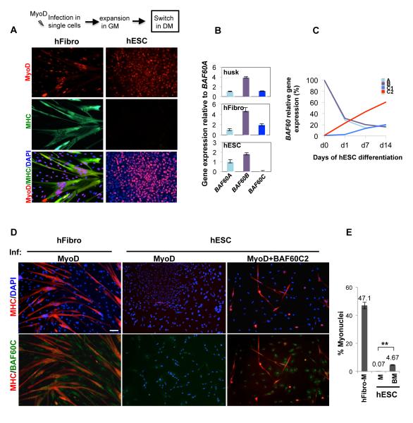
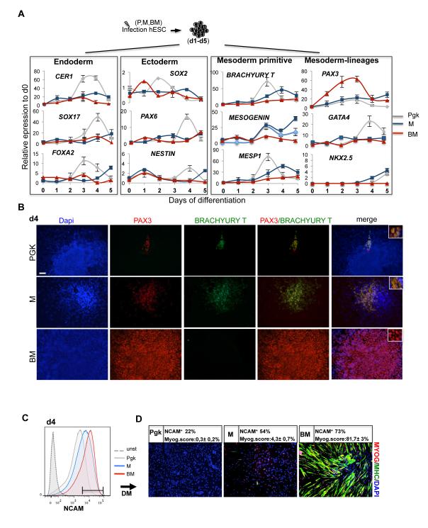
Direct generation of a homogeneous population of skeletal myoblasts from human embryonic stem cells (hESCs) and formation of three-dimensional contractile structures for disease modeling in vitro are current challenges in regenerative medicine. Previous studies reported on the generation of myoblasts from ESC-derived embryoid bodies (EB), but not from undifferentiated ESCs, indicating the requirement for mesodermal transition to promote skeletal myogenesis. Here, we show that selective absence of the SWI/SNF component BAF60C (encoded by SMARCD3) confers on hESCs resistance to MyoD-mediated activation of skeletal myogenesis. Forced expression of BAF60C enables MyoD to directly activate skeletal myogenesis in hESCs by instructing MyoD positioning and allowing chromatin remodeling at target genes. BAF60C/MyoD-expressing hESCs are epigenetically committed myogenic progenitors, which bypass the mesodermal requirement and, when cultured as floating clusters, give rise to contractile three-dimensional myospheres composed of skeletal myotubes. These results identify BAF60C as a key epigenetic determinant of hESC commitment to the myogenic lineage and establish the molecular basis for the generation of hESC-derived myospheres exploitable for "disease in a dish" models of muscular physiology and dysfunction.


Figures

Fig. 1
Fig. 1. BAF60C2 rescues MyoD-mediated myogenic conversion in hESCs
A) Immunofluorescence staining to detect Myosin heavy chain (MHC) (green) and MyoD (red) in human fibroblasts (H27) and hESCs (H9) infected with MyoD or a control virus (Pgk). Nuclei are visualized by DAPI staining (blue). Cells were cultured in monolayer and induced to differentiate by shifting medium from growing conditions (GM) – growth medium) to pro-myogenic DM (differentiation medium) (see Methods for details). B) Relative gene expression of BAF60 subunits (A,B,C) in hESCs (H9), human skeletal myoblasts (husk) and human fibroblasts (H27). C) Relative gene expression of BAF60 variants and human BAF60C isoforms C1 and C2 were measured at the indicated time points during H9 differentiation into EBs. D) Representative fluorescence staining to detect MHC (red) and Baf60c (green) in human fibroblasts (H27) infected with lentivirus expressing MyoD and in hESCs (H9) with lentiviruses expressing MyoD alone or together with BAF60C2. After infection cells were cultured in monolayer and induced to differentiate by shifting medium from GM to DM. E) Efficiency of myogenic conversion was calculated as a percentage of the nuclei contained within the myotubes (n=3; error bars represent ±S.D.).
Fig. 2
Fig. 2. Efficient generation of muscle cells from hESCBAF60c2/MyoD is achieved through BAF60C-dependent chromatin targeting of MyoD prior to differentiation into EBs
A) Protocol to derive muscle cells from undifferentiated hESCs (H9) infected sequentially with BAF60C2 (B) and MyoD (M) lentiviruses. Cells were collected at the pre-EB or post-EB stages, as indicated, for further analysis. B) Representative Images of hESCs infected with control (Pgk), BAF60C2-IRESGFP, Myod and BAF60C2/MyoD lentiviruses. Pre-EB cells were stained with antibodies against MyoD and GFP to reveal exogenous proteins. Post-EB cells were stained for Myosin heavy Chain (MHC) and Myogenin (Myog) to monitor the myogenic conversion. C) qPCR analysis performed at pre- and post-EB stage. D) ChIP analysis on MYOGENIN promoter and NKX2.5 enhancer. Chromatin from Pgk, M and BM cells at pre-EB and post-EB stages was immunoprecipitated with antibodies against MyoD, Baf60c and Brg1, IgG was used as a control. Protein recruitment is expressed as relative enrichment of each factor (black bars) compared to IgG (white bars) after normalization for total input control (n=3, error bars represent s.e.m.). E) FAIRE assay performed in pre- and post- EB cells to assess chromatin status on MYOGENIN promoter and NKX2.5 enhancer. A representative experiment is shown (n=3). Primer amplicons are depicted. F) Recruitment of Pol II ser5P and Pol II ser2P on MYOGENIN promoter or coding region performed on pre-EB and post-EB cells as described in D.
Fig. 3
Fig. 3. Direct generation of myogenic progenitors from hESC BAF60C2/MyoD
A) hESCs H9 infected with control (Pgk), MyoD (M) or BAF60C2+MyoD (BM) were collected for 5 consecutive days from the onset of differentiation into EB-like clusters and analyzed by qRT-PCR to monitor the expression of genes indicative of the three germ layers (mesoderm, endoderm, ectoderm). Values are expressed as relative to Pgk day-0. Error bars represent s.e.m. (n=3). B) EB-like clusters at day-4 (d4) were stained for early precursor markers BRACHYURY T and PAX3. Scale bars are 50μm. Insects show the higher magnification of nuclear staining for the indicated proteins. C-D) Isolation and characterization of CD56pos (expressing high levels of CD56) population sorted from d5 aggregates derived from hESCs infected with Pgk, MyoD or BAF60C2/MyoD. Histogram plots show the specific staining signal (Pgk, M, BM) versus unstained signal (unst) and the percentage of cells expressing CD56 (NCAM) (C). CD56pos cells were induced to differentiate and analyzed for the expression of skeletal muscle proteins myogenin and MHC by immunofluorescence (D).
Fig. 4
Fig. 4. Generation of skeletal myospheres from hESCBAF60C2/MyoD and electrophysiolagical properties
A) Schematic representation of the protocol used to generate 3D-skeletal myospheres. B) hESCs cultured in EB conditions to generate EBs according to the protocol depicted in (A) were subsequently sectioned and stained for the indicated muscle markers. C) Percentage of MYOG/MHC-positive cells within single EBs. EBs were classified based on the enrichment of myogenic fibers as follows: no myofibers (−), up to 10% (+), up to 30% (++), up to 100% (+++). D) Representative profile of gene expression of myogenic markers such as MYOG, embryonic MYHC (MYH3), perinatal MYHC (MYH8), MYOD1 and the cardiac marker GATA4. Values are expressed as relative to Pgk d0 (bars represent SD). E) Snapshot of movie showing contraction of myospheres (see supplementary movie 1). F) Representative calcium transient recordings in hESC-derived cardiomyocites (CM, black line), human skeletal myotubes derived from human fibroblasts converted by MyoD (SkM, blue line) and hESCBAF60C2/MyoD–derived myospheres (red line). G) Snapshot of the local calcium distribution inside skeletal muscle cells (see supplementary movie 2). H) Scheme of the three-step generation of myospheres form hESCBAF60C2/MyoD.

References

    1. Azuara V, Perry P, Sauer S, Spivakov M, Jorgensen HF, John RM, Gouti M, Casanova M, Warnes G, Merkenschlager M, et al. Chromatin signatures of pluripotent cell lines. Nat Cell Biol. 2006;8:532–538. - PubMed
    1. Barberi T, Bradbury M, Dincer Z, Panagiotakos G, Socci ND, Studer L. Derivation of engraftable skeletal myoblasts from human embryonic stem cells. Nat Med. 2007;13:642–648. - PubMed
    1. Berkes CA, Bergstrom DA, Penn BH, Seaver KJ, Knoepfler PS, Tapscott SJ. Pbx marks genes for activation by MyoD indicating a role for a homeodomain protein in establishing myogenic potential. Mol Cell. 2004;14:465–477. - PubMed
    1. Boyer LA, Plath K, Zeitlinger J, Brambrink T, Medeiros LA, Lee TI, Levine SS, Wernig M, Tajonar A, Ray MK, et al. Polycomb complexes repress developmental regulators in murine embryonic stem cells. Nature. 2006;441:349–353. - PubMed
    1. Buckingham M, Relaix F. The role of Pax genes in the development of tissues and organs: Pax3 and Pax7 regulate muscle progenitor cell functions. Annu Rev Cell Dev Biol. 2007;23:645–673. - PubMed

Publication types

MeSH terms

Substances