. 2013 May 15;591(10):2563-78.
doi: 10.1113/jphysiol.2012.247171. Epub 2013 Mar 11.

Cellular effects of acute direct current stimulation: somatic and synaptic terminal effects

Affiliations

Cellular effects of acute direct current stimulation: somatic and synaptic terminal effects

Asif Rahman et al. J Physiol. .

Abstract

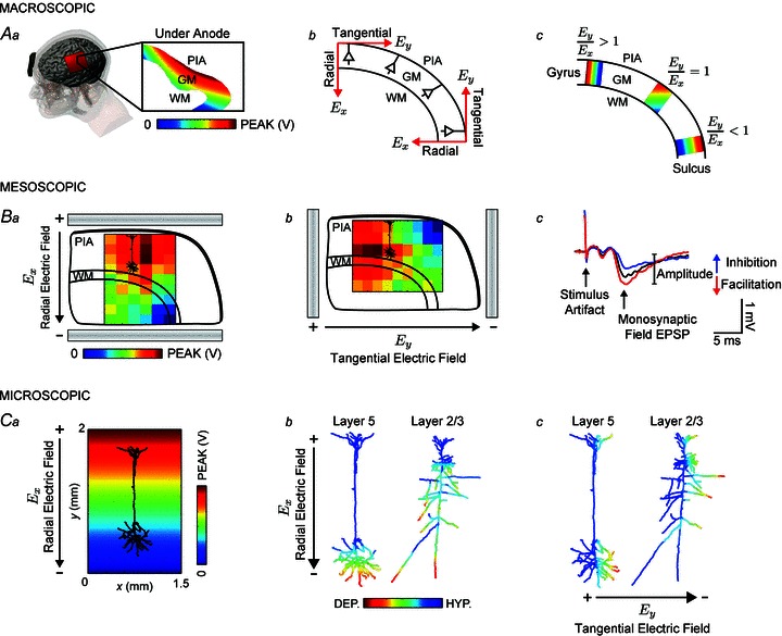
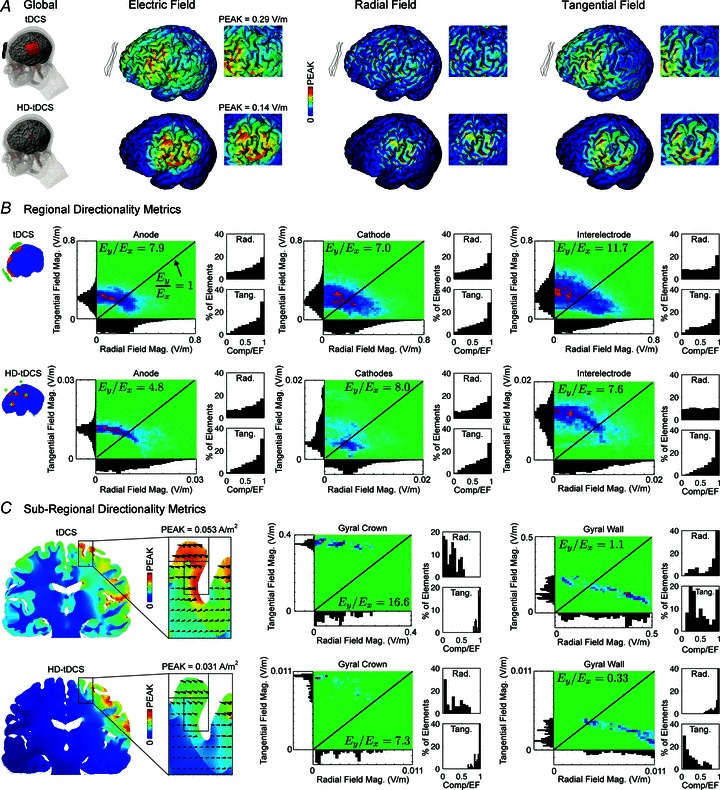
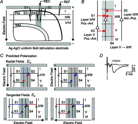
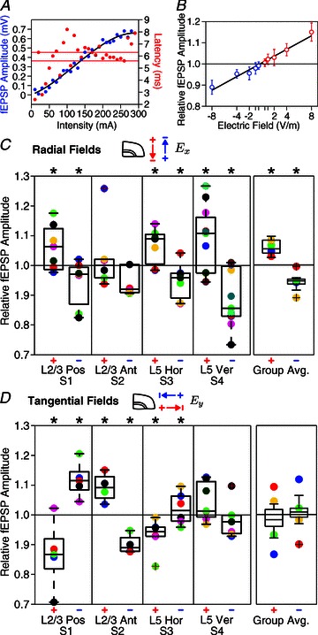
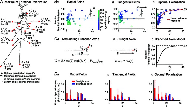
Transcranial direct current stimulation (tDCS) is a non-invasive brain stimulation technique to modulate cortical excitability. Although increased/decreased excitability under the anode/cathode electrode is nominally associated with membrane depolarization/hyperpolarization, which cellular compartments (somas, dendrites, axons and their terminals) mediate changes in cortical excitability remains unaddressed. Here we consider the acute effects of DCS on excitatory synaptic efficacy. Using multi-scale computational models and rat cortical brain slices, we show the following. (1) Typical tDCS montages produce predominantly tangential (relative to the cortical surface) direction currents (4-12 times radial direction currents), even directly under electrodes. (2) Radial current flow (parallel to the somatodendritic axis) modulates synaptic efficacy consistent with somatic polarization, with depolarization facilitating synaptic efficacy. (3) Tangential current flow (perpendicular to the somatodendritic axis) modulates synaptic efficacy acutely (during stimulation) in an afferent pathway-specific manner that is consistent with terminal polarization, with hyperpolarization facilitating synaptic efficacy. (4) Maximal polarization during uniform DCS is expected at distal (the branch length is more than three times the membrane length constant) synaptic terminals, independent of and two-three times more susceptible than pyramidal neuron somas. We conclude that during acute DCS the cellular targets responsible for modulation of synaptic efficacy are concurrently somata and axon terminals, with the direction of cortical current flow determining the relative influence.


Figures

Figure 1
Figure 1. Multi-scale methods and outcome measures of uniform EF directionality and effects
Aa, gyri-precise FEMs of current flow during tDCS indicate a uniform voltage gradient in cortical grey matter (GM) directly under the anode. Ab, the induced EF in the cortex can be decomposed into a radial component (Ex) that is parallel to the somatodendritic axis, and a tangential component (Ey) that is orthogonal to the somatodendritic axis. Ac, we quantified the relative occurrence of radial and tangential fields in cortical GM expressed as the ratio of the average of the field magnitude in the tangential direction to the average of the field magnitude in the radial direction (Ey/Ex). Ba and b, the brain slice preparation was used to study the change in synaptic efficacy during a uniform radial or tangential field by recording evoked field potentials. The voltage gradient between parallel Ag–AgCl wires is superimposed on a schematic of a sagittal slice of the rat primary motor cortex. From the macroscopic to the mesoscopic scale we can approximate a uniform EF along the length of a neuron (compare voltage gradients in Aa and Ba). Bc, the field (f)EPSP provides a measure of synaptic efficacy through facilitation or inhibition of the response amplitude. Ca, compartment model simulations of morphologically reconstructed neocortical pyramidal neurons were used to provide a description of axon terminal polarization in a uniform EF. Cb, the polarization profile of a layer V pyramidal neuron in a radially directed uniform EF indicating soma depolarization (red) corresponds to apical dendrite hyperpolarization (blue). Layer II/III neurons have a more complex polarization profile with long processes reaching maximal depolarization independent of the neuronal body. Cc, neurons in a uniform EF directed tangential to the somatodendritic axis preferentially affect processes that are oriented along the tangential field. WM, white matter.
Figure 2
Figure 2. Forward model of transcranial direct current stimulation (tDCS) and high-definition (HD)-tDCS quantifying electric field (EF) direction metrics
During tDCS, current may be dominantly tangential (along the cortical surface) rather than radial, even in brain regions directly under the electrodes. A, MRI-derived FEMs of current distribution in a gyri-precise head model are used to quantify the relative occurrence of radial (normal to the cortical surface) and tangential (along the cortical surface) components of the EF. Both conventional (top) and HD (bottom) tDCS montages produce radial (Ex, normal to the cortical surface) and tangential current (Ey, along the cortical surface) indicated by the global EF distribution (V m−1) across the head. In the HD-tDCS montage, current is focalized within the ring configuration (inset) with radial currents under the centre electrode and tangential currents between the surround electrodes. Qualitative comparison of the EF components indicates greater radial field magnitudes in the gyral wall and greater tangential field magnitudes in the gyral crown (compare insets). B, regionally, the distribution of field component magnitudes indicates prevailing tangential currents under the anode, cathode and between electrodes as described by the ratio of tangential to radial field magnitude (Ey/Ex ratio, see Methods). However, most elements have both radial and tangential components, and the isolated highest EFs are radial. The tangential and radial component for individual elements is shown for each subregion in false colour density plots, which show relative occurrence (relative density from absent (green) to maximal (red)). Axis histograms show the relative distribution of elements with a given tangential or radial component EF. Inset histograms describe the distribution of the percentage of elements in a region as a function of the normalized component magnitude (such that 1 indicates elements with dominant radial or tangential component). C, cortical folding further influences the distribution of the EF, therefore, subregional field component distributions are indicated for a gyral crown and wall. Tangential fields are dominant in magnitude in the gyral crown but are weaker in the walls where radial magnitudes are stronger, as observed in A.
Figure 3
Figure 3. Electrophysiology of direction-specific uniform DC EFs in synaptic pathways of the rat motor cortex
A, schematic of electrophysiology setup where uniform extracellular EFs were generated in all experiments by passing constant current across parallel Ag–AgCl wires positioned in the bath across the slice. Activity was monitored in layer II/III or layer V with a glass microelectrode. An additional field electrode (REF) was positioned in an iso-potential to remove the uniform field artifact. Activity was evoked with a bipolar stimulating electrode (S1–S4) positioned 500 μm from the recording electrode in either layer II/III or layer V targeting one of four distinct synaptic pathways corresponding to different orientations of afferent axons: posterior horizontal layer II/III (S1); anterior horizontal layer II/III (S2); posterior horizontal layer V (S3); and vertical layer V to II/III (S4). B, diagram summarizing the primary synaptic circuits in this study. Line thicknesses and diameters of the filled circles, which represent synapses, are correlated with the strength of the synaptic input. C, schematic of the expected polarization in distinct synaptic pathways exposed to radial and tangential fields. Somas, dendrites, axons and axon terminals are depolarized (red), hyperpolarized (blue) or not affected (black) by DC fields. D, characteristic fEPSP and field spike waveforms from the layer V pathway. The fEPSPs, but not earlier field spike, were suppressed by the non-NMDA receptor antagonist 6,7-dinitroquinoxaline-2,3-dione (DNQX). WM, white matter.
Figure 4
Figure 4. Modulation of pathway-specific synaptic efficacy by radial and tangential DC fields
Application of DC currents in cortical slice demonstrates that tangential currents are as effective in modulating pathway-specific synaptic efficacy as radial currents, though pathway-average effects result only for radial EFs. A, input–output curve of field (f)EPSP response amplitude and peak latency in the horizontal layer V pathway. Horizontal grey bars indicate the 25th and 75th percentiles of fEPSP peak latency. B, relative fEPSP amplitude in the vertical layer V to II/III pathway at different radially oriented EF intensities (correlation coefficient R2 of linear fit = 0.96). The fEPSP waveform inset shows a characteristic change in fEPSP amplitude with positive (+8 V m−1, red) and negative radial fields (−8 V m−1, blue) from control (no field, black). C, fEPSP responses are significantly (*P < 0.05) facilitated with +8 V m−1 fields (left) and reduced with −8 V m−1 (right) in three pathways. In each pathway, individual slice averages are indicated with coloured circles. Grouped average fEPSP amplitudes across all synaptic pathways indicate a 7% polarity-specific modulation of synaptic efficacy with 8 V m−1 radial fields. Circles in the grouped average represent the across slice means of distinct pathways (blue, red, green and yellow are S1, S2, S3 and S4 pathways, respectively). D, fEPSP amplitude was significantly modulated by tangential EFs in all three horizontal pathways but with direction sensitivity and not in the vertical pathway, all consistent with terminal polarization. Although tangential fields affected individual pathways, the grouped average of fEPSP amplitudes across all pathways was not significant.
Figure 5
Figure 5. Terminal polarization by uniform DC EFs using neuron compartment model and analytical/hybrid approximations
The maximum terminal polarization (Vt) depends on the length (ℓ) of the last axonal branch and becomes uncoupled from the bend point at distant terminals (ℓ > 3λ); however, for short branches the terminal membrane potential is coupled with the membrane potential at the bend (V0). In all cases, numerical simulations applied 1 V m−1 EFs. A, for a typical cortical pyramidal neuron, the maximum terminal polarization (Vt) is plotted with the corresponding optimal polarization angle (θ) of the branch relative to the EF and the length (ℓ) of the terminating axon branch. Ba and b, relative terminal polarization (Vt normalized by the axonal length constant and by the EF) as a function of branch electrotonic length and angle (circle colour). Bc, considering the optimal polarization angle, the relative polarization asymptotically approaches magnitude 1 with branch length (equivalently, terminal polarization reaches the maximal polarization Eλ for increasing lengths). Ca and b, schematic of a branched and straight axon in a uniform EF with analytical solutions (see Methods). The straight axon is a special case of the branched axon with infinite final branch length. For long branches, where ℓ≫λ, the terminal membrane potential becomes independent of the branch point and approaches Eλ cos θ. Cc, the branched axon model approaches maximal terminal polarization Eλ for ℓ > 3λ. D, error of approximations (analytical versus numerical estimates) for branched (blue) and straight (red) axons.

Similar articles

Cited by

References

    1. Anastassiou CA, Montgomery SM, Barahona M, Buzsaki G, Koch C. The effect of spatially inhomogeneous extracellular electric fields on neurons. J Neurosci. 2010;30:1925–1936. - PMC - PubMed
    1. Andreasen M, Nedergaard S. Dendritic electrogenesis in rat hippocampal CA1 pyramidal neurons: functional aspects of Na+ and Ca2+ currents in apical dendrites. Hippocampus. 1996;6:79–95. - PubMed
    1. Antal A, Varga ET, Kincses TZ, Nitsche MA, Paulus W. Oscillatory brain activity and transcranial direct current stimulation in humans. Neuroreport. 2004;15:1307–1310. - PubMed
    1. Arlotti M, Rahman A, Minhas P, Bikson M. Axon terminal polarization induced by weak uniform DC electric fields: a modeling study. Conf Proc IEEE Eng Med Biol Soc. 2012;2012:4575–4578. - PubMed
    1. Aroniadou VA, Keller A. The patterns and synaptic properties of horizontal intracortical connections in the rat motor cortex. J Neurophysiol. 1993;70:1553–1569. - PubMed

Publication types