. 2013 May;33(10):2016-28.
doi: 10.1128/MCB.00038-13. Epub 2013 Mar 11.

Distinct phospholipase C-β isozymes mediate lysophosphatidic acid receptor 1 effects on intestinal epithelial homeostasis and wound closure

Affiliations

Distinct phospholipase C-β isozymes mediate lysophosphatidic acid receptor 1 effects on intestinal epithelial homeostasis and wound closure

Sei-Jung Lee et al. Mol Cell Biol. 2013 May.

Abstract

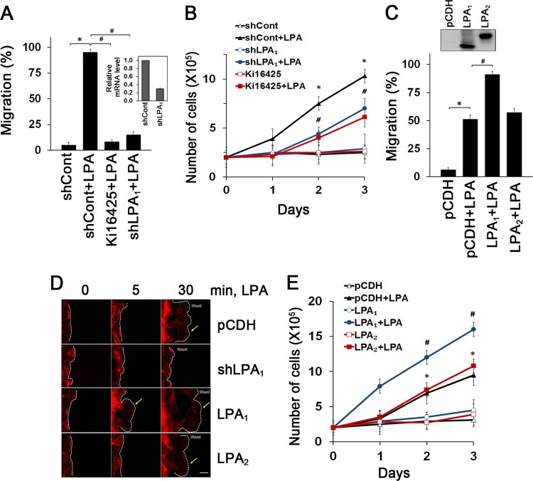
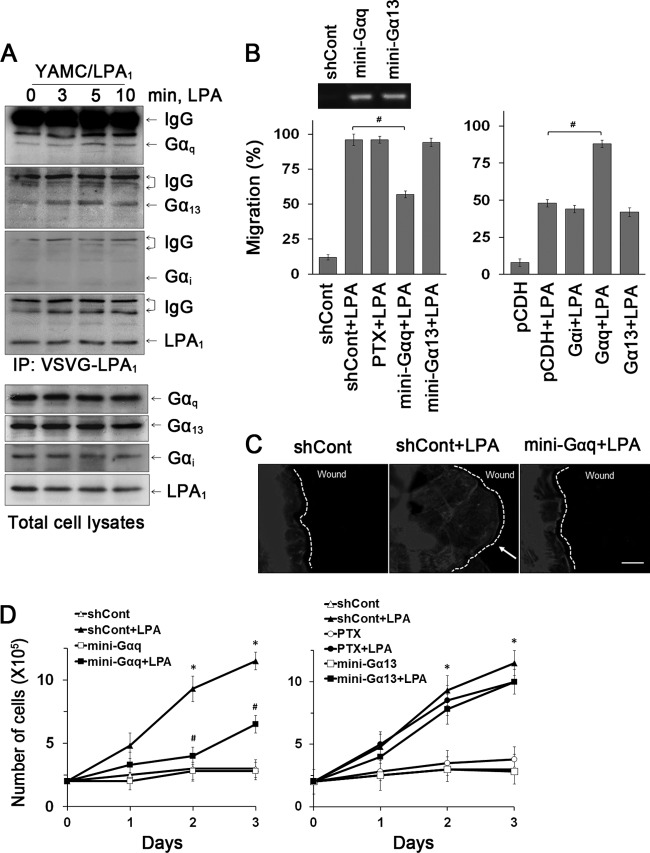
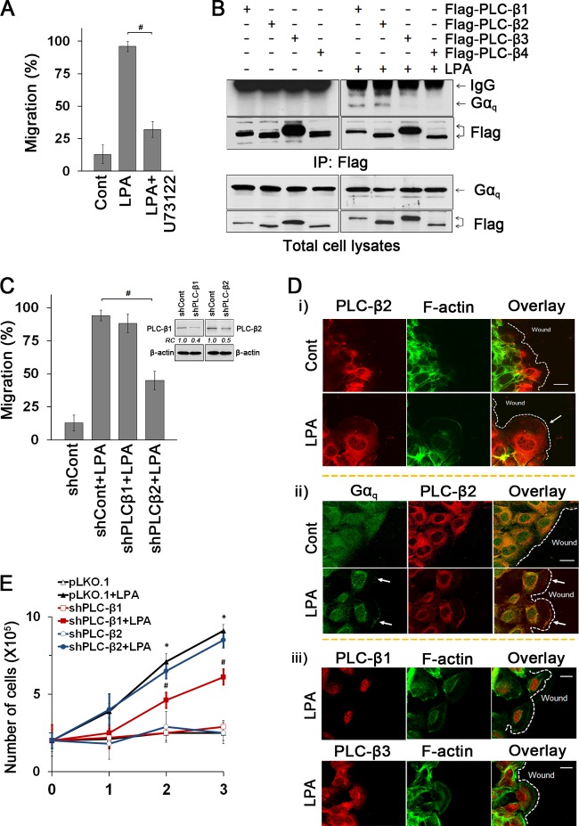
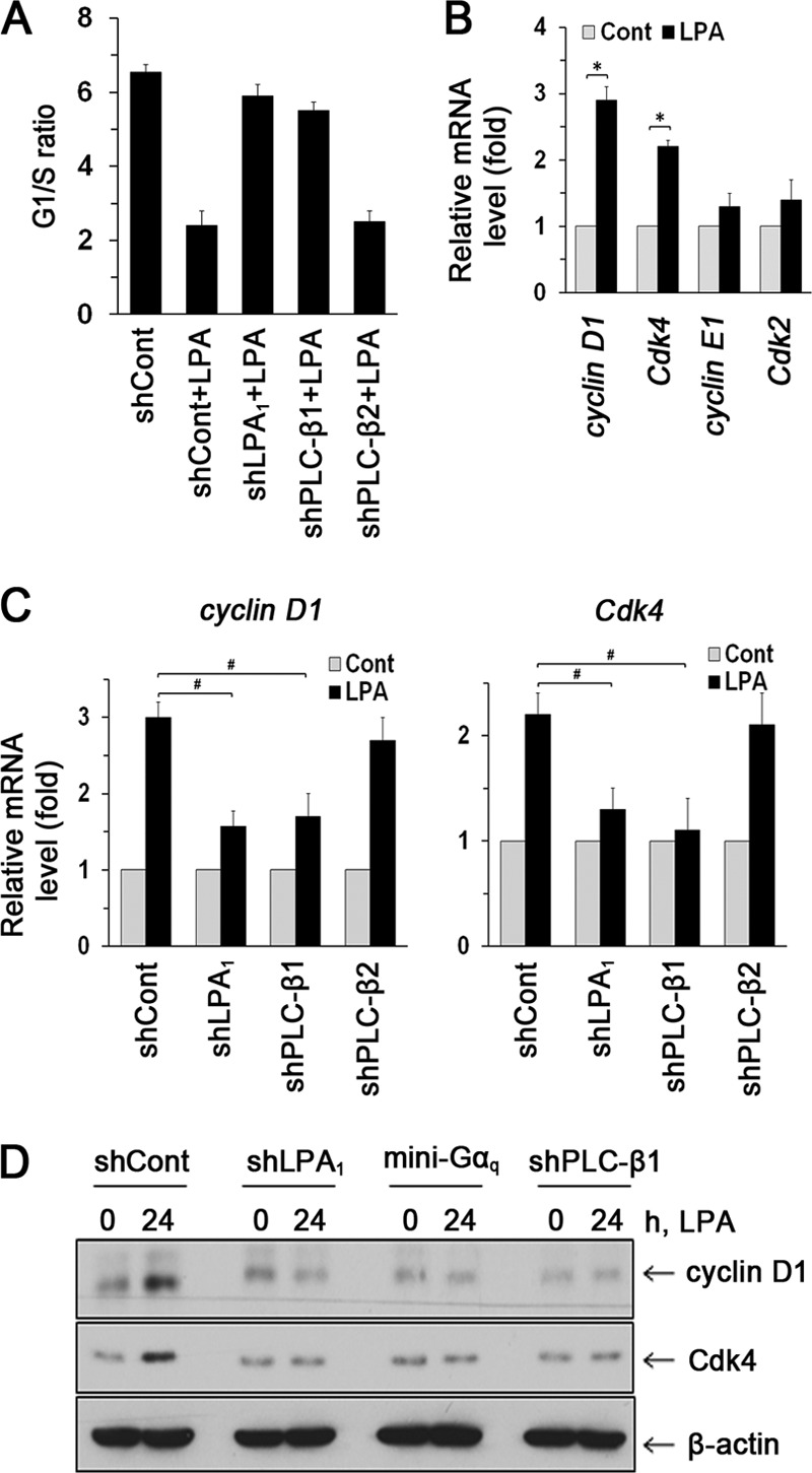
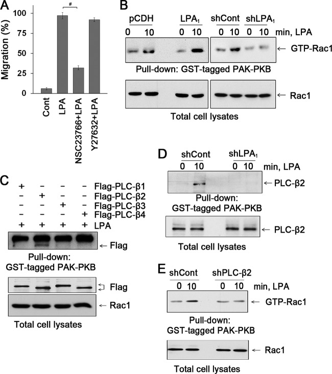
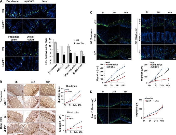
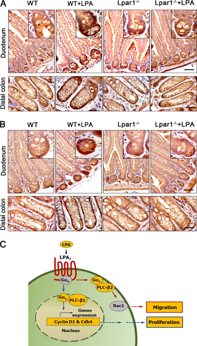
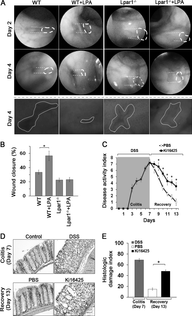
Maintenance of the epithelial barrier in the intestinal tract is necessary to protect the host from the hostile luminal environment. Phospholipase C-β (PLC-β) has been implicated to control myriad signaling cascades. However, the biological effects of selective PLC-β isozymes are poorly understood. We describe novel findings that lysophosphatidic acid (LPA) regulates PLC-β1 and PLC-β2 via two distinct pathways to enhance intestinal epithelial cell (IEC) proliferation and migration that facilitate wound closure and recovery of the intestinal epithelial barrier. LPA acting on the LPA1 receptor promotes IEC migration by facilitating the interaction of Gαq with PLC-β2. LPA-induced cell proliferation is PLC-β1 dependent and involves translocation of Gαq to the nucleus, where it interacts with PLC-β1 to induce cell cycle progression. An in vivo study using LPA1-deficient mice (Lpar1(-/-)) shows a decreased number of proliferating IECs and migration along the crypt-luminal axis. Additionally, LPA enhances migration and proliferation of IECs in an LPA1-dependent manner, and Lpar1(-/-) mice display defective mucosal wound repair that requires cell proliferation and migration. These findings delineate novel LPA1-dependent lipid signaling that facilitates mucosal wound repair via spatial targeting of distinct PLC-βs within the cell.


Figures

Fig 1
Fig 1
Enhanced intestinal epithelial cell migration and proliferation are LPA1 dependent. (A) Migration of YAMC cells into the nude area at 24 h of 1 μM LPA treatment is shown. Cells were pretreated with 10 μM Ki16425 or transfected with shLPA1 prior to LPA treatment. The extent of wound closure was quantified. Full recovery of the wound was considered 100%. *, P < 0.001; #, P < 0.001. n = 3. BSA (0.1%) was used as a control. The inset shows LPA1 knockdown efficacy determined by real-time RT-PCR (shLPA1; 69% decrease). (B) LPA-induced cell proliferation was determined by counting cells for up to 3 days. Cells were pretreated with Ki16425 or transfected with shLPA1 as indicated. *, P < 0.01 versus shCont; #, P < 0.01 versus shCont plus LPA. Error bars represent the means ± SEM from three independent experiments involving triplicates. (C) The extent of migration at 12 h of LPA treatment was determined. *, P < 0.01; #, P < 0.01. n = 4. The inset shows overexpression of LPA1 and LPA2 in YAMC cells. (D) Lamellipodial extrusions in cells transfected with pCDH, shLPA1, LPA1, or LPA2 are shown. Cells were fixed and labeled with phalloidin-Alexa Fluor 568 to identify the leading edge of lamellipodia (arrows). Dashed lines indicate the leading edges. n = 3. Bar, 20 μm. (E) Numbers of proliferating cells transfected with LPA1 or LPA2 were determined. *, P < 0.01 versus pCDH; #, P < 0.01 compared with pCDH plus LPA. n = 3.
Fig 2
Fig 2
Gαq is essential for LPA1-mediated cell migration and proliferation. (A) The interaction of Gα subunits with LPA1 in YAMC/LPA1 was determined by coimmunoprecipitation of LPA1 and Gα subunits (top four panels). Expression of Gα subtypes in cell lysates is shown in the bottom panels. n = 3. (B) The role of Gα subtypes in cell migration was determined. YAMC cells pretreated with PTX or transfected with Gα minigenes were treated with LPA for 24 h (left panel). The inset shows the presence of Gα minigenes in transfected cells. The right panel shows the effect of Gα subunit overexpression on migration during 12 h of LPA treatment. #, P < 0.01. n = 3. Full recovery of the wound was considered 100%. #, P < 0.01. n = 3. (C) Lamellipodial extrusions labeled with phalloidin-Alexa Fluor 568 are shown. Dashed lines indicate the leading edges. n = 3. Bar, 20 μm. (D) Effects of inhibition of G proteins on proliferation are determined. (Left) Inhibition of Gαq. (Right) Inhibition of Gαi or Gα13. n = 3. *, P < 0.01 compared with shCont; #, P < 0.01 versus shCont plus LPA. n = 3.
Fig 3
Fig 3
LPA1 mediates cell migration and proliferation via distinct PLC-β subtypes. (A) The PLC inhibitor U73122 blocked LPA-induced migration. #, P < 0.001. n = 3. (B) Gαq coimmunoprecipitated with Flag-PLC-β1 and Flag-PLC-β2. Expression of Flag-PLC-βs and Gαq in cell lysates is shown in bottom panels. (C) Effects of PLC-β1 and PLC-β2 knockdown on cell migration were compared. #, P < 0.01. n = 3. The inset shows the efficacy of PLC-β1 or PLC-β2 knockdown as determined by Western blotting: shPLC-β1, 61% decrease; shPLC-β2, 51% decrease. (D) Cellular distributions of PLC-β2 and F-actin (i), PLC-β2 and Gαq (ii), and PLC-β1 and PLC-β3 (iii) in cells treated with LPA or carrier for 30 min are shown. Arrows indicate lamellipodia. Dashed lines show the leading edges. n = 3. Bars, 20 μm. (E) Effects of PLC-β knockdown on proliferation were determined. *, P < 0.01 versus shCont; #, P < 0.01 versus shCont plus LPA. n = 3.
Fig 4
Fig 4
LPA1 regulates cell cycle progression. (A) G1/S ratios in cells treated with LPA are shown. n = 3. (B) Expression levels of cyclin D1, Cdk4, cyclin E1, and Cdk2 were determined by real-time RT-PCR. n = 3. *, P < 0.01. (C) Expression levels of cyclin D1 and Cdk4 mRNA are shown. n = 3. #, P < 0.01. (D) Expression levels of cyclin D1 and Cdk4 were determined by Western blotting. Representative blots from three independent experiments are shown.
Fig 5
Fig 5
LPA increased the nuclear abundance of Gαq and PLC-β1. (A) Expression of Gαq (green) and PLC-β1 (red) in the nuclei of YAMC cells was assessed by confocal immunofluorescence microscopy. TO-PRO iodide was used for nuclear counterstaining (blue). n = 3. Bars, 20 μm. Graphs represent Pearson's coefficient of colocalization of Gαq and TO-PRO (left), PLC-β1 and TO-PRO (middle), and Gαq and PLC-β1 (right) from 10 independent fields of cells. #, P < 0.05. (B) Western blot of Gαq and PLC-β1 in nuclear and total cell extracts. The transcription factor Oct-1 was used as a loading control for nuclear proteins. n = 3.
Fig 6
Fig 6
LPA induces the interaction between PLC-β2 and Rac1. (A) Effects of NSC23766 and Y-27632 on cell migration were determined by wound closure assays. n = 3. #, P < 0.01. (B) The effect of LPA on activation of Rac1 was determined. Cells were treated with LPA for 10 min, and activated Rac1 was isolated with GST-PAK-PBD domain. n = 3. (C) The interaction of PLC-β isozymes with activated Rac1 was determined. Flag-PLC-βs were copurified with activated Rac1 in GST-PAK-PBD pulldown assays. The bottom panels show PLC-βs and Rac1 in cell lysates. n = 4. (D) Knockdown of LPA1 attenuated the interaction between PLC-β2 and activated Rac1. n = 3. (E) Knockdown of PLC-β2 attenuated activation of Rac1 by LPA. n = 3.
Fig 7
Fig 7
Lpar1−/− mice display decreased numbers of proliferating cells and impeded cell migration. (A) Proliferating IECs of WT and Lpar1−/− mice were identified by EdU staining (green). DAPI (4′,6-diamidino-2-phenylindole) was used for nuclear counterstaining (blue). The mean numbers of EdU-labeled cells per crypt are shown in the bar graph. n = 8 per group. Bars, 100 μm. *, P < 0.05. (B) Migration of proliferating IECs along the crypt-villus axis was determined by BrdU pulse-chase for indicated times in duodenum (top) and in distal colon (bottom). The extent of cell migration was quantified by measuring the distance between the crypt base and the highest labeled cell along the crypt-villus axis. Bars, 100 μm. n = 8. *, P < 0.01 versus the WT. (C) Migration of IECs was determined in mice given an i.p. injection of Ki16425 or LPA orally for 5 days. Time-dependent migration of BrdU-positive cells (green) in WT duodenum (left) and distal colon (right) are shown. n = 6. DAPI was used for nuclear counterstaining (blue). Bars, 50 μm. *, P < 0.01 versus the WT. (D) IEC migration in Lpar1−/− duodenum treated with LPA or not is shown. Bars, 50 μm. n = 8.
Fig 8
Fig 8
LPA stimulates the expression of Gαq and PLC-β1 in the proliferating crypt compartment of the intestinal tract. Intestinal sections of WT and Lpar1−/− mice treated with LPA were stained with anti-Gαq (A) or anti-PLC-β1 (B) antibody. n = 5. Bars, 50 μm. (C) A model of LPA1-dependent effects on IECs is shown.
Fig 9
Fig 9
LPA1 requires epithelial mucosal wound repair. (A) Colonic mucosal wounds were induced in WT and Lpar1−/− mice by mouse colonoscopy biopsy. Mice were given LPA or carrier by gavage for 4 days. Representative colonoscopy images of colonic mucosal wound healing at day 2 and day 4 after biopsy-induced injury are shown. Close-up images of colonic mucosa wounds at day 4 are shown below. (B) Quantification of wound repair (means ± SEM) relative to original wound size is shown in the graph below. n = 6 per group. *, P < 0.01. (C) WT mice were subjected to DSS for 7 days to induce acute colitis. At day 7, mice were divided into 2 groups (n = 8), with one group receiving Ki16425 (20 mg/kg) every day by i.p. injection and the other receiving PBS. Clinical disease activity indexes of mice are shown. n = 8. *, P < 0.01 versus PBS. (D) Representative colonic tissues stained with H&E are shown. n = 8. Bars, 100 μm. (E) Histologic damage index scores from whole mouse colons are shown. *, P < 0.01 versus PBS. n = 8.

References

    1. Crosnier C, Stamataki D, Lewis J. 2006. Organizing cell renewal in the intestine: stem cells, signals and combinatorial control. Nat. Rev. Genet. 7:349–359 - PubMed
    1. Choi JW, Herr DR, Noguchi K, Yung YC, Lee CW, Mutoh T, Lin ME, Teo ST, Park KE, Mosley AN, Chun J. 2010. LPA receptors: subtypes and biological actions. Annu. Rev. Pharmacol. Toxicol. 50:157–186 - PubMed
    1. Mutoh T, Rivera R, Chun J. 2012. Insights into the pharmacological relevance of lysophospholipid receptors. Br. J. Pharmacol. 165:829–844 - PMC - PubMed
    1. Sturm A, Sudermann T, Schulte KM, Goebell H, Dignass AU. 1999. Modulation of intestinal epithelial wound healing in vitro and in vivo by lysophosphatidic acid. Gastroenterology 117:368–377 - PubMed
    1. Lin S, Yeruva S, He P, Singh AK, Zhang H, Chen M, Lamprecht G, de Jonge HR, Tse M, Donowitz M, Hogema BM, Chun J, Seidler U, Yun CC. 2010. Lysophosphatidic acid stimulates the intestinal brush border Na(+)/H(+) exchanger 3 and fluid absorption via LPA(5) and NHERF2. Gastroenterology 138:649–658 - PMC - PubMed

Publication types

MeSH terms