. 2013 May;33(10):2004-15.
doi: 10.1128/MCB.01603-12. Epub 2013 Mar 11.

Human DEAD box helicase 3 couples IκB kinase ε to interferon regulatory factor 3 activation

Affiliations

Human DEAD box helicase 3 couples IκB kinase ε to interferon regulatory factor 3 activation

Lili Gu et al. Mol Cell Biol. 2013 May.

Abstract

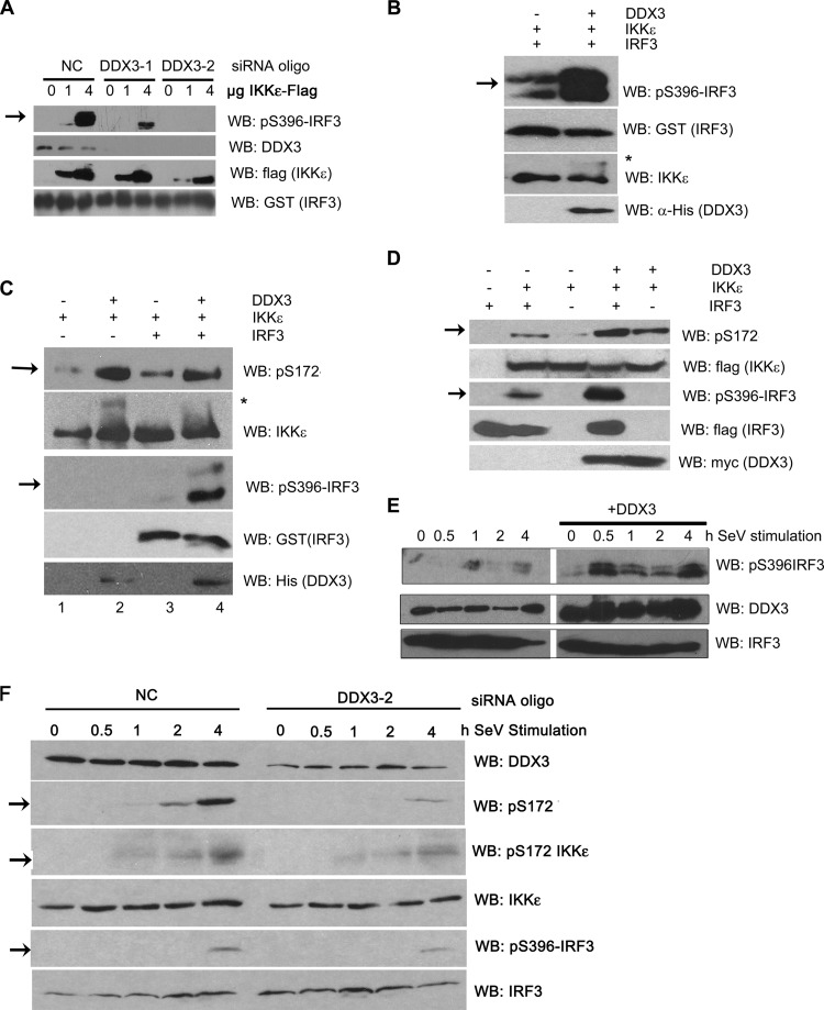
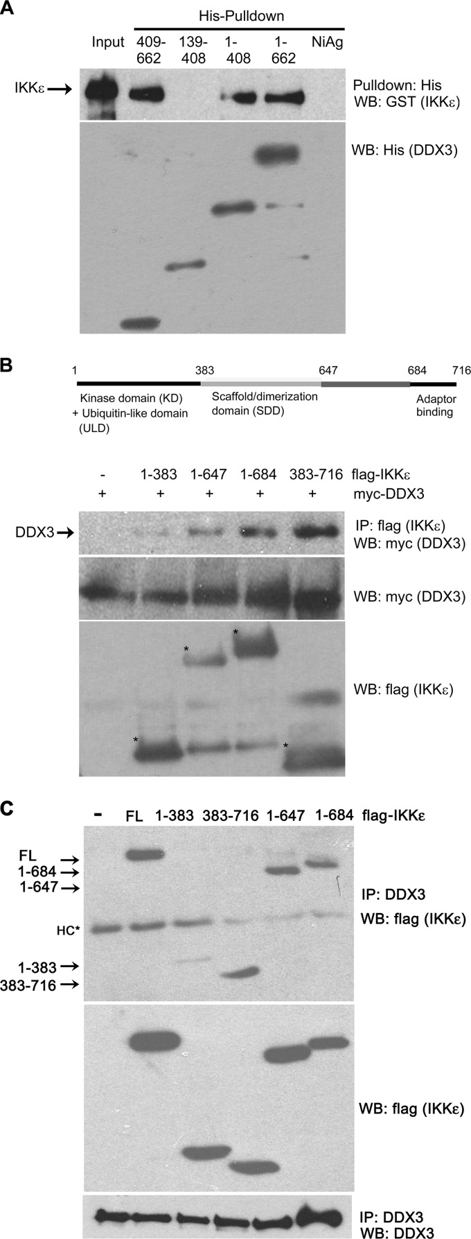
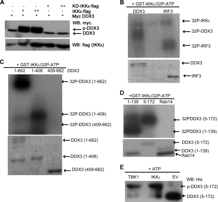
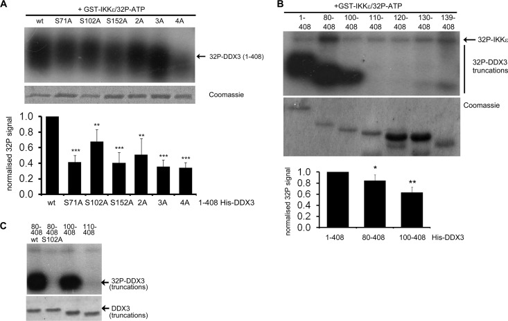
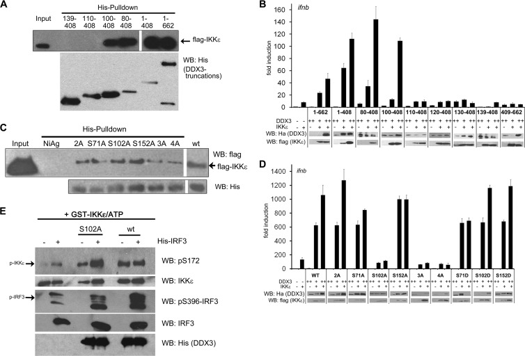
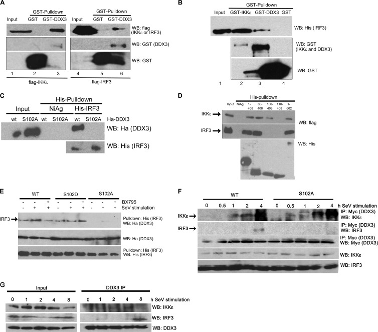
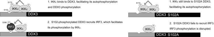
The human DEAD box protein 3 (DDX3) has been implicated in different processes contributing to gene expression. Interestingly, DDX3 is required as an essential host factor for the replication of HIV and hepatitis C virus (HCV) and is therefore considered a potential drug target. On the other hand, DDX3 interacts with IκB kinase ε (IKKε) and TANK-binding kinase 1 (TBK1) and contributes to the induction of antiviral type I interferons (IFNs). However, the molecular mechanism by which DDX3 contributes to IFN induction remains unclear. Here we show that DDX3 mediates phosphorylation of interferon regulatory factor 3 (IRF3) by the kinase IKKε. DDX3 directly interacts with IKKε and enhances its autophosphorylation and activation. IKKε then phosphorylates several serine residues in the N terminus of DDX3. Phosphorylation of DDX3 at serine 102 (S102) is required for recruitment of IRF3 to DDX3, facilitating its phosphorylation by IKKε. Mutation of S102 to alanine disrupted the interaction between DDX3 and IRF3 but not that between DDX3 and IKKε. The S102A mutation failed to enhance ifnb promoter activation, suggesting that the DDX3-IRF3 interaction is crucial for this effect. Our data implicates DDX3 as a scaffolding adaptor that directly facilitates phosphorylation of IRF3 by IKKε. DDX3 might thus be involved in pathway-specific activation of IRF3.


Figures

Fig 1
Fig 1
DDX3 enhances activation of IKKε and IRF3. (A) HEK293T cells were transfected with siRNAs against DDX3 (DDX3-1 and DDX3-2) or a negative-control oligonucleotide (NC). After 24 h, cells were transfected with a plasmid vector for Flag-IKKε or with an empty vector. Flag-IKKε was immunoprecipitated, and in vitro kinase assays were performed using recombinant GST-IRF3 (aa 380 to 427) as a substrate. IRF3 phosphorylation was detected with a phospho-specific antibody against serine 396 by Western blot (WB) analysis. (B and C) In vitro kinase assays were carried out with recombinant GST-IKKε and IRF3. Recombinant His-DDX3 was added where indicated. Phosphorylation of IKKε and IRF3 was detected with phospho-specific antibodies against S172 in IKKε and S396 in IRF3. The asterisk indicates a higher-molecular-weight band of IKKε. (D and E) HEK293T cells were transfected with expression constructs for myc-DDX3 and Flag-IRF3. (D) Cells were cotransfected with a Flag-IKKε construct or empty vector. (E) Cells were stimulated with SeV 24 h after transfection for the indicated length of time. (F) A549 cells were transfected with an siRNA oligonucleotide against DDX3 (DDX3-2) or a negative-control oligonucleotide (NC). After 48 h, cells were stimulated with SeV for the indicated length of time. Cell lysates were subjected to SDS-PAGE and WB analysis.
Fig 2
Fig 2
DDX3 directly interacts with the SDD domain of IKKε. (A) Purified recombinant full-length His-DDX3 or the indicated His-DDX3 truncation mutants were incubated with recombinant GST-IKKε. Following pulldown with nickel (Ni)-agarose, interacting proteins were subjected to SDS-PAGE and WB analysis. (B and C) HEK293T cells were transfected with constructs for Flag-IKKε or the indicated Flag-IKKε truncation mutants and Myc-DDX3 (panel B only). Cell lysates were subjected to immunoprecipitation (IP) with an anti-Flag (B) or anti-DDX3 (C) antibody, followed by SDS-PAGE and WB analysis. Asterisks mark bands of the correct molecular weight for the IKKε truncations (B). HC, antibody heavy chain band (C).
Fig 3
Fig 3
The N terminus of DDX3 is a direct phosphorylation target for IKKε. (A) HEK293T cells were transfected with constructs for Myc-DDX3 and 100 ng (+) or 250 ng (++) of a Flag-IKKε construct or 1,000 ng (+) or 1,800 ng (++) of a construct for a kinase-dead (KD) Flag-IKKε mutant (K38A). Cell lysates were subjected to SDS-PAGE and WB analysis. (B to D) For in vitro kinase assays, recombinant GST-IKKε was incubated with recombinant substrate in the presence of [γ-32P]ATP. Samples were then subjected to SDS-PAGE and autoradiograph analysis showing incorporation of [γ-32P]ATP. Total amounts of recombinant proteins were visualized by Coomassie staining. Substrates for IKKε were full-length DDX3 or full-length IRF3 (B), full-length His-DDX3 or the indicated His-DDX3 truncation constructs (C), or recombinant His-DDX3(1–139) or His-DDX3(5–172) or the unrelated control protein His-Rab14 (D). (E) In vitro kinase assays were carried out as described above but in the presence of unlabeled ATP. This was followed by SDS-PAGE and WB analysis with an anti-His antibody.
Fig 4
Fig 4
DDX3 is phosphorylated by IKKε on multiple sites. (A) Recombinant GST-IKKε was incubated with recombinant wild-type (wt) His-DDX3(1–408) or the indicated DDX3(1–408) alanine mutants in the presence of [γ-32P]ATP. Samples were then subjected to SDS-PAGE and autoradiograph analysis. Total amounts of His-DDX3 mutants were visualized by Coomassie staining. (B) The assay was carried out as described for panel A but using the N-terminal truncation mutants of DDX3 as substrates. (A and B) The top panel shows results of one representative experiment out of four. For each repeat experiment, the intensity of autoradiograph and Coomassie-stained bands was quantified using ImageJ software. Values for autoradiograph bands were normalized to the intensity of the corresponding Coomassie-stained bands to account for differences in protein loading. The normalized signal for DDX3(1–408) was set to 1 in each case. Data in the lower panel are presented as means and standard deviations from four independent experiments. *, P < 0.05; **, P < 0.01; ***, P < 0.001 [all compared to wt DDX3(1–408)] by an unpaired Student's t test. (C) The assay was carried out as described for panel B but including the S102A mutant of DDX3(80–408).
Fig 5
Fig 5
IKKε binding and serine 102 are required for the effect of DDX3 on the ifnb promoter. (A) Recombinant His-DDX3 or His-DDX3 truncation mutants were incubated with cell lysates containing Flag-IKKε. Following pulldown, interacting proteins were subjected to SDS-PAGE and Western blot (WB) analysis. The black border indicates images originate from the same autoradiography film and can be compared in intensity. (B) HEK293 cells were transfected with an ifnb promoter reporter gene construct and expression constructs for Flag-IKKε and HA-DDX3 or HA-DDX3 truncations. Expression of HA-DDX3 and Flag-IKKε was confirmed by WB analysis. Data for reporter gene assays are expressed as mean fold induction relative to control levels and standard deviations. Shown are results of one representative experiment out of four, performed in triplicate. (C) The assay was performed as described for panel A but using His-tagged alanine mutants of DDX3(1–408). Black borders indicate images that originated from the same autoradiography film and can be compared in intensity. (D) The assay was performed as described for panel B but testing full-length HA-tagged alanine mutants of DDX3. (E) Recombinant GST-IKKε was incubated with ATP, GST-IRF3 (aa 380 to 427), and His-DDX3(1–408) or the S102A mutant of His-DDX3(1–408). Samples were then subjected to SDS-PAGE and WB analysis. Phosphorylation of IKKε and IRF3 was detected with phospho-specific antibodies against pS172 (IKKε) and pS396 (IRF3).
Fig 6
Fig 6
DDX3, but not the S102A mutant, interacts directly with IRF3. (A) Recombinant GST-DDX3 or GST was incubated with cell lysates containing Flag-IKKε or Flag-IRF3. Following pulldown, interacting proteins were subjected to SDS-PAGE and Western blot (WB) analysis. (B) Recombinant GST-DDX3, GST-IKKε, or GST was incubated with recombinant His-IRF3. Following pulldown, interacting proteins were subjected to SDS-PAGE and WB analysis. (C) Recombinant His-IRF3 was incubated with cell lysates containing HA-DDX3 or HA-DDX3 S102A. Following pulldown, interacting proteins were subjected to SDS-PAGE and WB analysis. (D) Recombinant His-DDX3 or His-DDX3 truncations were incubated with cell lysates containing Flag-IRF3 or Flag-IKKε. Following pulldown, interacting proteins were subjected to SDS-PAGE and WB analysis. (E) HEK293T cells were transfected with constructs for wild-type (WT), S102D, or S102A HA-DDX3. After 24 h, cells were stimulated with SeV for 2 h. BX795 was added 1 h before SeV stimulation (50 ng/ml). Cell lysates were used for pulldowns with recombinant His-IRF3 and complexes were subjected to SDS-PAGE and WB analysis. (F) A549 cells were transfected with constructs for either Myc-DDX3 (WT) or the Myc-DDX3 S102A mutant and stimulated with SeV as indicated. Myc-DDX3 was immunoprecipitated (IP), followed by SDS-PAGE and WB analysis. (G) A549 cells were stimulated with SeV for the indicated length of time; cell lysates were then subjected to IP with an anti-DDX3 antibody, followed by SDS-PAGE and WB analysis. (A, B, and D) Black borders indicate images originating from the same autoradiography film, which can be compared in intensity.
Fig 7
Fig 7
DDX3 acts as a scaffolding adaptor for IKKε and IRF3. Our model for the sequential mechanism by which DDX3 facilitates IKKε (1) and IRF3 (2) activation is depicted here. DDX3 first recruits IKKε, which stimulates IKKε autophosphorylation and activation. In this step, the N-terminal tail of DDX3 is phosphorylated by IKKε on multiple sites, including serine 102. The phosphorylation of S102 mediates recruitment of IRF3 to the DDX3-IKKε complex. IRF3 can subsequently be phosphorylated by IKKε and then contributes to ifnb promoter activation. On the right, the process is shown for the DDX3 mutant lacking S102. In this case, the physical and functional interaction with IKKε proceeds normally (1). However, due to the lack of S102 phosphorylation, IRF3 is not recruited to DDX3 and its phosphorylation is disrupted (2).

Similar articles

Cited by

References

    1. Schröder M. 2010. Human DEAD-box protein 3 has multiple functions in gene regulation and cell cycle control and is a prime target for viral manipulation. Biochem. Pharmacol. 79:297–306 - PubMed
    1. Ariumi Y, Kuroki M, Abe K, Dansako H, Ikeda M, Wakita T, Kato N. 2007. DDX3 DEAD-box RNA helicase is required for hepatitis C virus RNA replication. J. Virol. 81:13922–13926 - PMC - PubMed
    1. Randall G, Panis M, Cooper JD, Tellinghuisen TL, Sukhodolets KE, Pfeffer S, Landthaler M, Landgraf P, Kan S, Lindenbach BD, Chien M, Weir DB, Russo JJ, Ju J, Brownstein MJ, Sheridan R, Sander C, Zavolan M, Tuschl T, Rice CM. 2007. Cellular cofactors affecting hepatitis C virus infection and replication. Proc. Natl. Acad. Sci. U. S. A. 104:12884–12889 - PMC - PubMed
    1. Yedavalli VS, Neuveut C, Chi YH, Kleiman L, Jeang KT. 2004. Requirement of DDX3 DEAD box RNA helicase for HIV-1 Rev-RRE export function. Cell 119:381–392 - PubMed
    1. Oshiumi H, Sakai K, Matsumoto M, Seya T. 2010. DEAD/H BOX 3 (DDX3) helicase binds the RIG-I adaptor IPS-1 to up-regulate IFN-beta-inducing potential. Eur. J. Immunol. 40:940–948 - PubMed

Publication types

MeSH terms

LinkOut - more resources